Vertebrate Zoology: A Textbook - T. A. Dauda 2014
Multicellular Animals
Chordates
Subphylum Acrania
Acrania (cephalochordates) comprise a small group of primitive marine benthic chordate animals. The Main Features of this phylum are retained throughout their entire lifespan.
Representatives of Acrania lack a Skull, their neural tube is not differentiated into a Brain AND SPINAL cord, Sensory Organs are poorly developed, and the Circulatory system is closed, yet they lack a Heart. The Skeleton is represented by a notochord, which, along with the overlying neural tube, is enclosed in a Connective Tissue sheath. Paired limbs are absent.
The simple Structure of acraniates is directly related to their habitat and lifestyle. Most modern species inhabit shallow coastal marine waters. The majority of their life is spent burrowed into sandy bottom sediments, with only the anterior end of the body—bearing the oral hood—exposed. They are passive filter feeders, consuming plankton consisting of small swimming animals and plant organisms.
Class="center">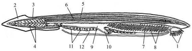
Fig. 3 Internal Structure of the lancelet (Branchiostoma lanceolatum):
1 — oral hood surrounded by tentacles; 2 — caudal fin; 3 — dorsal fin; 4 — Muscle segments (myomeres); 5 — neural tube; 6 — notochord; 7 — gill slits; 8 — gills; 9 — intestine; 10 — hepatic caecum; 11 — Gonads; 12 — atriopore / atrial cavity.
A typical representative of acraniates is the lancelet (Branchiostoma lanceolatum) (Fig. 3). The lancelet is a translucent animal up to 8 cm in length, possessing a fish-like body shape. A fin fold—the dorsal fin—runs along the dorsal side and extends around the posterior end to form a lanceolate caudal fin. At the anterior end, on its ventral side, lies the oral hood surrounded by 10–20 pairs of tentacles.
The integument of the lancelet is primitive, consisting of a single-layered outer epithelium and a thin layer of connective tissue, the corium.
The Skeleton of the lancelet is a notochord extending along the longitudinal axis of the body. The musculature is weakly differentiated and runs along the sides of the body in two bands, each divided by thin connective tissue partitions into a series of muscle segments.
The Central Nervous System is represented by a longitudinal, undifferentiated neural tube, from which dorsal and ventral nerves emerge in pairs. Photosensitive structures are distributed along the neural tube.
Thus, the entire neural cord Functions as a light-sensitive organ. The sensory Organs of the lancelet are primitive: true eyes are absent, meaning it cannot form visual images, and it also lacks auditory and equilibrium organs. Tactile Cells are scattered across the entire body.
The Digestive System begins with the oral hood surrounded by tentacles. At its base lies the Mouth, which leads into the Pharynx. The inner surface of the pharynx features dorsal and bottom ciliated grooves. Ciliary action moves food particles toward the intestine. The intestine is not subdivided into distinct regions. A large blind pouch—commonly referred to as the hepatic caecum—diverges from the ventral side of the anterior intestine.
The respiratory organs of the lancelet are primitive: gas exchange occurs via thin-walled branchial Blood Vessels bathed in Water. The gill slits open not directly to the exterior, but into a specialized peribranchial (atrial) cavity formed by the fusion of lateral Skin folds. Only at the posterior end do their margins remain free, forming an opening (atriopore) that connects the atrial cavity to the external environment. Ciliary action of the oral tentacles draws water into the pharynx, which then passes through the gill slits, bathes the gills, enters the atrial cavity, and is expelled to the outside through the atriopore.
The CIRCULATORY SYSTEM OF the lancelet is closed yet primitive, consisting of vessels carrying colorless blood. There is no heart. A ventral aorta pulsates beneath the pharynx, giving rise to branchial Arteries (Fig. 4). These pass through the walls between the gill slits, where gas exchange takes place. Oxygenated blood then flows via paired epibranchial vessels into the main arterial trunk—the dorsal aorta—which runs beneath the notochord along the body. Along its path, the dorsal aorta branches into vessels supplying Internal Organs, while anteriorly it gives off carotid arteries supplying the anterior region of the body. Blood from the front of the body is collected into paired jugular Veins running posteriorly, whereas blood from the posterior region is carried forward by paired cardinal veins.

Fig. 4 Diagram of the lancelet circulatory system:
1 — ventral aorta; 2 — bulbi of afferent branchial arteries; 3 — branchial arteries; 4 — roots of dorsal aorta; 5 — carotid arteries; 6 — dorsal aorta; 7 — caudal vein; 8 — subintestinal vein; 9 — hepatic portal system; 10 — peribranchial vein; 11 — anterior cardinal vein; 12 — posterior cardinal vein; 13 — Cuvierian duct.
The excretory system is represented by numerous tubular nephridia (up to 90 pairs) located in the pharyngeal region. One end of a nephridium opens into the peribranchial cavity, while the other features a series of openings connecting the excretory tubule to the coelom (body cavity). Within the walls of the body cavity, in close proximity to the nephridial tubules, lies a dense network of blood capillaries through which fluids containing Metabolic waste products are filtered.
Fluid is extracted from the lancelet's body cavity by the nephridial tubules and excreted to the exterior.
The reproductive organs (Ovaries and Testes) are located in the pharyngeal region in both males and females. Mature Gametes are released into the peribranchial cavity via temporary genital ducts and subsequently expelled to the outside through the atriopore. Fertilization is external, taking place in the bottom water layers, typically in the evening. Larval development lasts about three months.
About 30 species of lancelets are known. They find optimal living conditions primarily in temperate and warm seas of the Atlantic, Indian, and Pacific Oceans. Lancelets are also found in the Black Sea and the Sea of Japan, preferring shallow areas with water temperatures ranging from 17 to 30 °C.
Significance for humans. The lancelet body contains up to 80% protein and about 2% fat. Along the coasts of Southeast Asia, harvesting has long been practiced (up to 35 tons annually) by sifting sand carefully collected from the bottom surface. Lancelets are consumed boiled, fried, or dried. Although their practical economic value is minor, acraniates are exceptionally valuable as a model Organism for resolving scientific questions regarding THE ORIGIN OF vertebrates.
QUESTIONS FOR SELF-assessment and review:
1. What are the Characteristic Features of the Organization OF THE phylum Chordata?
2. What are the general Organizational features of acrania associated with their environmental conditions and lifestyle?
3. What is the external and internal structure of amphioxus?
4. What is The Significance of acrania for humans? ORIGIN AND EVOLUTION of lower Chordates
Lower chordates — tunicates and acrania — have not been preserved in the fossil record. However, comparative anatomical and embryological studies, particularly the works of A. O. Kovalevsky and A. N. Severtsov, provide strong evidence to suggest that the ancestors of lower chordates were restricted in mobility, crawling, bottom-dwelling, bilaterally symmetrical animals with a notochord spanning the entire length of the body, an unsegmented nervous system, and segmented musculature. They fed passively by filtering water.
Their further evolution apparently followed three distinct pathways. Individuals of one branch of primitive acrania, increasing their mobility and acquiring the capacity for active feeding, transitioned to a nektonic lifestyle and gave rise to vertebrates.
Representatives of the second branch retained a bottom-dwelling lifestyle, but developed adaptations for burrowing into the substrate and, consequently, underwent morphological simplification while maintaining passive feeding. As a result, this branch has survived to the present day in the form of acrania, which have successfully colonized sandy-bottom biocoenoses.
Finally, some primitive acrania apparently transitioned, already in the Cytology/cytology/16.html">Early stages of evolution, to a benthic yet sessile lifestyle on hard substrates. The formation of a tough tunic on the body surface protected these animals from most predators, while The Development of an efficient filtering apparatus ensured a food supply despite a stationary lifestyle. These adaptations arose through regressive evolution, resulting in the simplification of the adult anatomy (disappearance of the notochord and neural tube, reduction of Sense Organs, etc.). The presence of a motile, structurally more complex larva enabled ascidians to disperse, while the capacity for asexual reproduction (budding) allowed them to rapidly colonize newly available habitats. The success and proliferation of this ancient group are evidenced by the large number of living ascidians today.
Last update: 13/08/2026
Editorial and Educational Adaptation: This material has been compiled based on the primary/original source text. The project team performed an editorial review, corrected technical inaccuracies, structured sections, and adapted the content for an educational format.
What was processed:
- elimination of formatting defects (OCR errors, structural breaks, corrupted characters);
- editorial organization of content;
- standardization of terminology in accordance with academic sources;
- verification of factual statements against the original source text.
All mentions of the author, publication year, and origin of the primary text have been preserved in accordance with the source.