Pediatric Medical Genetics - S.I. Smiian 2003
Congenital malformations and hereditary diseases of the bronchopulmonary system in children
Congenital malformations of the lungs
Lung agenesis, aplasia, and hypoplasia. Agenesis (Fig. 14) is the absence of lung tissue with a rudimentary main bronchus present. By 1972, 200 cases of unilateral lung agenesis or aplasia (Fig. 15) had been described. Hypoplasia (Fig. 15) is a condition in which the main and lobar Bronchi terminate in a functionally imperfect rudiment, and the lung tissue remains underdeveloped.
Class="center">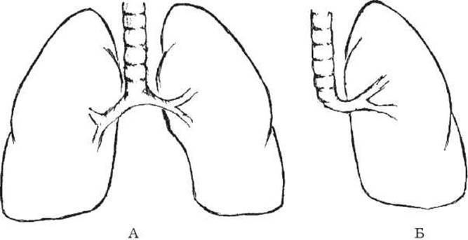
Fig. 14. Lung agenesis.
Diagram of a normal (A) and agenetic (B) lung.
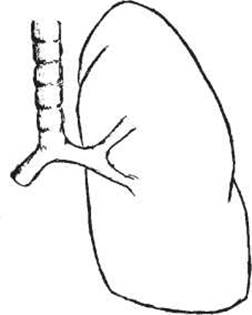
Fig. 15. Diagram of right lung aplasia.

Fig. 16. Diagram of right lung hypoplasia.
Clinical presentation. This pathology rarely runs an asymptomatic course. Children lag behind in physical development, exhibiting respiratory failure characterized by dyspnea and varying degrees of cyanosis. Cough and purulent sputum production are associated with The Development of an inflammatory process. Chest pain occurs occasionally. Other observations include flattening of one half of the chest and Scoliosis with convexity toward the healthy side. In the area of the defect, Percussion sound is shortened, and respiratory sounds are absent or diminished. The Heart is displaced toward the affected side to such an extent that dextrocardia may be erroneously diagnosed. In pronounced Hypoxia, clubbing of the terminal Phalanges of the fingers («drumstick» fingers) is observed. Hypoplasia presents with a less vivid clinical picture. The process proceeds similarly to chronic Pneumonia, with decreased VC and TLC. Radiography reveals a decreased chest volume on the affected side, accompanied by intense opacification, high standing of the Diaphragm, and Displacement of the heart and mediastinal Organs such that the spine appears exposed. Alongside this, a «lung hernia» may occur, where the healthy lung herniates through the anterior Mediastinum to the opposite side. Bronchography may indicate the absence of a bronchus (agenesis), a rudimentary bronchus (aplasia), or filling of bronchi in the absence of their branching (hypoplasia).
Treatment is generally conservative, and rarely surgical.
Prognosis is unfavorable.
Lung polycystic disease (cystic hypoplasia) (Fig. 17) is an antenatal underdevelopment of the pulmonary parenchyma, Blood Vessels, and bronchial tree, resulting in The formation of cavities distal to the subsegmental bronchi. This bronchopulmonary malformation is the most common. Undifferentiated cartilaginous, muscular, and Connective Tissue elements are found in the cyst walls. The volume of the respiratory part of the Lungs is reduced, and the lung tissue exhibits varying degrees of differentiation.
Clinical manifestations are caused by secondary infection. Boys are affected more frequently. The disease manifests in early childhood. Affected children lag in physical development and present with a cough producing purulent sputum and respiratory failure. The condition is accompanied by sternal protrusion, depression of the chest wall on the affected side, clubbed fingers («drumsticks»), oral crepitation, «drumroll» sounds, and multiple moist rales of varying calibers. In the presence of large cavities, amphoric breathing is auscultated. Obstructive and restrictive changes develop. Radiological examination reveals multiple thin-walled, rounded radiolucencies overlapping one another. Affected areas of the lung are reduced in volume. Unilateral localization predominates, most frequently in the lower left lobe, lingular segments, and the upper lobe of the right lung. Bronchoscopy reveals endobronchitis and displacement of the Trachea and bifurcation toward the affected side; bronchography determines deformation and dilation of bronchi terminating in club- and spherical-shaped cavities («bunch of grapes»). Among the complications of lung polycystic disease, cyst suppuration and Hemorrhage are the most frequent, while Spontaneous pneumothorax occurs less often.

Fig. 17. Three types of cystic adenomatoid malformations according to classical concepts.
Differential Diagnosis must be made with Bronchiectasis and destructive pneumonia.
Treatment is generally surgical. Conservative treatment is identical to the Management of chronic pneumonia.
Prognosis for life is favorable.
Pulmonary sequestration (Fig. 18) is a condition in which a segment of the lung is located within or outside the pulmonary lobe and does not participate in gas exchange. Blood supply to this area is provided by an aberrant vessel originating from the thoracic or Abdominal Aorta, or from intercostal Arteries. A total of 540 cases (1979) of pulmonary sequestration have been described, accounting for approximately 1-2% of all bronchopulmonary malformations.
Clinical manifestations are driven by inflammatory changes: cough, fever, dyspnea, and fine moist rales auscultated over the lesion focus.
Three forms of the pathological process are distinguished:
1) bronchiectatic, in which recurrent inflammations lead to The breakdown of lung tissue and secondary communication with the bronchial tree;
2) pseudotumorous, characterized by sparse clinical manifestations;
3) abscess formation or local empyema.
Pulmonary sequestration is diagnosed based on angiographic examination, whereas bronchography is of lesser importance in detecting this pathological process. Radiographic examination may reveal opacification of the inflamed sequestered segment.
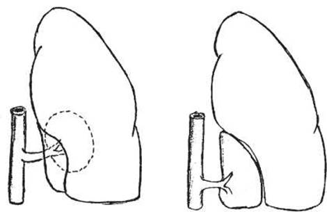
Fig. 18. Pulmonary sequestration. Diagram of the anomaly and its localization within the lung and outside the visceral Pleura.
Surgical treatment is indicated, involving resection of the sequestered segment.
The prognosis is favorable.
Last update: 11/08/2026
Editorial and Educational Adaptation: This material has been compiled based on the primary/original source text. The project team performed an editorial review, corrected technical inaccuracies, structured sections, and adapted the content for an educational format.
What was processed:
- elimination of formatting defects (OCR errors, structural breaks, corrupted characters);
- editorial organization of content;
- standardization of terminology in accordance with academic sources;
- verification of factual statements against the original source text.
All mentions of the author, publication year, and origin of the primary text have been preserved in accordance with the source.