Medical Genetics - V. M. Zaporozhan 2005
Congenital Malformations
Classification of Congenital Malformations
Congenital Malformations are classified according to their Etiology, chronological sequence of development, anatomical localization, and prevalence in the body (Table 9.2).
Table 9.2. Classification of Congenital malformations (G. I. Lazyuk, 1991)
|
Classification principles |
Groups of malformations |
|
By etiological feature |
Hereditary Exogenous (teratogenic) Multifactorial Malformations of unknown etiology |
|
Depending on The sequence of occurrence |
Primary Secondary |
|
By extent of involvement and prevalence in the body |
Isolated Systemic Multiple |
|
Depending on the time of onset in ontogeny |
Gametopathies Blastopathies Embryopathies Fetopathies |
1. Based on etiology, the following groups of congenital malformations are distinguished:
— hereditary malformations — caused by Mutations (Gene, chromosomal, or genomic); they can also be caused by uniparental isodisomy or disomy, or Genomic Imprinting. Chromosomal and genomic mutations lead to Chromosomal diseases, which manifest as syndromes of multiple congenital malformations. Monogenic malformations can be isolated, systemic, or multiple. Examples of monogenic malformations with various inheritance patterns are presented in Table 9.3. Genetic heterogeneity is characteristic of many malformations (mutations in different genes can lead to The formation of the same malformation);
— exogenous (teratogenic) malformations — caused by the direct action of teratogenic factors on the embryo or fetus (teratogenic factors are environmental agents that disrupt embryonic development and lead to the formation of malformations);
— multifactorial malformations — caused by the combined action of genetic and exogenous factors, none of which individually is the cause of the malformation (Table 9.4);
— malformations of unknown etiology (those whose exact cause cannot be established).
According to various authors, hereditary malformations account for approximately 20-30%, exogenous (teratogenic) for 2-5%, multifactorial for 30-40%, and those of unclear etiology for 25-50% (N. P. Bochkov, 2001).
However, the actual role of the genotype in the formation of malformations is significantly greater. Many malformations of unclear etiology are likely caused by genetic factors such as new dominant mutations, microdeletions, uniparental disomy or isodisomy, and loss of imprinting. Clarifying the genetic Diagnosis of such malformations requires The Use of specialized diagnostic Methods.
2. Depending on the sequence of occurrence, Primary and secondary congenital malformations are distinguished. Primary malformations arise directly from the action of an etiological factor (mutations and/or teratogenic factors). Secondary malformations are complications of primary malformations and are always pathogenetically related to them, essentially representing "malformations of malformations." For example, the consequence of a primary developmental malformation such as a diaphragmatic hernia (Fig. 9.8) is a secondary malformation — pulmonary hypoplasia. Genetic risk is always calculated for the primary malformation.
Table 9.3. Monogenic congenital malformations with various inheritance patterns
|
Examples of monogenic malformations |
Inheritance pattern |
|
Isolated malformations |
|
|
— Hydrocephalus caused by stenosis of the cerebral aqueduct; |
XR |
|
— microcephaly |
AR |
|
Eye: |
|
|
— aniridia; |
AD |
|
— cataract; |
AD/AR |
|
— microphthalmia; |
AD/AR |
|
— anophthalmia |
AR |
|
Limbs: |
|
|
— brachydactyly; |
AD |
|
— polydactyly; |
AD |
|
— ectrodactyly; |
AD |
|
Other: |
|
|
— adult-onset Polycystic Kidney Disease — manifests after 30 years of age |
AD |
|
Systemic malformations |
|
|
AD |
|
|
— Osteogenesis Imperfecta (increased bone fragility, blue sclerae, otosclerosis) |
AD/AR |
|
Multiple congenital malformations |
|
|
— Apert syndrome (acrocephaly, microcephaly, Syndactyly of hands and feet, intellectual disability); |
AD |
|
— Meckel syndrome (encephalocele, polydactyly, polycystic kidney disease) (Fig. 9.1); |
AR |
|
— Smith-Lemli-Opitz syndrome |
AR |
|
(prenatal growth retardation, microcephaly (Fig. 9.2), syndactyly, polydactyly, Heart, kidney, and lung defects, intellectual disability); |
(mutation of a gene involved in METABOLISM) |
|
— Lenz syndrome (unilateral anophthalmia or microphthalmia (Fig. 9.3), microcephaly, syndactyly, polydactyly, heart, gastrointestinal, and kidney defects, etc.) |
XR |
Class="center">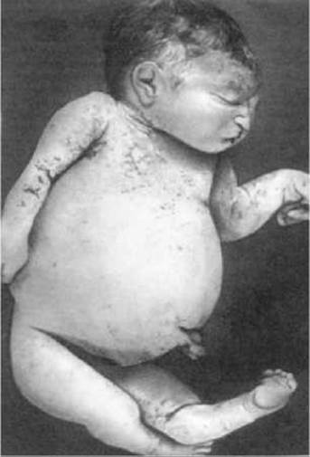
Fig. 9.1. Meckel syndrome (enlarged abdomen due to polycystic kidney disease, polydactyly, encephalocele)
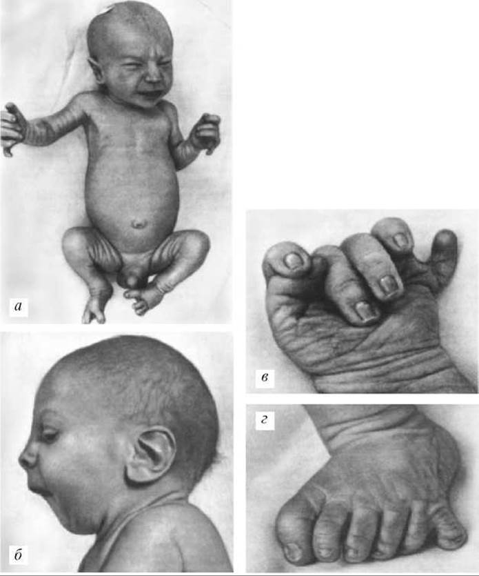
Fig. 9.2. Smith-Lemli-Opitz syndrome: a — general appearance of the patient, sandal gap on the FOOT; b — specific facial features: short Nose with upturned nostrils, ptosis, long philtrum, micrognathia, macrotia; c, d — postaxial polydactyly of the hand and foot

Fig. 9.3. Lenz syndrome (anophthalmia, prominent auricles, narrow face)
Note. AD — Autosomal dominant inheritance pattern; AR — Autosomal Recessive Inheritance pattern; XR — X-linked recessive inheritance pattern.
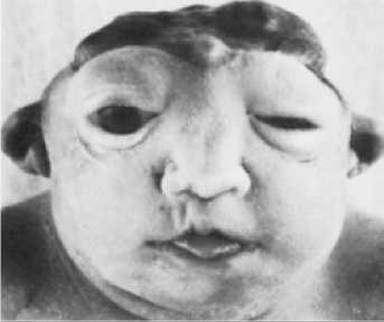
Fig. 9.4. Anencephaly

Fig. 9.5. Craniofacial hernia
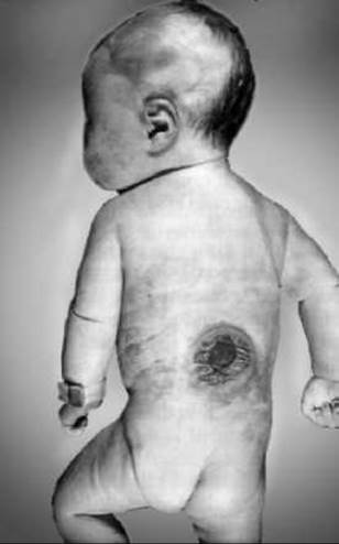
Fig. 9.6. Spina bifida
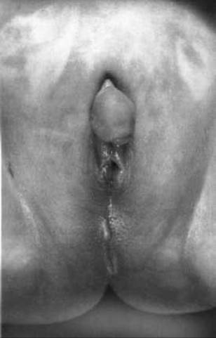
Fig. 9.7. Hypospadias (the Urethra in a boy opens on the Perineum)
Table 9.4. Examples of multifactorial malformations (isolated and systemic)
|
Organ system |
Examples of malformations |
|
Atrial septal defect Ventricular septal defect Tetralogy of Fallot Patent ductus arteriosus |
|
|
Central Nervous System — neural tube closure defects |
Anencephaly (Fig. 9.4) Encephalocele (Fig. 9.5, 11.3) Meningomyelocele (Fig. 9.6) Spina bifida |
|
Hypospadias (Fig. 9.7) Renal agenesis |
|
|
Gastrointestinal tract |
Hypertrophic pyloric stenosis |
|
Other |
Cleft lip and/or palate (see Fig. 3.4) Congenital hip dislocation Clubfoot |
3. According to the scope of involvement and prevalence in the body, malformations are classified as follows:
— isolated (single) — localized in a single organ (pyloric stenosis, polydactyly, etc.);
— systemic — malformations within a single organ system (Tetralogy of Fallot);
— multiple (MCMA / multiple congenital malformations) — a complex of two or more non-induced developmental malformations in different systems. Multiple congenital malformations may be causally (or pathogenetically) related or occur entirely by chance.
When identifying a group of multiple congenital malformations, primary malformations are taken into account. For example, Down syndrome involves microcephaly, heart defects, digestive tract defects, and sometimes Urinary System defects. This is a multiple congenital malformation syndrome.
At the same time, a complex of diaphragmatic hernia, pulmonary hypoplasia, and Liver lobulation disorders should not be considered multiple malformations, because the diaphragmatic hernia caused The Development of corresponding secondary malformations (pulmonary hypoplasia, liver lobulation disorders). Such a complex of malformations caused by a single primary malformation is called an anomalad. Another example is the Pierre Robin anomalad (Fig. 9.9). The primary malformation, microgenia, causes a secondary malformation — cleft palate.
Isolated and systemic malformations are classified according to the Anatomical and physiological principle. The following groups of malformations are distinguished:
1. Malformations of the central nervous system and Sense Organs.
2. Malformations of the face and neck.
3. Malformations of The Cardiovascular System.
4. Malformations of the Respiratory system.
5. Malformations of the digestive organs.
6. Malformations of The Musculoskeletal System.
7. Malformations of the urinary system.
8. Malformations of the genital organs.
9. Malformations of the Endocrine glands.
10. Malformations of the Cytology/cytology/66.html">Skin and its Appendages.
11. Malformations of the placenta and fetal membranes.
12. Other malformations.
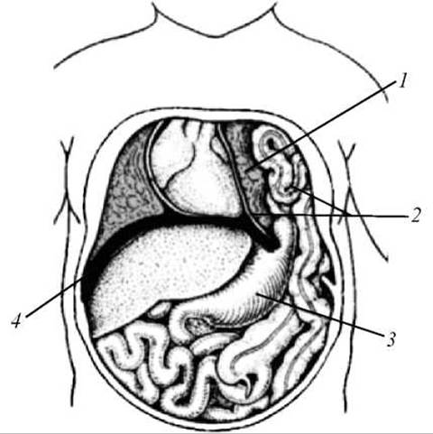
Fig. 9.8. Diaphragmatic hernia: 1 — left lung; 2 — bowel loops in the thoracic cavity; 3 — Stomach; 4 — Diaphragm
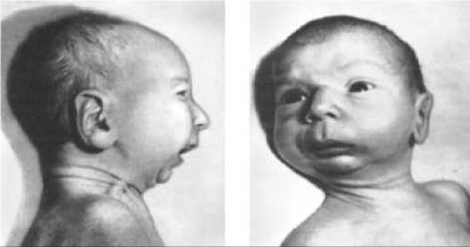
Fig. 9.9. Pierre Robin sequence (marked hypoplasia of the Mandible)
The frequencies of individual groups of birth defects are summarized in Table 9.5.
Multiple congenital anomalies are classified according to their etiological principle.
1. Chromosomal syndromes.
2. Gene syndromes.
3. Syndromes caused by teratogenic factors.
4. Syndromes of unknown etiology.
5. Multiple congenital anomalies of unknown etiology.
Currently, the international congenital defect monitoring program lacks the category "multiple congenital anomalies" because modern diagnostic methods allow for the precise genetic diagnosis of a specific MCA syndrome.
Table 9.5. Frequency of individual groups of birth defects in the population
|
Groups of defects |
Frequency, % |
|
Multiple congenital anomalies |
7.9-18.2 |
|
Isolated and systemic birth defects: — neural tube defects; |
8.4-22.3 (CNS defects — 30% of all defects) |
|
— cardiovascular system defects |
10.9-21.0 |
|
— limb defects |
7.4-24.5 |
|
— genital defects |
2.4-7.5 |
In foreign literature, isolated and systemic defects are classified as malformations, disruptions, deformations, and dysplasias.
Malformations are congenital anomalies that arise from the abnormal formation of embryonic structures. This means the organ's primordium is initially anomalous, and its development cannot proceed normally. Examples of malformations include cardiac defects such as ventricular septal defect, atrial septal defect, cleft lip and/or cleft palate, and neural tube defects (anencephaly, spina bifida), among others. Most isolated malformations are multifactorial. They can also result from gene, chromosomal, and genomic mutations, or teratogenic effects.
Disruptions are structural breakdowns that occur in normally developing organs under The Influence of infectious agents, mechanical damage (amniotic bands), or Circulatory Disorders. An example of a disruption is finger amputation caused by amniotic bands.
Deformation refers to the abnormal shape or position of a body part resulting from external mechanical (non-disruptive) forces. Examples include developmental Dysplasia of the hip and clubfoot, which can develop due to oligohydramnios, uterine tumors or malformations, Multiple Pregnancy, and other causes. Typically, deformations occur during the later stages of pregnancy.
Dysplasia encompasses systemic defects resulting from abnormal tissue architecture. The effects of dysplasia are observed in all organs containing that specific tissue. An example is anhidrotic ectodermal dysplasia, which affects multiple derivatives of the ectoderm (hair, Sweat Glands, Teeth, nails), as well as osteogenesis imperfecta (brittle bone disease). Most dysplasias are caused by Gene Mutations.
In foreign literature, multiple congenital anomalies are classified as syndromes, sequences, and associations.
A syndrome is a constellation of multiple congenital anomalies united by a single etiological factor. Examples include Chromosomal Disorders manifested by multiple congenital anomaly syndromes, monogenic MCA syndromes, and syndromes caused by teratogenic factors.
A sequence represents multiple congenital anomalies that form a "cascade" resulting from a single primary defect (a pathogenetic rather than causal relationship). For instance, a primary defect like spina bifida can lead to a sequence including lower limb paralysis, Muscle atrophy, and clubfoot. Another example is the Pierre Robin sequence. The primary defect is micrognathia (mandibular hypoplasia), which results in a sequence of subsequent anomalies: a reduced Oral Cavity, impaired palate formation, and cleft palate.
Associations are non-random combinations of several congenital anomalies occurring in different individuals, not yet recognized as a syndrome or sequence. The causes of associations are generally poorly understood. An example is the VATER association (vertebral defects, anal atresia, tracheoesophageal fistulas, radial hypoplasia, and thumb anomalies, etc.).
VATER association is an acronym formed from the first letters of the following English words:
Vertebral (V) anomalies — vertebral abnormalities
Anal (A) atresia — anal atresia
Tracheo (T)
Esophageal (E) fistula
Radial (R) and/or Renal (R) anomalies.
4. Classification of Congenital Malformations According to the Time of Ontogenetic Origin
Depending on which Structure is exposed to the damaging agent, malformations are classified into gametopathies, blastopathies, embryopathies, and fetopathies.
Gametopathies are hereditary congenital malformations caused by mutations in the germ Cells of the proband's parents (regardless of whether these mutations are inherited or arise de novo).
Blastopathies are malformations that develop within the first 15 days following Fertilization. This developmental stage is known as blastogenesis (hence the term blastopathies). This timeframe represents the first critical period of embryonic development, during which teratogenic factors operate on an "all-or-none" principle: either the embryo dies, or (thanks to the high reparative capacity of its cells) it continues to develop without malformations. The rate of embryonic loss During the first 15 days post-fertilization is at its highest, reaching 35–50% of all fertilized ova.
Nevertheless, certain developmental anomalies do arise during this period. These include:
— Hydatidiform Mole (abnormal development of trophoblastic derivatives coupled with impaired embryoblast formation), which is typically a consequence of polyploidy, uniparental diploidy, or occasionally other Chromosomal aberrations;
— conjoined twinning (fully or partially separated twins)—craniopagous, thoracopagous (Fig. 9.10), ischiopagous twins, etc.;
— sirenomelia — fusion of the lower extremities (Fig. 9.11);
— cyclopia — a malformation of the Brain and face, one of the KEY FEATURES OF which is the presence of a single eye (Fig. 9.12);
— implantation defects;
— hypoplasia or aplasia of extraembryonic structures (amnion, yolk sac), among others.
Blastopathies also include mosaic forms of chromosomal disorders caused by nondisjunction of Chromosomes during the early Cleavage Stages of the zygote.
Embryopathies are malformations that form between the 16th day post-fertilization and the end of the 8th week. This period marks the formation of embryonic Tissues and organs (histogenesis and Organogenesis); consequently, the vast majority of malformations, regardless of etiology, originate during this window. Some teratologists restrict the term embryopathy to teratogen-induced malformations, even though hereditary defects also become morphologically evident during this timeframe.
Throughout this period, the embryo exhibits maximum sensitivity to teratogenic agents. Weeks 3 to 8 post-fertilization constitute the second critical period of embryonic development (the most hazardous regarding malformation genesis). More than 10% of all registered pregnancies terminate in spontaneous abortions during this interval.
Fetopathies are malformations that develop from the 9th week of intrauterine life until birth (the fetal period). During this stage, primary structural malformations can arise only if an organ has not yet completed its development. Such organs include the brain, Lungs, teeth, and genitalia. Concurrently, secondary defects, tissue dysplasias, and hypoplasia of organs or the fetus as a whole may develop. By the 5th to 6th month of gestation, the fetus acquires the capacity for inflammatory reactions, which can lead to malformations driven by inflammatory processes (e.g., hydrocephalus in Toxoplasmosis). Fetopathies also encompass abnormalities associated with specific endocrine disorders, such as Diabetes Mellitus.
For every organ, one can define a terminal teratogenic period—the threshold period of intrauterine development during which an etiologic factor can induce a structural defect in that organ. For example, the upper limb bud appears on the 24th day, and the lower limb bud on the 28th. Limb morphogenesis continues until the 56th day, when the Formation of the nail Phalanges takes place. Consequently, the terminal teratogenic period for most limb malformations spans days 24 to 56.
Last update: 11/08/2026
Editorial and Educational Adaptation: This material has been compiled based on the primary/original source text. The project team performed an editorial review, corrected technical inaccuracies, structured sections, and adapted the content for an educational format.
What was processed:
- elimination of formatting defects (OCR errors, structural breaks, corrupted characters);
- editorial organization of content;
- standardization of terminology in accordance with academic sources;
- verification of factual statements against the original source text.
All mentions of the author, publication year, and origin of the primary text have been preserved in accordance with the source.