Anatomy and Physiology of Children and Adolescents - M. R. Sapin 2007
Structure of the Human Body
Cells: Structure, Chemical Composition, and Functions
The Human Body is an integrated, complex system that performs numerous and diverse Functions. It consists of Cells, Tissues, and Organs. Organs formed from tissues constitute Organ Systems and apparatuses, which together form the complete human Organism. At the foundational level of the organism, its organs, and tissues, lie cells.
The Cell is the smallest Structural and functional unit of living matter. Cells of All living organisms, including humans, share a similar Structure. Studying Cell Structure, functions, and intercellular interactions is fundamental to understanding such a complex organism as a human being. The cell actively responds to stimuli, carries out GROWTH AND REPRODUCTION, is capable of self-Replication and transmitting Genetic information to descendants, and possesses capacities for regeneration and environmental adaptation.
Structure. The adult human body contains approximately 200 cell types, which vary in shape, structure, chemical composition, and metabolic profile. Despite this vast diversity, each individual cell within any organ represents an integrated living system. The key Structural components of a cell are the cytolemma, Cytoplasm, and nucleus (Fig. 5).
Cytolemma. Every cell is enclosed by a membrane—the cytolemma (Plasma Membrane)—which separates the cellular contents from the external (extracellular) environment. The cytolemma not only delimits the cell externally but also ensures its direct communication with the environment. It performs protective and transport functions and perceives environmental stimuli. Through the cytolemma, various molecules (particles) enter the cell and exit into the surrounding medium.
Class="center">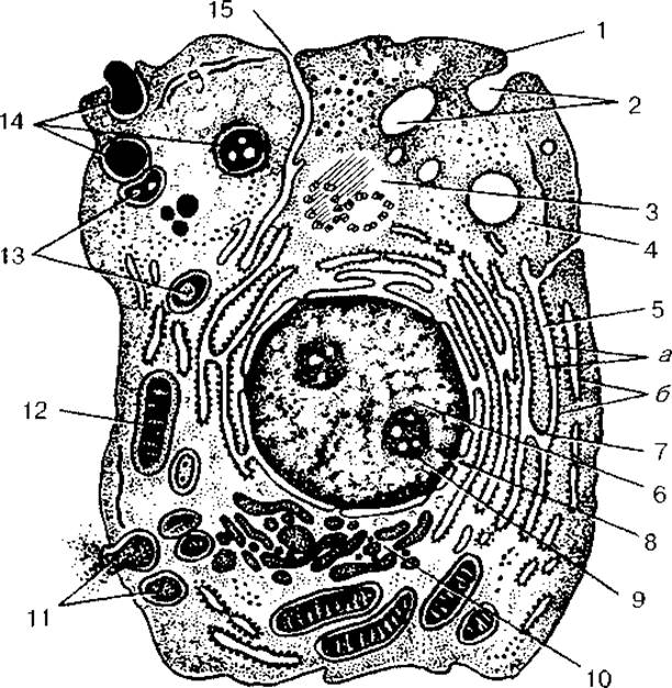
Fig. 5. Ultramicroscopic structure of a cell:
1 — cytolemma (plasma membrane); 2 — pinocytotic vesicles; 3 — centrosome (cell center, cytocenter); 4 — hyaloplasm; 5 — Endoplasmic reticulum (a — membranes of The endoplasmic reticulum, 6 — Ribosomes); 6 — nucleus; 7 — connection between the perinuclear space and the cavities of the endoplasmic reticulum; 8 — nuclear pores; 9 — nucleolus; 10 — intracellular reticular apparatus (Golgi apparatus); 11 — secretory vacuoles; 12 — Mitochondria; 13 — Lysosomes; 14 — three consecutive stages of phagocytosis; 15 — connection of The Cell wall (cytolemma) with the membranes of the endoplasmic reticulum
The cytolemma consists of lipid and protein molecules held together by complex intermolecular interactions, which maintain the structural integrity of the membrane. The foundation of the cytolemma is also formed by lipoprotein sheets (Lipids in complex with Proteins). With a thickness of about 10 nm, the cytolemma is the thickest of all biological membranes. As a semipermeable biological membrane, the cytolemma exhibits a three-layered structure (Fig. 6, see color insert). The outer and inner hydrophilic layers are formed by lipid molecules (lipid bilayer) and have a thickness of 5—7 nm. These layers are impermeable to most Water-soluble molecules. Situated between the outer and inner layers is the intermediate hydrophobic layer of lipid molecules. Membrane Lipids include a large group of organic substances that are poorly soluble in water (hydrophobic) and highly soluble in organic Solvents. Cellular membranes contain Phospholipids (glycerophosphatides), steroidal lipids (Cholesterol), and others.
Lipids account for approximately 50% of the mass of The Plasma Membrane.
Lipid molecules possess hydrophilic (water-loving) heads and hydrophobic (water-fearing) tails. They are arranged within the cytolemma such that the outer and inner layers (The Lipid Bilayer) are formed by the hydrophilic heads, while the intermediate layer is formed by their hydrophobic tails.
Membrane Proteins do not form a continuous layer within the cytolemma. Instead, they are embedded within the lipid layers to varying depths. Protein molecules have an irregular, globular shape and are formed from polypeptide helices. Non-polar regions of proteins—devoid of charge and rich in non-polar Amino Acids (Alanine, valine, Glycine, leucine)—are embedded in the region of the lipid membrane where the hydrophobic tails of the lipid molecules reside. Polar, charged regions of proteins, which are also rich in amino acids, interact with the hydrophilic heads of the lipid molecules.
Proteins constitute nearly half of the plasma membrane's mass. Membrane proteins are classified as transmembrane (integral), semi-integral (peripheral-integral), and peripheral proteins. Peripheral proteins are located on the membrane surface, whereas integral and semi-integral proteins are embedded within the lipid layers. Integral protein molecules span the entire lipid layer of the membrane, whereas semi-integral proteins are only partially embedded. Based on their biological roles, membrane proteins are subdivided into carrier proteins (transport proteins), enzymatic proteins, and receptor proteins.
Membrane CARBOHYDRATES are represented by polysaccharide chains attached to membrane proteins and lipids, known as Glycoproteins and Glycolipids. The carbohydrate content in the cytolemma and other biological membranes is relatively small, ranging from 2 to 10% of the membrane mass. Carbohydrates are located on the external surface of The cell membrane, which does not contact the cytoplasm. Together, they form a supra-membrane layer known as the glycocalyx, which participates in intercellular recognition processes and has a thickness of 3—4 nm. Chemically, the glycocalyx is a glycoprotein complex comprising various carbohydrates linked to proteins and lipids.
Functions of the plasma membrane. One of the most critical functions of the cytolemma is transport. It ensures the influx of nutrients and energetic substances into the cell, the removal of Metabolic waste products and biologically active Materials (secretions), regulates the passage of various ions into and out of the cell, and maintains the appropriate intracellular pH.
Several mechanisms exist for the uptake and release of substances by the cell: diffusion, Active Transport, and exocytosis or endocytosis.
Diffusion is the movement of molecules or ions from an area of higher concentration to an area of lower concentration, i.e., down the concentration gradient. Through diffusion, molecules of oxygen (O2) and carbon dioxide (CO2) are transported across membranes. Ions, molecules of glucose, amino acids, and Fatty acids diffuse across membranes relatively slowly.
The direction of ion diffusion is determined by two factors: concentration and electrical charge. Ions typically move toward areas of opposite charge while being repelled from areas of like charge, diffusing from regions of high concentration to regions of low concentration.
Active transport involves the movement of molecules or ions across membranes against a concentration gradient, requiring the expenditure of energy. Energy, derived from the Cleavage of adenosine triphosphate (ATP), is necessary to drive substances from a lower to a higher concentration environment. A classic example of active transport is the sodium-potassium pump (Na+, K+-pump). Na+ ions and ATP approach the membrane from the cytoplasmic side, while K+ ions approach from the extracellular side. For every two K+ ions entering the cell, three Na+ ions are pumped out. As a result, the intracellular environment becomes negatively charged relative to the extracellular medium, establishing a potential difference across the two surfaces of the membrane.
The Transmembrane Transport of large molecules such as NUCLEOTIDES and amino acids is mediated by membrane transport proteins, which include carrier proteins and channel-forming proteins. Carrier proteins bind to the target molecule and transport it across the membrane through a process that can be either passive or active. Channel-forming proteins create narrow, tissue-fluid-filled pores that span the lipid bilayer. These channels feature gates that open transiently in response to specific events occurring at the membrane.
The cytolemma also participates in the uptake and release of various macromolecules and large particles. The inward transport of such particles across the membrane is termed endocytosis, while their expulsion is called exocytosis. During endocytosis, the plasma membrane forms invaginations or protrusions that pinch off to become vesicles, carrying trapped particles or fluid into the interior of the cell. Endocytosis is divided into two main types: phagocytosis and pinocytosis. Phagocytosis (from Greek phagos, meaning "eating") is the Uptake and Transport of large particles into the cell, such as cellular debris or Bacteria. Pinocytosis (from Greek pino, meaning "drinking") is the uptake of fluid and macromolecular solutes. Most particles or molecules internalized by the cell end up in lysosomes, where they are digested. Exocytosis is the reverse process of endocytosis, wherein the contents of transport or secretory vesicles are released into the extracellular space. During this process, the vesicles fuse with the plasma membrane, rupture at the surface, and discharge their contents into the extracellular milieu.
The Receptor Functions of the cell membrane are carried out by numerous specialized structures called receptors located on the cytolemma surface. These receptors are capable of perceiving various chemical and physical stimuli and are chemically represented by cytolemma glycoproteins and glycolipids. Receptors are distributed uniformly across the cell surface or concentrated in specific Regions of the cell membrane. Specialized receptors exist for recognizing Hormones, Neurotransmitters, Antigens, and various proteins.
Intercellular junctions are formed by the close apposition and sealing of the cytolemmas of adjacent cells. They ensure the transmission of chemical and electrical signals between cells and mediate mutual cellular interactions. Intercellular junctions include tight, adhering, gap, and synaptic junctions. Simple junctions occur when the cytolemmas of two neighboring cells are merely in close contact. At tight junctions, the Plasma Membranes of two cells are brought into extreme proximity and locally fuse to form what appears to be a single membrane. Gap Junctions (nexuses) feature a very narrow intercellular space (2—3 nm) between the two cytolemmas. Synaptic junctions (synapses) are characteristic of contacts between Nerve Cells, where a signal (Nerve Impulse) is transmitted unidirectionally from one neuron to another.
Functionally, intercellular junctions can be grouped into three categories: occluding junctions, anchoring junctions, and communicating junctions. Occluding junctions bind cells tightly together, preventing even small molecules from passing between them. Anchoring junctions mechanically link cells to neighboring cells or extracellular structures. Communicating junctions facilitate the transmission of chemical and electrical signals between cells, with gap junctions and synapses serving as the primary types.
REVIEW AND SELF-Control Questions:
1. What chemical compounds (molecules) make up the cytolemma? How are these molecules arranged within the membrane?
2. Where are membrane proteins located, and what role do they play in cytolemma functions?
3. Name and describe the types of substance Transport Across the membrane.
4. How does active Transport of substances across membranes differ from passive transport?
5. What are Endocytosis and Exocytosis? How do they differ from each other?
6. What types of intercellular junctions (connections) do you know?
Cytoplasm. Inside the cell, beneath its cytolemma, lies the cytoplasm, which consists of a homogeneous, semi-fluid portion—the hyaloplasm—along with Organelles and inclusions suspended within it.
The hyaloplasm (from the Greek hyalmos, meaning transparent) is a complex colloidal system filling the space between cell organelles. Proteins are synthesized in the hyaloplasm, and it serves as the cell's energy reservoir. The hyaloplasm integrates various cell structures and facilitates their chemical interactions, forming the matrix—the internal environment of the cell. Externally, the hyaloplasm is enclosed by the cell membrane, or cytolemma. Water accounts for up to 90% of the hyaloplasm's composition. Proteins essential for the cell's viability and functioning are synthesized here. It stores energy reserves in the form of ATP molecules, fat droplets, and Glycogen. The hyaloplasm contains general-purpose structures—organelles present in all cells—as well as non-permanent formations known as cytoplasmic inclusions. Organelles include the granular and agranular endoplasmic reticulum, the internal reticular apparatus (Golgi complex), the cell center (centrosome), ribosomes, and lysosomes. Inclusions include glycogen, proteins, fats, Vitamins, pigment granules, and other substances.
Organelles are cell structures that perform specific vital functions. They are divided into membranous and non-membranous organelles. Membranous organelles are closed, isolated or interconnected regions of the cytoplasm separated from the hyaloplasm by membranes. These include the endoplasmic reticulum, the internal reticular apparatus (Golgi complex), mitochondria, lysosomes, and Peroxisomes.
The endoplasmic reticulum consists of groups of cisternae, vesicles, or tubules bounded by a membrane 6–7 nm thick. Together, these structures form a network-like appearance. The structure of the endoplasmic reticulum is heterogeneous, comprising two types: granular (rough) and agranular (smooth). In the granular endoplasmic reticulum, numerous small, rounded bodies—ribosomes—are attached to the tubular membranes. The membranes of the agranular endoplasmic reticulum lack ribosomes on their surface. The primary function of the granular endoplasmic reticulum is Protein Synthesis, whereas the membranes of the agranular endoplasmic reticulum synthesize lipids and Polysaccharides.
The internal reticular apparatus (Golgi complex) is typically located near the Cell Nucleus. It consists of flattened, membrane-bound cisternae accompanied by numerous small vesicles. The Golgi complex is involved in Processing and concentrating products synthesized in the Endoplasmic reticulum and exporting these substances out of the cell. Additionally, it is responsible for The formation of cellular lysosomes and peroxisomes.
Lysosomes are spherical membranous vesicles (0.2–0.4 µm in diameter) filled with active chemical substances—hydrolytic Enzymes (Hydrolases)—that break down proteins, carbohydrates, fats, and Nucleic Acids. They are responsible for intracellular Digestion of Biopolymers.
Peroxisomes are small, oval vacuoles measuring 0.3–1.5 µm that contain the enzyme catalase. Catalase breaks down hydrogen peroxide, which is produced as a byproduct of the Oxidative Deamination of amino acids.
Mitochondria serve as the power plants of the cell. These ovoid or spherical organelles are approximately 0.5 µm in diameter and 1–10 µm in length. Unlike other organelles, mitochondria are bounded by two membranes rather than one. The outer membrane has a smooth contour and separates the mitochondrion from the hyaloplasm. The inner membrane encloses the mitochondrial contents—its finely granular matrix—and folds inward to form numerous shelves known as cristae. The primary function of mitochondria is The oxidation of Organic compounds and the utilization of the released energy for ATP synthesis. This synthesis requires oxygen and takes place on the mitochondrial membranes, specifically on the cristae. The liberated energy is used to phosphorylate ADP (adenosine diphosphate) molecules, converting them into ATP.
Non-membranous cell organelles include the cell's supporting framework (Cytoskeleton)—comprising microfilaments, microtubules, and Intermediate filaments—as well as the cell center and ribosomes.
The supporting apparatus, or cytoskeleton, enables the cell to maintain a defined shape and perform directed movements. It is formed by protein filaments that permeate the entire cytoplasm, filling the space between The Nucleus and the cytolemma.
Microfilaments are protein threads 5–7 nm in thickness, located predominantly in the peripheral regions of the cytoplasm. They contain contractile proteins such as Actin, Myosin, and Tropomyosin. Thicker microfilaments, measuring about 10 nm in diameter, are called intermediate filaments or microfibrils. Intermediate filaments are organized into bundles and vary in composition across different cell types: in Muscle cells, they are composed of desmin; in epithelial cells, of Keratins; and in nerve cells, they form neurofibrils.
Microtubules are hollow cylinders about 24 nm in diameter, composed of the protein tubulin. They serve as the primary structural and functional elements of Cilia and flagella, which are cytoplasmic projections. Their main function is structural support. Microtubules ensure the motility of the cells themselves, as well as the beating of cilia and flagella found on certain cells (such as the epithelium of the respiratory tract and other organs). They are also a key component of the cell center.
The cell center (centrosome) consists of centrioles surrounded by a dense matrix known as the centrosphere, and is typically located near the cell nucleus. Centrioles are shaped like hollow cylinders about 0.25 µm in diameter and up to 0.5 µm in length. Their walls are built of microtubules arranged in nine triplets (9x3 structure).
A non-dividing cell typically contains a pair of centrioles positioned at right angles to each other, forming a diplosome. During preparation for Cell Division, the centrioles duplicate, resulting in four centrioles prior to mitosis. Surrounding the centrioles (the diplosome) is the centrosphere, appearing as a structureless halo with radially oriented fibrils. In dividing cells, the centrioles and centrosphere participate in forming the mitotic spindle and occupy its poles.
Ribosomes are granules measuring 15–35 nm in diameter, composed of approximately equal weight ratios of proteins and RNA molecules. They lie freely in the cytoplasm or are attached to the membranes of the granular endoplasmic reticulum. Ribosomes participate in protein synthesis, assembling amino acids into chains strictly according to METABOLISM/28.html">The Genetic Code carried by DNA. In addition to single ribosomes, cells frequently contain groups of ribosomes forming Polysomes, or polyribosomes.
Cytoplasmic inclusions are non-essential cell components that appear and disappear depending on the cell's functional state. They are primarily located in the cytoplasm, where they accumulate as droplets, granules, or crystals. Inclusions are classified into trophic, secretory, and pigmentary. Trophic inclusions include glycogen granules in Liver cells, protein granules in egg cells, and fat droplets in adipocytes; they serve as nutrient reserves for the cell. Secretory inclusions are produced by glandular epithelial cells during their metabolic activity and contain BIOLOGICALLY ACTIVE SUBSTANCES stored as secretory granules. Pigment inclusions may be endogenous (synthesized within the organism itself, such as Hemoglobin, lipofuscin, or melanin) or exogenous (such as Dyes and other foreign substances) in origin.
Review and Self-Control Questions:
1. Name the main Structural elements of the cell.
2. What properties characterize The Cell as the elementary unit of life?
3. What are cell organelles? Describe their Classification.
4. Which organelles participate in the synthesis and Transport of substances within the cell?
5. Describe the structure and Functional Significance of the Golgi apparatus.
6. Describe the STRUCTURE AND FUNCTIONS of mitochondria.
7. Name the non-membranous cell organelles.
8. Define cell inclusions and provide Examples.
The cell nucleus is an essential element of the cell. It contains genetic (hereditary) information and regulates protein synthesis. Genetic information is stored in molecules of deoxyribonucleic acid (DNA). During cell division, this information is transmitted in equal amounts to the daughter cells. The nucleus possesses its own protein-synthesizing apparatus and controls synthetic processes in the cytoplasm. Various types of ribonucleic acid—messenger, transfer, and ribosomal—are synthesized on DNA templates.
The nucleus typically has a spherical or ovoid shape. Certain cells (such as leukocytes) are characterized by bean-shaped, rod-shaped, or segmented nuclei. The nucleus of a non-dividing (interphase) cell consists of a nuclear envelope, nucleoplasm (karyoplasm), Chromatin, and a nucleolus.
The nuclear envelope (karyotheca) separates the Contents of the nucleus from the Cell Cytoplasm and regulates The transport of substances between the nucleus and the cytoplasm. The karyotheca consists of outer and inner membranes separated by a narrow perinuclear space. The outer nuclear membrane is in direct contact with the cell cytoplasm and the membranes of the endoplasmic reticulum cisternae. Numerous ribosomes are located On the surface of the nuclear membrane facing the cytoplasm. The nuclear envelope features nuclear pores closed by a complex Diaphragm formed by interconnected protein granules. Metabolic exchange between the cell nucleus and cytoplasm occurs through these nuclear pores. Ribonucleic acid (RNA) molecules and ribosomal subunits exit the nucleus into the cytoplasm, while proteins and nucleotides enter the nucleus.
Beneath the nuclear envelope lie the homogeneous nucleoplasm (karyoplasm) and the nucleolus. In the nucleoplasm of a non-dividing nucleus, within its nuclear protein matrix, there are granules (clumps) of so-called heterochromatin. Areas of more loosely packed chromatin located between the granules are called euchromatin. Loosened chromatin is referred to as decondensed chromatin, and synthetic processes occur most intensively within it. During cell division, chromatin compacts, condenses, and forms Chromosomes.
The chromatin of a non-dividing nucleus and the chromosomes of a dividing nucleus have the same chemical composition. Both chromatin and chromosomes consist of DNA molecules associated with RNA and proteins (Histones and non-histones). Each DNA molecule consists of two long, right-handed coiled polynucleotide chains (a double helix). Each nucleotide consists of a nitrogenous base, a sugar, and a phosphoric acid residue. Furthermore, the bases are located on the inside of The Double Helix, while the sugar-phosphate backbone is on the outside.
Hereditary information in DNA molecules is encoded in the linear sequence of its nucleotides. The elementary unit of heredity is the Gene. A gene is a segment of DNA with a specific nucleotide sequence responsible for the synthesis of one specific, defined protein.
DNA molecules in the chromosomes of a dividing nucleus are packed compactly. For instance, a single DNA molecule containing 1 million nucleotides in a linear arrangement has a length of 0.34 mm. The extended length of a single human chromosome is about 5 cm. DNA molecules associated with histone proteins form nucleosomes, which serve as the structural units of chromatin. Nucleosomes appear as beads 10 nm in diameter. Each nucleosome consists of histones around which a DNA segment comprising 146 Base Pairs is wrapped. Linear DNA segments consisting of 60 base pairs are located between nucleosomes. Chromatin is represented by fibrils that form loops about 0.4 µm long, containing from 20,000 to 300,000 base pairs.
As a result of compaction (Condensation) and supercoiling of deoxyribonucleoproteins (DNP) in the dividing nucleus, chromosomes appear as elongated, rod-shaped formations with two arms separated by a constriction known as the centromere. Depending on THE POSITION OF the centromere and the length of the arms, Three types of chromosomes are distinguished: metacentric, which have approximately equal arms; submetacentric, which have arms of unequal length; and acrocentric chromosomes, which have one long arm and one very short, barely noticeable arm.
The surface of chromosomes is covered with various molecules, primarily ribonucleoproteins (RNPs). Somatic cells contain two copies of each chromosome. These are called homologous chromosomes; they are identical in length, shape, and structure, and carry the same genes located at the same loci. The structural features, number, and size of chromosomes are referred to as the karyotype. A normal Human Karyotype includes 22 pairs of somatic chromosomes (autosomes) and one pair of sex chromosomes (XX or XY). Human somatic cells (diploid) have a doubled chromosome number—46. Germ Cells contain a haploid (single) set of 23 chromosomes. Therefore, germ cells contain half as much DNA as diploid somatic cells.
One or more nucleoli are present in all non-dividing cells. They appear as intensely staining, rounded bodies whose size is proportional to the intensity of protein synthesis. The nucleolus consists of an electron-dense nucleolonema (from the Greek nema, meaning thread), in which filamentous (fibrillar) and granular parts are distinguished. The filamentous part consists of numerous intertwined RNA threads about 5 nm thick. The granular part is formed by granules about 15 nm in diameter, which represent ribonucleoprotein particles—the precursors of ribosomal subunits. Ribosomes are formed within the nucleolus.
Chemical composition of the cell. All Cells of the human body are similar in their chemical composition, comprising both inorganic and organic substances.
Inorganic substances. More than 80 chemical elements are found within the cell composition. At the same time, six of these—carbon, hydrogen, nitrogen, oxygen, phosphorus, and sulfur—account for about 99% of the total cell mass. Chemical elements exist in the cell in the form of various compounds.
Water occupies the primary place among cell substances, making up about 70% of the cell mass. Most reactions occurring in the cell can proceed only in an aqueous environment. Many substances enter the cell in aqueous solution. Metabolic waste products are also removed from the cell in aqueous solution. Thanks to the Presence of water, the cell maintains its volume and turgor. In addition to water, the inorganic substances of the cell include salts. For cellular vital processes, the most important cations are K+, Na+, Mg2+, Ca2+, as well as anions such as H2PO-4, Cl-, and HCO-3. The concentration of cations and anions inside and outside the cell differs. For instance, the concentration of potassium ions is always quite high inside the cell and sodium ions are low. Conversely, in the surrounding extracellular environment (tissue fluid), there are fewer potassium ions and more sodium ions. In a living cell, these concentration gradients of potassium and sodium ions between the intracellular and extracellular environments are maintained at a constant level.
Organic substances. Almost all molecules of the cell belong to carbon compounds. Due to the presence of four electrons in its outer shell, the carbon atom can form four strong covalent bonds with other atoms, creating large and complex molecules. Other atoms widely represented in the cell that readily bond with carbon are hydrogen, nitrogen, and oxygen. Like carbon, they are small in size and capable of forming very strong covalent bonds.
Most organic compounds form large molecules known as macromolecules (from the Greek makros, meaning large). Such molecules consist of repeating, structurally similar interconnected units called monomers (from the Greek monos, meaning single). A macromolecule formed by monomers is called a polymer (from the Greek polý, meaning many).
Proteins constitute the bulk of the cell cytoplasm and nucleus. All proteins contain hydrogen, oxygen, and nitrogen atoms. In addition, many proteins contain sulfur and phosphorus atoms. Each protein molecule consists of thousands of atoms. There is a vast number of different proteins built from amino acids.
Over 170 Amino acids are found in the Cells and Tissues of animal and plant organisms. Each amino acid possesses a carboxyl group (—COOH) with acidic properties and an amino group (—NH2) with basic properties. The portions of the molecules not occupied by the carboxyl and Amino groups are called radicals (R). In the simplest case, the radical consists of a single hydrogen atom, whereas in more complex amino acids, it can be an intricate structure consisting of many carbon atoms.
The most important amino acids include alanine, glutamic acid, aspartic acid, Proline, leucine, and Cysteine. The bonds connecting amino acids to one another are called peptide bonds. The resulting compounds of amino acids are called Peptides. A peptide composed of Two amino acids is called a dipeptide; of three, a tripeptide; and of many, a polypeptide. Most proteins comprise 300 to 500 amino acids. There are also larger protein molecules consisting of 1,500 or more amino acids. Proteins differ in the composition, number, and sequential order of amino acids in the polypeptide chain. It is precisely this Amino Acid Sequence that is of paramount importance for the existing Diversity of proteins. Many protein molecules have a large length and high molecular weight. For instance, the Molecular Weight of Insulin is 5,700, that of hemoglobin is 65,000, whereas the molecular weight of water is only 18.
The polypeptide chains of proteins are not always extended in length. On the contrary, they can be coiled, bent, or folded in A wide variety of ways. The Diversity of the physical and Chemical properties of proteins ensures the specific functions they perform: structural, motor, transport, protective, and energetic.
Carbohydrates that are part of cells are also organic substances. Carbohydrates contain carbon, oxygen, and hydrogen atoms. Simple and complex carbohydrates are distinguished. Simple carbohydrates are called Monosaccharides. Complex carbohydrates are polymers in which monosaccharides play The Role of monomers. Two monomers form a disaccharide, three form a trisaccharide, and many form a polysaccharide. All monosaccharides are colorless substances that are highly soluble in water. The most common monosaccharides in animal cells are glucose, ribose, and deoxyribose.
Glucose serves as the primary source of cellular energy. During breakdown, it is converted into carbon dioxide and water (СО2 + Н2О). This reaction releases energy (The breakdown of 1 g of glucose yields 17.6 kJ of energy). Ribose and deoxyribose are essential Components of nucleic acids and ATP.
Lipids consist of the same chemical elements as carbohydrates—carbon, hydrogen, and oxygen. They are insoluble in water. The most common and well-known lipids are fats, which act as an energy source. The breakdown of fats releases twice as much energy as the breakdown of carbohydrates. Because lipids are hydrophobic, they form an integral part of cell membranes.
Cells contain nucleic acids, namely DNA and RNA. The term "nucleic acids" originates from the Latin word *nucleus*, referring to the cell nucleus where they were first discovered. Nucleic acids are polymers formed by chains of interconnected nucleotides. A nucleotide is a chemical compound consisting of a single sugar molecule and a single organic base molecule. When interacting with acids, organic bases can form salts.
Each DNA molecule consists of two strands twisted around each other in a double helix. Each strand is a polymer whose monomers are nucleotides. Every nucleotide contains one of four bases: adenine, cytosine, guanine, or thymine. During the Formation of the double helix, the nitrogenous bases of one strand "dock" with those of the opposite strand. The bases approach each other so closely that Hydrogen Bonds form between them. There is a precise pattern in how these nucleotides pair: adenine (A) on one strand is always paired with thymine (T) on the other, while guanine (G) on one strand always pairs with cytosine (C). In each of these pairs, the two nucleotides Complement one another. The Latin word for "addition" is *complementum*. Therefore, it is standard to say that guanine is complementary to cytosine, and thymine is complementary to adenine. Thus, knowing The sequence of nucleotides in one strand immediately reveals The nucleotide sequence in the other strand via THE PRINCIPLE OF complementarity.
Within DNA polynucleotide chains, every three consecutive nucleotides form a triplet (a three-component unit). Each triplet is not merely a random group of three nucleotides, but a codagene (derived from Greek, denoting a segment that forms a codon). Each codon encodes (specifies) a single amino acid. The sequence of codogenes contains the primary genetic information defining The amino acid sequence in proteins. DNA possesses a unique property—The ability to replicate—not shared by any other known molecule.
RNA molecules are also polymers composed of nucleotide monomers. RNA consists of a single-stranded molecule structured in the same manner as a single DNA strand. Ribonucleic acid, much like DNA, contains triplets—combinations of three nucleotides, or informational units. Each triplet directs the incorporation of a specific amino acid into a protein. The order of synthesized amino acids is determined by the sequence of RNA triplets. The information contained in RNA is transcribed from DNA, with information transfer governed by the familiar principle of complementarity.
A complementary RNA triplet pairs with each DNA triplet. This RNA triplet is called a codon. The sequence of codons holds the blueprint for the amino acid sequence in proteins, copied directly from the codogene sequence within the DNA molecule.
Unlike DNA, the content of which remains relatively constant in the cells of specific organisms, RNA content fluctuates depending on the cell's synthetic activity.
Based on their functions, several types of ribonucleic acid are distinguished. Transfer RNA (tRNA) is found mainly in the cell cytoplasm. Ribosomal RNA (rRNA) constitutes a substantial structural part of ribosomes. Messenger RNA (mRNA) is found in both the cell nucleus and cytoplasm, carrying Protein Structure information from DNA to the ribosomes where protein synthesis occurs. All types of RNA are synthesized on DNA templates.
Adenosine triphosphate (ATP) is present in every cell. Structurally, ATP belongs to the nucleotide family. Each ATP molecule—as well as standard nucleotides—contains one organic base (adenine), one carbohydrate molecule (ribose), and three phosphoric acid molecules. ATP differs significantly from conventional nucleotides by possessing three phosphoric acid residues rather than one.
Adenosine monophosphate (AMP) is a component of all RNA molecules. Upon The addition of two more phosphoric acid molecules (Н3РО4), it converts into ATP, becoming a source of cellular energy. Specifically, the bond between the second and third phosphate groups is rich in chemical energy. The chemical energy of this phosphate bond can be readily transferred to other cellular compounds. When one phosphoric acid molecule is cleaved, ATP is converted into ADP (adenosine diphosphate). If Two molecules are cleaved, ATP turns into AMP (adenosine monophosphate). The cleavage of each phosphoric acid molecule releases 419 kJ/mol of energy.
Enzymes act as catalysts for reactions within living cells. Without enzymes, organic compound reactions proceed at extremely low rates. The word "enzyme" derives from the Latin *fermentum* (leaven). Enzymes that break down carbohydrates are called saccharases, those removing hydrogen are dehydrogenases, and those breaking down fats are lipases.
Review and Self-Control Questions:
1. Name the chemical elements known to you that make up cells.
2. Name the primary inorganic substances found in cells.
3. List the bases that form part of the organic molecules in cells.
4. Describe the Structural Features of proteins as polymers.
5. Characterize the biological roles of Introduction/36.html">Carbohydrates and lipids.
6. What Types of Nucleic acids are found in cells?
7. What is the Biological Role of DNA and RNA?
8. Compare the structures of DNA and RNA.
9. Which structural features determine the primary function of ATP?
Cellular Functions. Every cell exhibits all the characteristics of living matter. These include metabolism, responsiveness to external stimuli (irritability), excitability, growth, reproduction (the ability for self-replication and transmission of genetic information), regeneration (repair), and adaptation.
Metabolism in a living cell involves the uptake of substances from the environment and The excretion of metabolic waste products. All reactions occurring within a cell can be divided into two groups: anabolic and catabolic. Anabolic reactions involve the synthesis of large molecules from smaller, simpler precursors. These processes require an energy input. Using glucose, amino acids, organic acids, and nucleotides entering the cell, proteins, carbohydrates, lipids, and nucleic acids are continuously synthesized. These substances form cell membranes, organelles, and other structures. Synthesis is particularly intense in young, growing cells. The chemical composition of a cell is repeatedly renewed throughout its lifespan. Imported substances participate in Biosynthesis (the formation of biological structures—proteins, fats, and carbohydrates—from simpler molecules). Biosynthesis yields substances necessary for cellular life and functioning. For instance, in Skeletal Muscle fibers and smooth muscle myocytes, proteins are synthesized to ensure contraction. Processes that result in the formation of living matter are termed anabolism (assimilation).
Simultaneously with biosynthesis, the breakdown and degradation of organic compounds occur within cells. This breakdown yields structurally simpler substances (water, carbon dioxide, urea, etc.). Most degradative reactions proceed with the participation of oxygen and the release of energy. Processes involving the breakdown of large organic molecules are called Catabolism (dissimilation). Catabolic reactions typically release energy. Certain reactions associated with detoxifying the cell require an expenditure of energy. The totality of Catabolic and anabolic reactions taking place in a cell at any given moment constitutes its metabolism. Organic substances entering the cell serve as building blocks for cellular components as well as a source of chemical energy. Nutrient breakdown releases energy, a significant portion of which the cell uses to sustain its vital processes. These include biosynthesis, cell division, active transport, and—in specialized cells—Muscle contraction, electrical impulses, etc. Chemical energy is the most suitable form for cellular use because it can rapidly spread from one part of the cell to another, pass between cells, and be expended economically in precisely measured portions. The energy source for any cellular function is adenosine triphosphate (ATP). ATP is present in all living cells, earning it the title of universal energy carrier. However, the cellular reserve of ATP is modest. (For example, in Muscle tissue, ATP stores suffice for only 20–30 contractions.) Consequently, alongside ATP consumption, continuous ATP synthesis occurs. The collective reactions providing cells with energy are referred to as Energy Metabolism.
The primary source of adenosine triphosphate is the oxidation of organic compounds such as carbohydrates, fats, and proteins (cellular Respiration). Most cells preferentially utilize carbohydrates for oxidation, which are hydrolyzed into glucose. Fats serve as the primary reserve and are mobilized mainly when carbohydrate stores are depleted. Proteins are utilized only after all carbohydrate and fat reserves have been exhausted, such as during prolonged starvation. The breakdown of glucose, which drives ATP synthesis, occurs in two sequential stages.
The First stage is anaerobic glucose breakdown, or Glycolysis. The Second Stage is known as aerobic oxidation. Glycolysis is a sequence of enzymatic reactions in which a single glucose molecule is split into two molecules of pyruvic acid, yielding two molecules of ATP. This reaction takes place in the cell cytoplasm rather than the mitochondria and does not require oxygen. While ATP Synthesis in the cytoplasm does not depend on membranes, the aerobic process strictly requires mitochondrial membranes. The primary fuel for oxidative metabolism in mitochondria consists of Fatty Acids and pyruvic acid. The complete cellular breakdown of 1 molecule of glucose into СО2 and Н2О drives the synthesis of 38 molecules of ATP. Of these, 2 molecules are produced during the anaerobic stage, and 36 molecules during the aerobic stage.
Cellular irritability refers to a cell's ability to actively respond to external and internal stimuli. Cells respond to stimuli by altering their metabolism, contracting, or generating nerve impulses, among other reactions. Factors that trigger changes in cellular function are called stimuli. Excitation is a specific form of cellular response to stimuli. Excitation is a complex biological reaction whose hallmark feature is A change in Membrane Potential. In resting cells, a potential difference, or electrical charge, is maintained across the two surfaces of the cytoplasmic membrane. The Structure and function of the cytoplasmic membrane in all cells are such that its inner surface is negatively charged relative to the outer surface. The potential difference between the outer and inner surfaces of a resting cell membrane is termed the membrane potential, or Resting Potential. Depending on the cell type or organism, the resting potential typically ranges from — 20 to — 200 мВ. An effective stimulus alters the membrane potential (causing depolarization) and triggers an Action Potential. However, depolarization of the cytoplasmic membrane and the generation of an action potential are characteristic only of nerve, muscle, and glandular cells. These biological structures are capable of rapid responses to stimulation. Upon excitation, the rates of anabolic and catabolic reactions change, allowing cells to perform their specific functions: glandular cells synthesize and secrete substances, muscle cells contract, and nerve cells generate nerve impulses.
Review and Self-Assessment Questions:
1. List the fundamental functions (vital properties) of the cell.
2. What is the role of ATP (adenosine triphosphate) in cells?
3. What is the energetic significance of carbohydrates, fats, and proteins in the body?
4. Describe the anaerobic and aerobic breakdown of carbohydrates.
5. What is cellular irritability, and what happens to the cell when it is exposed to stimuli?
6. What are the resting potential and the action potential, and in which cellular functions do they manifest?
Last update: 10/08/2026
Editorial and Educational Adaptation: This material has been compiled based on the primary/original source text. The project team performed an editorial review, corrected technical inaccuracies, structured sections, and adapted the content for an educational format.
What was processed:
- elimination of formatting defects (OCR errors, structural breaks, corrupted characters);
- editorial organization of content;
- standardization of terminology in accordance with academic sources;
- verification of factual statements against the original source text.
All mentions of the author, publication year, and origin of the primary text have been preserved in accordance with the source.