FUNDAMENTALS OF BIOCHEMISTRY. READER - G. A. Sevryukova - 2018
CHAPTER 3. PROTEIN BIOCHEMISTRY: STRUCTURE, CLASSIFICATION, FUNCTIONS
3.1. Structural Features of Proteins
Proteins are naturally occurring high-molecular-weight Organic compounds. In the 19th century, G. J. Mulder drew attention to the similarities in the coagulation reactions of heated egg white, Blood, and protoplasm. He concluded that a single foundational substance underlies all these Materials. J. J. Berzelius, from a purely philosophical standpoint, referred to this substance as the primary basis of all living things. Derived from Latin, the word protein means primary or principal.
Proteins are present in all Cells. The construction of every protein utilizes the same set of diverse Amino Acids, covalently linked to one another in a specific sequence unique to that particular protein. The first amino acid, asparagine, was discovered in 1806. Threonine turned out to be the last of the 20 amino acids found in proteins (1938).
All 20 amino acids occurring in proteins share a common structural feature: the presence of a carboxyl group and an amino group attached to the same carbon atom (Fig. 12).
Class="center">Fig. 12. General Structure of Amino acids

In the late 19th century, the German chemist Emil Fischer began studying natural substances, deciphering the molecular structures found in animal and plant cells. Understanding the structural basis of a substance is essential for comprehending its chemical properties.
Emil Fischer tackled the molecular structure of proteins step by step. First, by breaking down proteins into their natural components—amino acids—he hypothesized that the sequence in which Amino acids are joined determines their properties, given that proteins consist of several thousand amino acids with an unlimited number of possible combinations. Fischer created a model of a "protein-like" molecule and defined the bond formed between the carboxyl and amino groups with the elimination of Water as a peptide bond (Fig. 13), believing it to be the sole linkage connecting amino acids in a protein.
Fig. 13. Formation of a peptide bond

Peptide bonds are simple and entirely independent of side chains, allowing for the creation of diverse chains from various Amino acid sequences. Joining A large number of amino acids in this manner gives rise to a structure known as a polypeptide. Due to the Specific features of its side chain, each amino acid possesses chemical individuality, meaning the entire group of 20 Amino acids can be viewed as the alphabet of the Cell/13.html">Protein Structure "language".
R. Synge and A. Martin, having developed two-dimensional paper Chromatography technology, were able to determine the Amino Acid Sequence in a protein. When they first applied paper chromatography using an amino acid hydrolysate, they observed nothing on the paper because amino acids are colorless substances. Later, using a chemical indicator—ninhydrin, which forms colored compounds upon reacting with amino acids—they established a correlation between THE POSITION OF the "pink spot" on the paper and the Location of a particular amino acid within the chain sequence. Subsequently, this enabled the microanalytical composition of proteins to be analyzed.
Insulin became the first polypeptide whose Primary Structure was completely determined. For this work, F. Sanger was awarded the Nobel Prize in Chemistry in 1958. The empirical formula of insulin (C337N65O75S6) contains three disulfide bridges and consists of two chains: chain A (containing 21 amino acid residues) and chain B (containing 30 amino acid residues) (Fig. 14).
Fig. 14. STRUCTURE OF THE insulin molecule

The Classification of amino acids is based on characteristics inherent to their R-groups, namely the ability of R-groups to interact with water at physiological pH values close to 7.0.
The Functional Properties of a polypeptide are determined by its amino acid sequence.
Based on their degree of polarity, all amino acid R-groups can be arranged in a continuous spectrum, starting from completely nonpolar (hydrophobic) R-groups and ending with polar (hydrophilic) R-groups. Four main classes of amino acids are distinguished:
1) nonpolar or hydrophobic;
2) polar or hydrophilic (uncharged);
3) polar or hydrophilic (negatively charged);
4) polar or hydrophilic (positively charged).
Eight amino acids contain nonpolar R-groups (Table 2):
✔ Five amino acids with aliphatic R-groups;
✔ Two amino acids with aromatic rings;
✔ one amino acid containing sulfur.
Table 2. Amino acids containing nonpolar or hydrophobic R-groups (uncharged)

Seven amino acids contain polar uncharged R-groups. These amino acids are more soluble in water, i.e., they are more hydrophilic than nonpolar amino acids, and their functional groups form Hydrogen Bonds with water molecules (Table 3).
Table 3. Amino acids containing polar or hydrophilic R-groups (uncharged)

Two amino acids contain polar negatively charged (acidic) R-groups. Each of these amino acids features a second carboxyl group and carries a net negative charge at pH 7.
Three amino acids contain polar positively charged (basic) R-groups. These amino acids contain either a second amino group or an imidazole group, and carry a net positive charge at pH 7 (Tables 4; 5).
Table 4. Amino acids containing polar or hydrophilic R-groups (negatively charged)

Table 5. Amino acids with polar or hydrophilic R-groups (positively charged)

3.2. Major Representatives of Polypeptides (Proteins)
Proteins are classified into several major classes according to their biological Functions.
The most diverse and highly specialized class of proteins consists of Enzymes—proteins possessing catalytic activity. Almost all Chemical Reactions Involving organic Biomolecules are catalyzed by enzymes.
Transport proteins, such as Hemoglobin found in erythrocytes, bind oxygen (oxyhemoglobin) and deliver it to peripheral Tissues, where oxygen is released and used to oxidize food components—a process that generates energy. Upon binding CO2, hemoglobin becomes carbaminohemoglobin, facilitating the removal of carbon dioxide from the body via the Respiratory system.
The seeds of many plants (wheat, corn, rice) store nutritional proteins consumed during the Cytology/cytology/16.html">Early stages of embryonic development.
Certain proteins impart The ability to contract, change shape, or move to cells or organisms (contractile proteins). Actin and Myosin are Fibrous proteins functioning within the contractile system of Skeletal Muscle.
Many proteins form fibers (structural proteins). They perform a supportive function, holding biological structures together and providing them with strength. Collagen is a component of Cartilage and tendons, Elastin of ligaments, and keratin of Hair, Nails, and feathers.
Protective proteins. IMMUNOGLOBULINS possess the ability to recognize and neutralize foreign Bacteria and Viruses. Fibrinogen and Thrombin are blood-clotting proteins. They protect the body against blood loss upon damage to The Vascular System.
Regulatory proteins. Some proteins participate in regulating cellular or physiological activity. Insulin regulates glucose METABOLISM. Parathyroid hormone regulates The transport of calcium and phosphate ions.
Proteins can be divided into two broad classes according to their molecular shape: globular and fibrous (Fig. 15).
Fig. 15. Shapes of protein molecules

In Globular proteins, polypeptide chains are folded into a tight, compact, spherical structure. Globular proteins are soluble in aqueous systems.
Fibrous proteins are water-insoluble, long, thread-like molecules.
There is a correlation between The amino acid sequence, biological activity, and species Specificity of proteins. However, the characterization of proteins is by no means limited to their primary structure—the term commonly used to describe a protein's Covalent Structure and amino acid sequence. Proteins in their natural state are referred to as native proteins. The changes that occur in proteins upon heating are collectively called Denaturation, and proteins that have undergone this process are consequently known as denatured proteins.
As a result of denaturation, a protein loses its characteristic biological activity: it becomes insoluble and lacks catalytic activity.
It has been established that denaturation is not accompanied by the Cleavage of covalent bonds within the polypeptide chain, yet biological activity is lost. Consequently, In addition to primary structure, proteins possess higher LEVELS OF STRUCTURAL Organization.
The simplest conformation of a polypeptide chain containing rigid peptide bonds around which rotation is impossible is the helical structure, which L. Pauling and R. Corey named the α-Helix (Fig. 16).
Fig. 16. Structural organization of the α-helix

In this structure, the polypeptide backbone forms tight turns around the long axis of the molecule, while the R-groups of the amino acid residues project outward from the helical backbone. The pitch of the helix corresponds to 0,54 nm. This conformational structure of the polypeptide chain allows for The formation of hydrogen bonds between each hydrogen atom attached to the electronegative nitrogen atom of a peptide bond and the electronegative oxygen atom of the carboxyl group of the fourth amino acid residue. Thus, each successive turn is linked to the preceding one by multiple hydrogen bonds, conferring stability upon the entire structure.
The spatial arrangement, i.e., conformation, of adjacent amino acid residues in the polypeptide chain constitutes the Introduction/11.html">Secondary structure of a protein. In addition to the α-conformation, there is the β-conformation, which features a zigzag structure rather than a coiled helical form of the polypeptide chain (Fig. 17), with hydrogen bonds linking parallel-aligned polypeptide chains.
Fig. 17. Structural ORGANIZATION OF THE β-pleated sheet

The category of fibrous insoluble proteins (fibrous proteins) includes α- and β-Keratins, collagen, and elastin.
α-Keratin is the primary structural element of hair. The polypeptide chain of α-keratin has an α-helical conformation. Three α-helical chains form a coiled three-stranded rope, and 11 such ropes make up a hair microfibril.
An example of biochemical technology: α-keratins, which make up our hair, can stretch and assume a β-conformation when heated in a moistened state, but upon cooling they spontaneously return to the α-helical conformation. This property of α-keratins, along with their high content of disulfide cross-links, forms The basis of Permanent hair waving. First, the hair is wound onto curlers, then moistened with a reducing agent that, upon heating, breaks the hydrogen and Disulfide Bonds (reducing Cysteine to yield two cysteine residues)—causing the α-helix to unwind. After some time, the reducing solution is rinsed out, and the hair is treated with an oxidizing solution that promotes the formation of new disulfide bonds. Ultimately, the hair acquires the desired shape of curly strands.
β-Keratins, notably Fibroin (the protein of silk and spider webs), differ from α-keratins in having a different conformational periodicity, with structural elements repeating every 0,70 nm in the β-conformation.
The β-conformation is characterized by the absence of intra-chain hydrogen bonds. Instead, inter-chain hydrogen bonds form between the peptide groups of adjacent polypeptide chains.
In β-keratins, there are no cross-linking cysteine bonds between adjacent chains, and neighboring polypeptide chains generally run in opposite directions, i.e., they have an antiparallel orientation, whereas α-keratins are characterized by a parallel orientation.
Collagen, elastin, and Proteoglycans are the major fibrous proteins of Connective Tissues. Tendons, ligaments, cartilage, and the organic bone matrix are the most familiar elements of Connective Tissue.
Collagenous connective tissue is composed of fibers, which in turn consist of collagen fibrils characterized by a striated banding pattern. Collagen fibrils can withstand loads 10,000 times their own weight, meaning their tensile strength surpasses that of steel wire of equal cross-section.
Upon boiling in water, fibrous, insoluble collagen is converted into gelatin—a soluble mixture of polypeptides used in cooking to make aspics and jellies. Covalent cross-links in collagen are one of the main reasons why meat must be heat-treated, as the collagen in connective tissue and Blood Vessels makes meat tough.
Collagens contain about 35% Glycine residues, 11% Alanine, and roughly 21% Proline and 4-hydroxyproline.
Impaired collagen synthesis underlies disorders known as collagenoses. A hallmark manifestation of these conditions is damage to the ligamentous apparatus, cartilage, and Skeletal System. In addition to hereditary factors, collagenoses can be caused by a deficiency or "malfunction" of enzymes involved in collagen Biosynthesis—specifically, a deficiency in hydroxylating enzymes, which disrupts The conversion of proline into 4-hydroxyproline.
Many maritime voyages, particularly to polar regions, are fraught with accounts of the tragic deaths of sailors. Seafarers perished from a strange and terrifying disease: their Gums swelled and bled, their Teeth loosened and fell out, their joints swelled and ached, and their bodies became covered in dark spots. This disease was named scurvy, and it frequently led to secondary collagen disorders.
It was observed that the disease rapidly disappeared as soon as the ship reached land and the sailors eagerly devoured the abundant supply of fresh vegetables and fruits. In 1795, the British Parliament passed a law requiring a daily ration of lemon juice to be issued to ship crews. It turned out that the ascorbic acid found in citrus fruits plays an essential role in proline hydroxylation.
As a rule, every third position in the collagen polypeptide chain is occupied by glycine: gly-pro-ala-gly-pro-pro. With age, an increasing number of cross-links form within and between tropocollagen subunits, making collagen fibrils more rigid and brittle.
Collagen fibrils are non-extensible, whereas elastin fibrils exhibit a high degree of extensibility (Fig. 18).
Fig. 18. Structural conformation of Collagen and elastin

The MAIN TYPES OF connective tissue rich in elastin include the yellow elastic tissue of ligaments and the elastic connective tissue layer in the walls of large Arteries. The primary subunit of elastin fibrils is tropoelastin. Similar to collagen, elastin is rich in glycine and alanine, but differs in containing a high number of Lysine residues and very few proline residues. Four lysine R-groups come into close proximity and are enzymatically converted into desmosine. Through this mechanism, tropoelastin polypeptide chains can assemble into systems capable of stretching in all directions (Fig. 18).
Proteoglycans function as the ground substance in which the fibrous elements of connective tissue are embedded. Proteoglycans also act as intertissue spacers and serve as lubricants in joints.
Proteins whose polypeptide chains are folded into compact globules (globular proteins) include almost all currently known enzymes. Antibodies, Hormones, as well as membrane and ribosome components also belong to the class of globular proteins.
The polypeptide chains of globular proteins are compactly folded into a spherical globule (tertiary structure), which gives rise to their biological activity. Globular proteins differ in their three-dimensional structure corresponding to their completely diverse biological functions.
The first major breakthrough in elucidating the three-dimensional Structure of Globular proteins was achieved by J. Kendrew (England, 1950) through X-Ray Diffraction Analysis of Myoglobin.
Myoglobin is an oxygen-binding protein found in muscle cells. It stores bound oxygen and facilitates its transport to the Mitochondria. The myoglobin molecule consists of a single polypeptide chain of 153 amino acid residues and a single heme group—an iron-protoporphyrin complex (Fig. 19).
Fig. 19. Tertiary Structure of myoglobin

Proteins possessing a tertiary structure include cytochrome c, which participates in Electron transport along the Respiratory Chain, and Ribonuclease, an enzyme that catalyzes the Hydrolysis of specific bonds in ribonucleic acid molecules. X-Ray Structural Analysis of cytochrome c showed that 40% of its residues are organized into α-helices, whereas in myoglobin 80% of the amino acid residues are contained within α-helical segments. Overall, the tertiary structure of cytochrome c features various bends, turns, and irregular coils. The tertiary structure of ribonuclease contains very few α-helical regions, but has a large number of segments in the β-conformation and four cystine residues that form disulfide bonds between loops of the polypeptide chain.
There are several bonds that stabilize the tertiary structure of proteins:
✔ disulfide bonds between the side chains of two cysteine molecules (covalent cross-links);
✔ hydrogen bonds between polar (hydrophilic) R-groups;
✔ electrostatic forces of attraction between R-groups bearing oppositely charged ionizable groups (ionic bonds);
✔ hydrophobic interactions between nonpolar (hydrophobic) R-groups.
The secondary structure of a protein is determined by The sequence of R-groups in closely spaced Regions of the chain, whereas the tertiary structure depends on the amino acid sequence of distantly separated regions of the chain.
Oligomeric Proteins are those containing two or more polypeptide chains. The best-known oligomeric protein is hemoglobin, which contains four polypeptide chains and four prosthetic heme groups in which the iron atoms are in the ferrous state [Fe(II)].
The protein moiety of the molecule—globin—consists of two α-chains (141 residues each) and two β-chains (146 amino acid residues each) (Fig. 20).
Fig. 20. Quaternary Structure of the hemoglobin molecule

In oligomeric proteins, each polypeptide chain forming a subunit is characterized by its own secondary and tertiary Spatial Structure. Furthermore, the packing of the polypeptide chains that make up the individual subunits forms The quaternary structure of the native protein conformation.
The fundamental property of a protein that enables its function is its selective interaction with a specific substance, known as a Ligand. Protein molecules contain specific regions called active sites, to which ligands bind. The high specificity of this protein-ligand interaction (or "recognition") is ensured by the structural complementarity between the Active Site and the ligand.
Complementarity refers to the chemical and spatial correspondence between the Active Site of a protein and its ligand.
Oligomeric proteins are capable of interacting with multiple ligands at sites located away from the primary active site. These sites are termed allosteric sites, and the ligands that bind to them are called allosteric ligands.
A ligand that interacts with a protein and disrupts its biological function is called an inhibitor. Inhibitors compete with the natural ligand for the active site on the protein molecule or enzyme.
Hemoglobin consists of four protomers (polypeptide chains). The primary function of hemoglobin is to transport oxygen from the Lungs to the tissues.
Our body's cells require oxygen to generate energy. Because of the large distance between the external environment and individual cells, specialized transport systems are necessary to deliver oxygen (O2) to the cells and remove carbon dioxide (CO2), which is produced as a byproduct of oxidative metabolism.
The binding of oxygen to the iron atom of the first protomer causes the iron atom to shift into the plane of the heme group (Fig. 21). This shift, in turn, displaces the Histidine residue linked to the iron atom. The altered position of histidine disrupts several weak bonds within the first protomer, inducing a conformational change. Because all protomers are interconnected, this conformational change propagates to the second, third, and fourth protomers as well. This structural shift facilitates the binding of oxygen to the remaining protomers—a phenomenon known as the cooperative effect. As a result, the binding of the fourth oxygen molecule occurs roughly 300 times more readily than that of the first.
Fig. 21. Interaction of hemoglobin with oxygen: A - conformational change of a hemoglobin protomer upon interaction with O2, B - cooperative effect during hemoglobin-O2 binding

Upon the release of oxygen, the quaternary structure reverts to its initial state, which favors the binding of CO2 and H+ ions.
Peptides containing up to 10 amino acid residues are called oligopeptides (e.g., tripeptides, pentapeptides, octapeptides). Peptides consisting of more than 10 amino acid residues are referred to as polypeptides. Polypeptides comprising more than 50 amino acid residues are generally classified as proteins.
3.3. Structural Characteristics of Enzymes. Mechanism of enzyme Action
In 1897, E. Buchner pressed ground Yeast to obtain a cell-free extract that was nonetheless capable of inducing Alcoholic Fermentation. These experiments established the concept that living cells contain substances capable of catalyzing specific reactions, and that these substances can be extracted from cells and studied using chemical Methods.
J. Sumner entered biochemistry purely by chance after losing an arm in a sports accident. In 1926, given an assignment in a Cornell University biochemistry laboratory, he conducted the following experiment. He extracted the urea-splitting enzyme, urease, from soybean meal. Lacking the refined techniques for isolating enzymes via adsorption, Sumner simply mixed soybean meal with water in a large vessel. Through repeated filtration, he obtained an aqueous extract, poured cold acetone over it, and—being somewhat carefree—left the entire setup on ice before heading home. The next day, he witnessed a "magical sight": the solution was active and refracted light because crystals had formed, which turned out to be pure urease. Upon analyzing the substance, Sumner hypothesized that all enzymes are proteins.
Enzymes are a group of proteins endowed with the ability to accelerate chemical reactions. They differ from other catalysts by a set of unique properties, namely: high catalytic efficiency, reaction specificity, and regulatory capability.
The names of all enzymes end in the suffix "-ase", appended to the name of the substrate upon which the enzyme acts. For example, glutaminase is the enzyme that catalyzes the hydrolysis of glutamine. In addition to the substrate name, an enzyme's name may also reflect the specific action it performs; for instance, Glutamate dehydrogenase is the enzyme that catalyzes the dehydrogenation of glutamic acid. Certain enzymes have retained their traditional trivial names, such as Pepsin and Trypsin.
Based on the type of reactions they catalyze, all enzymes are divided into six major classes. Complex enzymes are assigned a code number in which the first digit indicates the class, the second digit the subclass, the third digit the sub-subclass, and the fourth digit the serial number within the sub-subclass. For example, hexose-6-phosphotransferase (which catalyzes The transfer of a phosphate group from ATP to a hexose) bears the classification number 2.7.1.1.
The six enzyme classes comprise: oxidoreductases, transferases, Hydrolases, lyases, isomerases, and ligases.
Oxidoreductases catalyze oxidation-reduction Reactions Involving Two substrates, forming the basis of Biological Oxidation. The main types of oxidoreductases include: aerobic dehydrogenases or oxidases, which catalyze the direct transfer of protons (electrons) to oxygen; anaerobic dehydrogenases, which facilitate the transfer of protons (electrons) to an intermediate substrate rather than oxygen; and Cytochromes, which catalyze the transfer of electrons exclusively. This class also includes the heme-containing enzymes catalase and peroxidase, which catalyze reactions involving hydrogen peroxide.
Transferases. This class includes enzymes that catalyze the intermolecular transfer of various atoms, atomic groups, and radicals. Transferases are distinguished by the specific group they transfer, such as single-carbon residues, acyl, glycosyl, aldehyde or ketone groups, nucleotide residues, nitrogenous groups, or phosphate and sulfate groups. Examples include methyl- and formyltransferases, acetyltransferases, aminotransferases, and phosphotransferases.
Hydrolases. This large group of enzymes catalyzes the cleavage of intramolecular bonds in organic compounds through the participation of water molecules. They include: esterases, which catalyze the hydrolysis and synthesis of esters; glycosidases, which accelerate the cleavage of glycosidic bonds; Phosphatases and peptidases, which catalyze the hydrolysis of phosphoanhydride and peptide bonds; and amidases, which accelerate the cleavage of non-peptide amide bonds.
Lyases. This class comprises enzymes that catalyze the non-hydrolytic cleavage of C-O, C-C, and C-N bonds, as well as the reverse Reactions Involving the removal of various groups from substrates. These reactions are accompanied by either the formation of a double bond or The addition of groups across an existing double bond. For example, fumarate hydratase catalyzes the reversible removal of a water molecule from malic acid to yield fumaric acid. Decarboxylases (carboxylases) and amidine-lyases also belong to this group.
Isomerases. This class includes enzymes that catalyze the mutual conversion of optical and geometric isomers. When isomerization involves the intramolecular transfer of a chemical group, the enzyme is typically designated as a "mutase".
Ligases (Synthetases). This class encompasses enzymes that catalyze the synthesis of organic molecules from two starting components, driven by The energy released from the cleavage of ATP (or another nucleoside triphosphate).
All enzymes are globular proteins. The activity of many enzymes is manifested only in the presence of non-protein compounds known as Cofactors. The molecular complex consisting of the protein moiety (the apoenzyme) and its cofactor is termed a holoenzyme (Fig. 22). Cofactors can be Metal Ions or organic molecules, the latter commonly referred to as Coenzymes.
Fig. 22. Structure of the holoenzyme

Cofactors (coenzymes) perform the following functions during biochemical reactions (Table 6):
✔ participate in forming the Tertiary Protein Structure and ensuring complementarity between the enzyme and the substrate;
✔ can directly participate in the reaction as an additional substrate, acting as a donor or acceptor of specific chemical groups.
Table 6. Coenzymes and Their main functions
Precursor coenzyme |
Function |
Vitamin |
NAD+; NADP+ |
Hydrogen (electron) transfer in oxidation-reduction reactions |
Nicotinic acid – Vitamin PP |
FAD |
Hydrogen (electron) transfer in oxidation-reduction reactions |
Riboflavin – vitamin B2 |
Coenzyme A |
Activation and transfer of acyl groups in Reactions Catalyzed by ligases and transferases |
Pantothenic acid |
Biotin |
Binding of CO2, activation, and incorporation into molecules (ligase class) |
Biotin – vitamin H |
Amino group transfer and AMINO ACID DECARBOXYLATION (transferase and lyase classes) |
Pyridoxine – vitamin B6 |
|
Tetrahydrofolic acid |
Transfer of single-carbon fragments (transferase class) |
The Mechanism of enzyme action includes the following stages (Fig. 23):
1. Specific binding of the enzyme to the ligand—the substrate—occurs within the active site, which is formed by the spatial convergence of appropriately oriented amino acid residues. In complex enzymes, the cofactor is located within the active site. Some R-groups of the active site participate in substrate binding, while others are involved in catalysis.
2. Substrate binding to the enzyme induces conformational changes, enhancing the Specificity of the enzyme-substrate interaction—known as induced fit.
3. Formation of reaction products.
4. Release of reaction products and return of the enzyme to its initial state.
Fig. 23. Mechanism of enzyme action

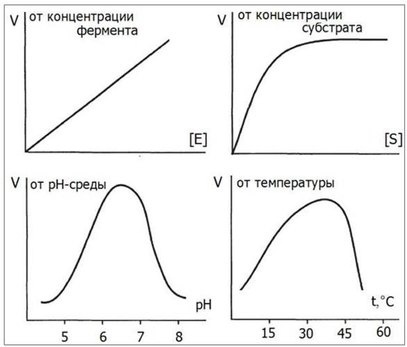
Maximum enzyme activity is observed under optimal reaction conditions. For example, gastric juice enzymes function only at a Temperature of 37 °C and in an acidic environment; therefore, during food intake, Hydrochloric acid is first secreted in The Stomach, followed by the activation of enzymes, whereas salivary enzymes are active in a slightly alkaline medium. A change in pH alters the ionization degree of the ionogenic groups of the enzyme, leading to a redistribution of inter-residue bonds. This alters the conformation of the enzyme and disrupts the complementary fit between the active site and the substrate.
Temperature changes produce a twofold effect: on the one hand, as the temperature rises to 37 °C, The rate of the enzymatic reaction increases due to the higher kinetic energy of the reacting molecules; on the other hand, at temperatures above 40 °C, enzyme denaturation begins, and the reaction rate decreases.
Activation energy is defined as The amount of energy, in calories, required for all molecules of 1 mole of a substance at a given temperature to reach the Transition State, which corresponds to the peak of the energy (activation) barrier. The peak of the activation barrier is the probability point at which substrate molecules enter a reaction to form the final reaction product. The rate of any chemical reaction is proportional to the concentration of molecules in the transition state.
There are two main ways to increase the rate of a chemical reaction. The first is increasing the temperature (a 10 °C temperature rise approximately doubles the rate of a chemical reaction), and the second is adding an enzyme with the appropriate catalytic activity.
Enzymes accelerate chemical reactions by finding "alternative pathways" that allow substrate molecules to overcome the activation barrier at a lower energy level (Fig. 24).
Fig. 24. Action of a catalytic enzyme during a chemical reaction: S – substrate, P – product

The Rate of Enzymatic reactions is measured by the depletion of the substrate or the accumulation of the product per unit of time. The change in the rate of an enzymatic reaction is directly proportional to the Enzyme Concentration (Fig. 25). If the enzyme concentration is constant, the dependence of the reaction rate on the Substrate Concentration appears as a hyperbola on the graph, resembling a protein-ligand saturation curve.
The graph also illustrates the dependence of the reaction rate on temperature and pH of the medium.
Fig. 25. Dependence of the biochemical reaction rate on substrate concentration, enzyme concentration, medium pH, and temperature
A ligand that interacts with an enzyme and disrupts its function is called an inhibitor.
There are two main types of inhibitors: irreversible and reversible.
Irreversible inhibitors destroy a functional group of the enzyme molecule that is essential for its catalytic activity. An example of an irreversible inhibitor is diisopropylfluorophosphate (DFP), which inhibits acetylcholinesterase, a key enzyme in the transmission of nerve impulses. DFP is a potent neurotoxic agent (nerve gas).
Reversible inhibitors are divided into two categories: Competitive and non-competitive.
A competitive inhibitor competes with the substrate for binding to the active site, but unlike the substrate, it does not undergo enzymatic conversion. The Effect of a competitive inhibitor can be overcome by increasing the substrate concentration. An example of a competitive inhibitor is ditilin (suxamethonium), which is used in medicine as a muscle relaxant during short-term surgical Procedures.
In non-competitive inhibition, the inhibitor binds to the enzyme not at the active site where the substrate binds, but at a different site, leading to a conformational change in the enzyme molecule and the inactivation of its catalytic activity.
In a living cell, the rate of enzymatic reactions is under strict control, allowing each metabolic pathway to continuously adjust its velocity in response to changing environmental conditions and The Cell's demand for the product.
3.4. Structural Features of Nucleic Acids and Their Role in the Transmission of Hereditary Information
Nucleic acids are complex macromolecular compounds found in all cells of living organisms. They play a leading role in the storage and transmission of hereditary information. Nucleic acids were discovered in the mid-1860s by the Swiss scientist Friedrich Miescher.
The term "nucleic acids" was proposed in 1889. They were named "nucleic" because they were first discovered in cell nuclei, and "acids" due to the presence of phosphoric acid residues in their composition. It was not until the late 1930s that the Chemical composition of Nucleic Acids was precisely determined, and it was established that there are two Types of Nucleic acids: deoxyribonucleic acid (DNA) and ribonucleic acid (RNA). Depending on their function in the cell, three main types of RNA are distinguished:
1) messenger or mRNA;
2) Transfer RNA, which form "cloverleaf" shaped molecules that bind an amino acid at one end and attach to mRNA at the other;
3) ribosomal RNA.
The Genome (the complete set of an Organism's cell DNA) encodes The structure of all proteins and molecules of that organism. During DNA Synthesis (Replication), the amount of genetic material is doubled and distributed to daughter cells upon division.
DNA Repair corrects alterations in the genetic material that occur during recombination (the EXCHANGE OF GENETIC material between Chromosomes) as well as structural damages in DNA.
Hereditary information is passed from DNA via RNA to protein (Fig. 26). The expression of this information in cells involves Transcription, or RNA Synthesis, and Translation, or Protein Synthesis.
Fig. 26. Transmission of hereditary information

During transcription in The Nucleus, messenger, transfer, and Ribosomal RNAs are synthesized, which are necessary for protein synthesis.
During translation, the information regarding protein structure, transcribed from DNA to mRNA, is translated into the amino acid sequence of proteins with the help of tRNA. The template-driven nature of nucleic acid and protein synthesis ensures The fidelity of information replication.
DNA and RNA are polymers constructed from NUCLEOTIDES. Each nucleotide consists of three components: a nitrogenous base, a pentose sugar, and a phosphoric acid residue (Fig. 27).
Fig. 27. Structure of a nucleotide

Nitrogenous bases are represented by two purine derivatives: adenine and guanine, and three pyrimidine derivatives: cytosine, thymine (in DNA), and uracil (in RNA) (Fig. 28).
Fig. 28. Structure of nitrogenous bases

The cyclic P-forms of nucleic acids contain deoxyribose (in DNA) and ribose (in RNA) (Figs. 29, 30).
Fig. 29. Structure of ribose and deoxyribose

Fig. 30. Fragment of an RNA polymer chain

The primary Structure of Nucleic Acids is the sequence of nucleotides in a polynucleotide chain linked by 3',5'-phosphodiester bonds. This results in the formation of polymers with a phosphate group at the 5'-end and a free pentose -OH group at the 3'-end.
The secondary structure of DNA is represented by a right-handed helix in which two polynucleotide chains run antiparallel and are held together by interactions between complementary nitrogenous bases. According to the base-pairing rules, adenine pairs with thymine, and cytosine pairs with guanine.
The tertiary structure of DNA is formed through its interaction with proteins, which allows DNA to be packaged into a discrete chromosome (Fig. 31).
Fig. 31. Tertiary structure of DNA (chromosome formation)

To concisely represent The nucleotide sequence in nucleic acids, a single-letter code is used. The sequence is written from left to right, such that the first nucleotide has a free 5'-phosphate end and the last one has a 3'-hydroxyl group on the ribose or deoxyribose. For example, the Primary structure of DNA can be represented as follows: CGNAAGTTCG...
Last update: 06/08/2026
Editorial and Educational Adaptation: This material has been compiled based on the primary/original source text. The project team performed an editorial review, corrected technical inaccuracies, structured sections, and adapted the content for an educational format.
What was processed:
- elimination of formatting defects (OCR errors, structural breaks, corrupted characters);
- editorial organization of content;
- standardization of terminology in accordance with academic sources;
- verification of factual statements against the original source text.
All mentions of the author, publication year, and origin of the primary text have been preserved in accordance with the source.