LEHNINGER PRINCIPLES OF BIOCHEMISTRY - VOL 1. THE FOUNDATIONS OF BIOCHEMISTRY: STRUCTURE AND CATALYSIS - 2011
PART I. STRUCTURE AND CATALYSIS
8. NUCLEOTIDES AND NUCLEIC ACIDS
8.2. Nucleic Acid Structure
The Discovery of the Introduction/20.html">DNA Structure by Watson and Crick in 1953 stands as a landmark scientific event that catalyzed The Development of entirely new fields of research and profoundly influenced existing ones. In this section, we focus primarily on The structure of DNA, some of the historical milestones surrounding its discovery, and more recent refinements that enhance our understanding of it. The Structural Organization of RNA molecules will also be examined here.
Much like Cell/13.html">Protein Structure (Chapter 4), it is useful to describe the architecture of NUCLEIC ACIDS through a hierarchy of structural organization (primary, secondary, and tertiary). The Primary Structure of a nucleic acid is defined by its covalent backbone and nucleotide sequence. Any regular, stable conformation adopted by some or all of the NUCLEOTIDES can be designated as the Secondary structure of the nucleic acid. All spatial arrangements discussed further in this chapter fall under secondary structure. Complex higher-order assemblies formed by large Chromosomes from eukaryotic Chromatin or bacterial nucleoids are generally classified as tertiary structures, which will be discussed in Chapter 24.
DNA Is a Double Helix That Stores Genetic information
DNA was first isolated and characterized by Friedrich Miescher in 1868, who referred to the phosphorus-rich substance as "nuclein." However, it was not until the 1940s—through the landmark experiments of Oswald T. Avery, Colin MacLeod, and Maclyn McCarty—that compelling evidence emerged demonstrating that DNA carries genetic information. Avery and his colleagues discovered that DNA extracted from a pathogenic strain of the bacterium Streptococcus pneumoniae (which causes severe disease in humans) could genetically transform a harmless strain of the same Organism into a virulent form. They concluded that the DNA isolated from the pathogenic strain carried the hereditary trait of virulence. In 1952, the experiments of Alfred D. Hershey and Martha Chase, which investigated the infection of bacterial Cells by Bacteriophages containing radioactively labeled DNA or protein, conclusively proved that DNA, rather than protein, harbors genetic information.
Another crucial clue to the structure of DNA came from the work of Erwin Chargaff and his colleagues in the late 1940s. They discovered that the four nucleotide bases of DNA occur in varying proportions in different organisms, yet the relative amounts of certain Base Pairs remain remarkably constant. These data, collected from a vast array of diverse species, led Chargaff to formulate several key empirical generalizations:
1. The base composition of DNA generally varies from one species to another.
2. DNA samples isolated from different Tissues of the same species have the same base composition.
3. The base composition of DNA in a given species does not change with age, nutritional state, or environmental conditions.
4. In all cellular DNAs, regardless of the species, the number of adenosine residues equals the number of thymidine residues (A = T), and the number of guanosine residues equals the number of cytidine residues (G = C). From this it follows that the sum of purine residues equals the sum of pyrimidine residues; that is, A + G = T + C.
These quantitative ratios, commonly known as "Chargaff's rules," were subsequently confirmed by numerous researchers. They played an instrumental role in unraveling the three-dimensional structure of DNA and shed vital light on how genetic information is encoded and transmitted across generations.
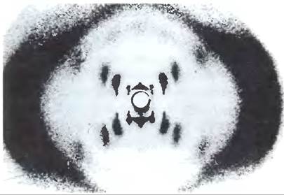
Rosalind Franklin and Maurice Wilkins employed the powerful technique of X-Ray Diffraction (Box 4-5) to investigate DNA fibers. In the early 1950s, they demonstrated that X-ray diffraction patterns of DNA exhibit distinct, characteristic features (Fig. 8-12). These diffraction photographs indicated that DNA molecules possess a periodic, helical structure with two prominent repeat intervals along their long axis: 3.4 Å and 34 Å. The challenge then was to construct a three-dimensional molecular model that accounted not only for these X-ray diffraction data, but also for Chargaff's rules of base equivalence (A = T and G = C) and other known Chemical properties of DNA.
Class="center">
Fig. 8-12. X-ray diffraction pattern of DNA. The reflections form an X-shaped pattern characteristic of a helical structure. The dark streaks along the sides are due to repeating nucleotide bases.
James Watson and Francis Crick synthesized the available data on DNA to deduce its molecular architecture. In 1953, they proposed a three-dimensional model for DNA that satisfied all the known experimental constraints. It consisted of two helical polynucleotide chains wound around a common axis to form a right-handed double helix (see Box 4-1 for an explanation of how right- and left-handed helices are defined). The hydrophilic backbones of alternating deoxyribose and phosphate groups are located on the outside of The Double Helix, exposed to the aqueous environment. Each deoxyribose furanose ring adopts a C-2' endo conformation. The purine and pyrimidine bases of both strands are stacked closely on the inside of the double helix, forming hydrophobic, nearly planar structures oriented perpendicular to the main axis of the DNA. This structural arrangement generates a major groove and a minor groove along The surface of the duplex (Fig. 8-13). Each nucleotide base on one strand pairs with a complementary base on the opposite strand within the same plane. Watson and Crick demonstrated that hydrogen-bonded base pairs of G with C and A with T, as illustrated in Fig. 8-11, fit best into their structural framework and elegantly account for Chargaff's rules that G = C and A = T in any DNA molecule. It is important to emphasize that three Hydrogen Bonds can form between G and C (denoted as G ≡ C), whereas only two form between A and T (denoted as A = T). This is one reason why separating paired DNA strands is more difficult when the proportion of G–C pairs is higher than that of A–T pairs. Non-standard base pairings lead to varying degrees of structural destabilization within the double helix.
Fig. 8-13. The Watson-Crick model of DNA structure. The original model proposed by Watson and Crick featured 10 base pairs per 34 Å (or 3.4 nm) helical turn; subsequent measurements revealed that a turn contains 10.5 base pairs and spans 36 Å (or 3.6 nm). (a) Schematic representation showing key dimensions of the helix. (b) Stick model illustrating the DNA backbone and stacked bases. (c) Space-filling model.

In constructing their model, Watson and Crick had to resolve whether the two chains of the DNA double helix were parallel or antiparallel—that is, whether their 5',3'-phosphodiester linkages ran in the same or opposite directions. The antiparallel orientation yielded a far more stereochemically plausible model, and subsequent studies on DNA polymerases (Chapter 25) provided experimental evidence confirming that the strands are indeed antiparallel, a fact later verified unequivocally by high-resolution X-ray crystallography.
To account for the periodicity observed in X-ray diffraction patterns of DNA fibers, Watson and Crick built a molecular model in which adjacent bases along the same strand within the double helix were spaced 3.4 Å apart, with the overall structure repeating every 34 Å, corresponding to 10 base pairs per complete helical turn. In aqueous solution, the structure of DNA deviates slightly from the crystalline state, possessing 10.5 base pairs per full turn of the helix (Fig. 8-13).
As shown in Fig. 8-14, the two antiparallel polynucleotide strands of DNA are identical neither in base sequence nor in composition; rather, they are complementary to one another. Whenever adenine appears in one strand, thymine is found opposite it in the other; similarly, guanine in one strand pairs exclusively with cytosine in the other.
Fig. 8-14. Complementarity of strands in the DNA double helix. The complementarity of antiparallel DNA strands is a direct consequence of the base-pairing rules postulated by Watson and Crick. Complementary antiparallel strands differ in base composition: for instance, if the left strand has the composition A3T2G1C3, the right strand will be A2T3G3C1. They also differ in sequence when read in the 5' -> 3' direction. Note, however, that the total number of paired bases in the double helix remains balanced according to A = T and G = C.

DNA is a double helix (duplex) held together by Two Types of forces described earlier: hydrogen bonding between base pairs (Fig. 8-11) and stacking interactions between the parallel planes of adjacent bases. The complementarity between DNA strands is ensured by Hydrogen bond formation between base pairs. The primary contribution to the Stability of the double helix comes from stacking interactions between adjacent bases in the same strand, which are less specific regarding base types. Important Features of the DNA double helix model are supported by a vast body of chemical and biological data. Furthermore, this model also explains the mechanism by which genetic information can be transferred. An essential characteristic of the model is the complementarity of the two DNA strands. Watson and Crick managed to deduce this feature logically, even before confirming data were obtained, predicting that such a structure could be replicated by (1) separating the two strands and (2) synthesizing a complementary strand for each helix. Because the nucleotides of each new strand are selected According to the base-pairing rules, each existing strand acts as a template for the synthesis of its complementary strand (Fig. 8-15). These explanations were subsequently confirmed experimentally, revolutionizing our understanding of biological heredity.
Fig. 8-15. Replication of DNA strands proposed by Watson and Crick. The pre-existing ("parental") DNA strands separate, and each serves as a template for the synthesis of a complementary ("daughter") strand (in red).

Example 8-1. Base Pairs in DNA
In DNA samples isolated from two unknown bacterial species (X and Y), adenine accounts for 32% and 17 of the total base composition, respectively. What is the relative content of guanine, thymine, and cytosine in the two DNA samples? What assumptions did you have to make to answer this question? One of the bacterial species was isolated from a hot spring with a Temperature of 64 °C. Which of the two species is most likely the one from the hot spring, and why?
Solution. In any DNA double helix, A = T and G = C. The DNA from bacterium X contains 32% A, and therefore it contains 32% T. Together, this accounts for 64%, meaning the remaining 36% is divided equally between G and C (18% each). The sample from bacterium Y contains 17% A, and thus 17% T, totaling 34%. The remaining 66% is distributed equally between G and C (33% each). This calculation is based on the assumption that both DNA molecules are double-stranded.
The higher the G + C content in a DNA molecule, the higher its melting temperature. It is more likely that bacterium Y, which contains a higher percentage of G+C pairs, was isolated from the hot spring. Its DNA has a higher melting temperature and, consequently, greater stability at the Water temperature of the hot spring.
DNA Can Adopt Different Spatial Configurations
DNA is a dynamic molecule. Rotation around certain bonds of the sugar-phosphate (phosphodeoxyribose) backbone is possible, and thermal motion can lead to the bending, stretching, and unwinding (melting) of the strands. A considerable variety of structures derived from the Watson-Crick DNA model have been found in cellular DNA, many of which may play important roles in DNA METABOLISM. These structural variations typically do not affect the key properties of DNA defined by Watson and Crick: strand complementarity and antiparallelism, and the fulfillment of the A = T and G = C rules.
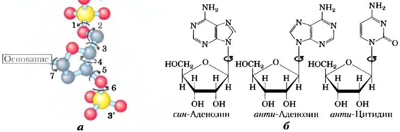
Changes in the Spatial Structure of DNA are associated with three factors: different Conformations of deoxyribose, rotation around adjacent bonds that make up the phosphodeoxyribose backbone (Fig. 8-16, a), and free rotation around the C-1'-N-glycosidic bond (Fig. 8-16, b). Due to steric constraints, Purines and purine nucleotides can exist in only two stable conformations relative to deoxyribose, termed syn and anti (Fig. 8-16, b). Pyrimidines can exist only in a single anti conformation due to steric hindrance between the sugar and the carbonyl oxygen at the C-2 atom of the pyrimidine ring.
Fig. 8-16. Variations in DNA structure. (a) The conformation of nucleotides in DNA depends on the torsion angles around seven different bonds. Free rotation is possible around six of them. Restricted rotation around the fourth bond alters the conformation of the pentose ring: one atom in the five-membered furanose ring lies out of the plane formed by the other four. This results in an endo or exo conformation, depending on whether the out-of-plane atom is on the same side of the plane as the C-5' atom or on the opposite side (Fig. 8-3, b). (b) For purine nucleotides relative to ribose, only two steric conformations are possible — anti or syn. Pyrimidines typically occur in the anti conformation.
The Spatial Organization of DNA proposed by Watson and Crick is also referred to as B-form DNA (B-DNA). The B-form is the most probable conformation for a random-sequence DNA molecule under physiological conditions, and thus B-DNA serves as the standard reference when studying any properties of DNA. Two other variants, well-characterized in crystals, are called the A- and Z-forms. These three DNA conformations are shown in Fig. 8-17, along with a brief summary of their main properties. The A-form is formed in environments with low water content. A-DNA is a right-handed double helix with 11 base pairs per turn (as opposed to 10.5 in B-DNA). The plane of the base pairs in A-DNA is tilted at an angle of approximately 20° to the helix axis. As a result of these structural changes, the major groove deepens, while the minor groove becomes almost imperceptible. Reagents used in DNA crystallization typically dehydrate it, which is why most short DNA molecules crystallize in the A-form.
Fig. 8-17. Comparison of the A-, B-, and Z-forms of DNA. Each molecule shown here consists of 36 base pairs. Bases are shown in gray, phosphorus atoms in yellow, and ribose and phosphate oxygen atoms in blue. In subsequent chapters, blue is used to depict DNA strands. The table summarizes The properties of the three DNA forms.

The Z-form of DNA differs markedly from the B-structure; The most significant difference is that it is a left-handed helix. It has 12 base pairs per turn, and the molecule itself is narrower and more elongated. The DNA backbone adopts a zigzag conformation. The left-handed helix is most commonly formed by specific nucleotide sequences. The best-known Examples of such sequences are alternating pyrimidine and purine bases, particularly alternating C and G or 5-methyl-C and G residues. Upon Formation of the left-handed helix in Z-DNA, purine residues adopt the syn conformation and alternate with pyrimidines in the anti conformation. The major groove in Z-DNA is barely noticeable, whereas the minor groove is narrow and deep.
Whether A-DNA occurs in living cells remains unknown, but evidence suggests that short fragments of Z-DNA may exist in both PROKARYOTES AND EUKARYOTES. These Z-DNA fragments may play a role (as yet undetermined) in The regulation of certain Gene expressions or in genetic recombination.
Specific DNA Sequences Can Adopt Unusual Spatial Architectures
A number of other three-dimensional DNA conformations have been found in large chromosomes, the shapes of which depend on The base sequence. This can influence the function and metabolism of adjacent DNA segments. For example, bends occur in DNA helices wherever four or more adenosine residues are strung together. Six consecutive adenosines introduce a bend of about -18°. Such curvature, generated by this or other sequences, may play a crucial role in the binding of DNA to certain Proteins.
A fairly common type of DNA sequence is the so-called palindrome. A palindrome is a word, phrase, or sentence that reads the same forwards and backwards, such as level or radar. This term is used in genetics to describe DNA regions with inverted repeats of base sequences that possess twofold rotational Symmetry across the two DNA strands (Fig. 8-18). Such a sequence is self-complementary and can therefore form hairpin or cruciform structures (two symmetric hairpins) (Fig. 8-19). If inverted repeats occur within a single DNA strand, these sequences are called mirror repeats. Mirror repeats are not self-complementary and are incapable of forming hairpins or cruciform structures. Sequences of this type are found in virtually all large DNA molecules and can range from a few base pairs to several thousand base pairs in length. The frequency with which palindromes adopt cruciform structures within The Cell is unknown, although such forms of DNA organization have occasionally been observed in vivo in Escherichia coli. Single strands of self-complementary DNA (or RNA) sequences isolate in solution as complex structures containing numerous hairpins.
Fig. 8-18. Palindromes and mirror repeats. Palindromes are sequences in a nucleic acid double helix that exhibit twofold rotational symmetry. Upon a 180° rotation around a horizontal axis followed by a 180° rotation around a vertical axis, such a structure superimposes onto itself, as indicated by the colored arrows. Mirror repeats are symmetric repeats within each individual strand. To superimpose this structure onto itself, only a single 180° rotation around the vertical axis is required.

Fig. 8-19. Hairpins and cruciforms. Palindromic DNA (or RNA) sequences can fold in various ways to form base-paired structures within a single strand. (a) When only a single DNA (or RNA) sequence is involved in the structure, the resulting conformation is called a hairpin. (b) A cruciform is a secondary structure formed by palindromic sequences involving both DNA strands. Asymmetric DNA regions that are complementary to sequences within the same or the other strand are shaded in blue.

Some unusual DNA structures are formed from three or four DNA strands. The nucleotides involved in Watson-Crick base pairing (Fig. 8-11) can form a number of additional hydrogen bonds, particularly with functional groups located in the major groove. For example, a protonated cytidine residue can bind to a guanosine residue involved in a G=C base pair, while thymidines can pair with adenosines in an A=T pair (Fig. 8-20). The N7, O6, and N6 atoms of purines can participate in hydrogen bonding within a DNA triplex; these atoms are sometimes referred to as Hoogsteen positions, and the non-Watson-Crick pairs as Hoogsteen pairs, named after Karst Hoogsteen, who first pointed out the possibility of this unusual pairing mode in 1963. These atypical hydrogen bonds give rise to triple-stranded DNA (triplexes). The triple-stranded fragments shown in Fig. 8-20 (a, b) are most stable at low pH because protonation of cytosine is required to form the G=C•C+ triplet. In the triple-stranded structure, the pKa of cytosine is >7.5, which differs from its normal value of 4.2. These structures also form more readily in long sequences containing exclusively pyrimidines or purines in one of the strands. Some triple-stranded DNA structures contain two pyrimidine strands and one purine strand, whereas others consist of two purine strands and one pyrimidine strand.
Four DNA strands can also associate to form a four-stranded structure (a tetraplex or quadruplex), but this occurs exclusively in DNA strands with a high guanosine content (Fig. 8-20, c, d). The guanosine four-stranded structure, or G-tetraplex, is quite stable over a wide range of conditions. The orientation of the strands within a tetraplex can vary, as shown in Fig. 8-20, e.
Fig. 8-20. Structures of three- and four-stranded DNA. (a) Schematic representation of base pairing in one of the well-characterized forms of triple-stranded DNA. Hoogsteen pairs are highlighted in red in each diagram. (b) DNA triple helix consisting of two pyrimidine strands (poly(T)) and one purine strand (poly(A)) (derived from PDB ID 1BCE). The dark blue and light blue strands run antiparallel and form standard Watson-Crick pairs. The third (all-pyrimidine) strand (lilac) runs parallel to the purine strand and is linked to the other strands via non-Watson-Crick hydrogen bonds. Bottom view of the triple-stranded structure, showing a stack of five base planes. The closest triplet is rendered in multiple colors, (c) Fragment of a four-stranded guanosine structure. (d) Two consecutive G-tetraplexes (derived from PDB ID 1QDG), bottom view, with the closest tetraplex rendered in multiple colors, (e) Possible strand orientation variants in G-tetraplexes.

In the DNA of living cells, the recognition sites for many specific DNA-binding proteins (Chapter 28) are represented by palindromes, whereas polypyrimidines and polypurines—which can form triple helices or even H-DNA—are found in regions involved in the regulation or expression of certain eukaryotic genes. In principle, artificial DNA strands synthesized to bind to these sequences and form triple-stranded DNA could disrupt Gene Expression. This is particularly significant given the strong commercial interest in regulating gene expression for various medical and agricultural Applications.
Messenger RNAs Contain Information for Polypeptide Chains
Having briefly examined DNA structure, we now turn to the genetic information it encodes. RNA, the second major type of nucleic acid in the cell, performs A wide variety of Functions. During gene expression, RNA acts as an intermediary, using the information encoded in DNA to direct the Amino Acid Sequence of a functional protein.
Given that eukaryotic DNA is confined to The Nucleus while Protein Synthesis takes place in Ribosomes in the Cytoplasm, genetic information must be carried from the nucleus to the cytoplasm by molecules other than DNA. Since the early 1950s, RNA has been considered the most likely candidate: these molecules were found in both the nucleus and the cytoplasm, and an increase in protein synthesis was accompanied by an increase in cytoplasmic RNA content and a higher turnover rate. These and other findings led some researchers to suggest that RNA transfers genetic information from DNA to the protein-synthesizing machinery, the ribosome. In 1961, François Jacob and Jacques Monod proposed a comprehensive (and fundamentally correct) picture of this process, introducing the term Messenger RNA, or mRNA, for the fraction of cellular RNA that carries genetic information from DNA to ribosomes, serving as a template for the assembly of a specific amino acid sequence. Although the length of mRNA can vary significantly between different genes, it has a definite, characteristic size for any given gene. The process by which mRNA is synthesized using a DNA template is called Transcription.
In Bacteria and archaea, a single mRNA molecule can encode one or more polypeptide chains. An mRNA encoding only a single polypeptide is called monocistronic; one encoding two or more different Polypeptides is called polycistronic. In eukaryotes, most mRNAs are monocistronic. (For simplicity, a Cistron is referred to here as a gene. The term itself has historical roots in genetics, and its formal definition goes beyond The Scope of this Discussion.) The minimum length of an mRNA is determined by the length of the polypeptide chain it encodes. For example, the synthesis of a polypeptide chain of 100 amino acid residues requires an RNA coding sequence of at least 300 nucleotides, since each amino acid is encoded by a nucleotide triplet (this and other details of protein synthesis are discussed in Chapter 27). However, the length of an mRNA synthesized from a DNA template is always slightly greater than strictly necessary to encode the polypeptide sequence(s). The additional, non-coding RNA sequence contains regions that regulate protein synthesis. Figure 8-21 illustrates the general structure of a bacterial mRNA.
Fig. 8-21. Bacterial mRNA. Schematic diagrams showing (a) monocistronic and (b) polycistronic bacterial mRNA molecules. Red segments represent the Regions of the RNA encoding a single gene; gray segments represent non-coding RNA regions. In the polycistronic transcript, non-coding RNA regions separate the three genes.

Many RNA Molecules Possess Highly Complex Three-Dimensional Structures
Messenger RNA is only one of several classes of cellular RNA. Transfer RNA acts as an adaptor molecule in protein synthesis; it is covalently linked to an amino acid while also being capable of complementary base-pairing with messenger RNA, ensuring that The amino acid is incorporated into the growing chain in the correct order. Ribosomal RNA functions as a Structural and functional component of ribosomes. There are also many Different types of RNA with specialized functions, including those with catalytic activity (ribozymes).
All classes of RNA are discussed in detail in Chapter 26. RNAs perform a multitude of functions, often several simultaneously, reflecting the fact that their three-dimensional structures are far more diverse than those of DNA molecules.
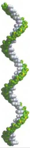
The primary product of DNA Transcription is always single-stranded RNA. This single strand typically adopts a right-handed helical conformation driven by base-stacking interactions (Fig. 8-22), which are much stronger between two purines than between a purine and a pyrimidine, or between two pyrimidines. The purine-purine interaction is so strong that a pyrimidine separating two purines may be displaced from the stack to allow direct contact between the purines. Any self-complementary sequence will form more complex structures. RNA can form base pairs with either DNA or RNA. These base pairs are virtually identical to those in DNA: G pairs with C, and A pairs with U (or with the rarely occurring residue T in certain RNAs). The only difference is that G-U base pairing is possible—which is unusual for DNA but quite common in RNA (see Fig. 8-24). Paired strands in double-stranded RNAs or RNA-DNA hybrids are antiparallel, just as in DNA.
Fig. 8-22. Typical right-handed fragment of single-stranded RNA. Bases are shown in gray, phosphorus atoms in yellow, and ribose residues along with phosphate oxygen atoms in green. In subsequent chapters, green is used to represent RNA strands, and blue is used to designate DNA strands.
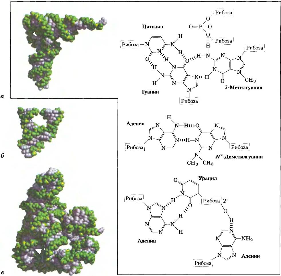
RNA lacks a regular secondary structure that could serve as a universal standard, unlike the DNA double helix. The three-dimensional structures of many RNAs, much like those of proteins, are complex and unique. Weak interactions, particularly base stacking, play a crucial role in stabilizing RNA structures, just as they do in DNA. In regions containing complementary sequences, the predominant double-stranded structure is the A-form right-handed double helix. Z-form helices have been produced under laboratory conditions (at high temperature and very high Ionic strength). The B-form of RNA has not been observed. Breaks in the regular A-form helix, caused by mismatched or unpaired bases in one or both strands, occur quite frequently, resulting in bulges or internal loops (Fig. 8-23). Nearby complementary RNA segments form hairpin loops. Because RNA inherently favors The formation of Double helices (Fig. 8-24), hairpins are the most frequently encountered secondary structure. Specific short base sequences (such as UUCG) are frequently found at the ends of RNA hairpins; these are known to form exceptionally tight and stable loops. Such sequences can serve as nucleation sites for the folding of an RNA molecule into its tertiary structure. An important additional contribution to molecular stability comes from hydrogen bonds that do not participate in standard Watson-Crick pairing. For example, hydrogen bonds can be formed by the 2'-hydroxyl group of ribose. Some of these features have been extensively characterized in the structure of Yeast phenylalanine transfer RNA (responsible for incorporating Phe residues into polypeptides) and in two RNA Enzymes, or ribozymes, whose functions—like those of protein enzymes—depend on their three-dimensional structures (Fig. 8-25).
Fig. 8-23. Secondary structure of RNA molecules. (a) Bulges, internal loops, and hairpins. (b) As shown here for a hairpin structure, double-stranded RNA regions typically adopt the A-form.

Fig. 8-24. Double-Helical structures in RNA. This illustration shows a proposed RNA secondary structure for the M1 component of RNase P from E. coli; this RNA molecule contains A large number of hairpins. RNase P, which also contains a protein component (not shown), participates in the maturation of transfer RNA molecules (Fig. 26-27). Two brackets highlight two other complementary sequences that can interact during tertiary folding. Blue dots indicate the formation of a non-Watson-Crick G=U base pair (inset). Note that G=U pairing can only occur after the RNA chains have been synthesized and fold back on themselves, or when they anneal to one another. There is no known RNA polymerase (the enzyme that synthesizes RNA molecules from a DNA template) that inserts a U opposite a G, or vice versa, during the synthesis of a new RNA molecule.

Fig. 8-25. Three-dimensional structure of RNA. (a) Three-dimensional architecture of yeast phenylalanine tRNA (PDB ID 1TRA). Some of the unusual base pairs found in this tRNA are highlighted. Note the hydrogen bonds (shown in red) between a phosphodiester bond oxygen of ribose and the 2'-hydroxyl group of another ribose. (b) A hammerhead ribozyme derived from a plant virus, featuring a hammerhead-like secondary structure (from PDB ID 1MME). Ribozymes, or RNA enzymes, catalyze a variety of reactions, primarily in RNA metabolism and protein synthesis. The intricate three-dimensional organization of these RNAs reflects their role in catalysis, much like proteins described in Chapter 6. (c) An mRNA segment identified as an intron (non-coding sequence) from the ciliated protozoan Tetrahymena thermophila (from PDB ID 1GRZ). This intron (ribozyme) catalyzes its own excision from the RNA chain region located between two exons (coding sequences) (described in Chapter 26).
Analyzing RNA architecture and uncovering the relationship between Structure and function represents an emerging research frontier, presenting challenges similar to those encountered in studying protein spatial organization. The Importance of understanding RNA Structure continues to grow as new functions of RNA molecules are discovered.
Summary of Section 8.2 Nucleic Acid Structure
■ A substantial body of diverse data indicates that DNA serves as the carrier of genetic information. In particular, the experiments by Avery, MacLeod, and McCarty demonstrated that DNA isolated from one bacterial strain can enter the cells of another strain and transform them into the first strain, conferring certain hereditary traits of the donor. Furthermore, the Hershey-Chase experiment showed that the DNA of a bacterial virus—rather than its protein coat—transfers the genetic information required for viral replication into the host cell.
■ Synthesizing a wealth of published data, Watson and Crick postulated that native DNA consists of two antiparallel strands wound into a right-handed helix. Hydrogen bonds form within the helix between complementary base pairs, A = T and G = C. These base pairs lie in planes perpendicular to the long axis of the double helix, with a distance of 3.4 Å between adjacent bases and 10.5 base pairs per helical turn.
■ DNA can exist in several conformational variants that differ in their spatial architecture. In addition to the standard Watson-Crick B-form, other principal conformations include A-DNA and Z-DNA. Certain structural variations depend on The nucleotide sequence, often resulting in bends within the DNA molecule. Furthermore, DNA strands with specific sequences can form hairpins, cruciform structures, as well as regions of triple-stranded and quadruple-stranded DNA.
■ Messenger RNA carries genetic information from DNA to ribosomes for protein synthesis. Transfer RNA and ribosomal RNA also play essential roles in Translation. The spatial structure of RNA is quite intricate; single strands can fold into hairpins, adopt double-helical regions, or form complex loops.
Last update: 06/08/2026
Editorial and Educational Adaptation: This material has been compiled based on the primary/original source text. The project team performed an editorial review, corrected technical inaccuracies, structured sections, and adapted the content for an educational format.
What was processed:
- elimination of formatting defects (OCR errors, structural breaks, corrupted characters);
- editorial organization of content;
- standardization of terminology in accordance with academic sources;
- verification of factual statements against the original source text.
All mentions of the author, publication year, and origin of the primary text have been preserved in accordance with the source.