LEHNINGER PRINCIPLES OF BIOCHEMISTRY - VOL. 1. THE FOUNDATIONS OF BIOCHEMISTRY: STRUCTURE AND CATALYSIS - 2011
PART I. STRUCTURE AND CATALYSIS
Class="center">This Structure is simply bound to exist.
James Watson, The Double Helix, 1968
8. NUCLEOTIDES AND NUCLEIC ACIDS
NUCLEOTIDES fulfill A wide variety of Functions in cellular METABOLISM. They carry chemical energy for biochemical transformations, serve as crucial chemical messengers (second messengers) in cellular responses to Hormones and other extracellular stimuli, and act as Structural components of several enzyme Cofactors and metabolic intermediates. Finally, nucleotides make up Nucleic Acids: deoxyribonucleic acid (DNA) and ribonucleic acid (RNA), which store Genetic information at THE MOLECULAR LEVEL. The structure of every protein—and ultimately of all Biomolecules and cellular components—is determined by the information encoded in The nucleotide sequence of nucleic acid molecules. The ability to store and transmit genetic information from one generation to the next is one of the fundamental prerequisites for life.
This chapter provides An Overview of The chemical properties of nucleotides and nucleic acids found in most Cell types; a detailed description of nucleic acid functions is presented in Part III.
8.1. Basic Concepts
Nucleotides are the Building Blocks of nucleic acids. The Amino Acid Sequence of every cellular protein and the nucleotide sequence of every RNA molecule are determined by the nucleotide sequence of the corresponding region of cellular DNA. A segment of a DNA molecule containing the information required to synthesize a biological product with a specific function, whether a protein or an RNA, is called a Gene. A cell typically contains many thousands of genes, and it is hardly surprising that DNA molecules are extremely large. Storing and transmitting biological information is the sole known function of DNA molecules.
RNA has a broader range of functions, and several distinct types of these molecules exist within The Cell. Ribosomal RNA (rRNA) is a component of Ribosomes—the complex assemblies where Protein Synthesis takes place. Messenger RNA (mRNA) acts as an intermediary, carrying genetic information from one or more genes to the ribosome, where the corresponding Proteins can be synthesized. Transfer RNA (tRNA) accurately translates mRNA information into the corresponding amino acid sequence. In addition to these three major classes, numerous types of RNA perform specialized functions, as described in more detail in Part III.
Nucleotides and nucleic acids contain characteristic bases and pentoses
Nucleotides consist of three characteristic components: (1) a nitrogenous (nitrogen-containing) base, (2) a pentose sugar, and (3) a phosphate group (Fig. 8-1). A molecule lacking the phosphate group is called a nucleoside. The nitrogenous bases are derivatives of two parent substances: pyrimidine and purine. In typical nucleotides, both the bases and the pentoses are heterocyclic compounds.
Fig. 8-1. Structure of Nucleotides, a) Standard numbering of pentose atoms in a ribonucleotide. In deoxyribonucleotides, the OH group at the 2' carbon atom (shown in red) is replaced by -H. b) Standard numbering of purine and pyrimidine ring atoms—compounds that serve as the foundation for the purine and pyrimidine bases of nucleotides and nucleic acids.

Key Conventions.
The carbon and nitrogen atoms in parent structures are numbered in a conventional manner to facilitate the naming and identification of numerous derivatives. The numbering of atoms in the pentose ring follows the rules given in Chapter 1, but in nucleotide pentoses, a prime symbol (') is added to the carbon numbers to distinguish them from the atoms of the nitrogenous bases. ■
The base of a nucleotide is covalently linked (via the N-1 atom of Pyrimidines and the N-9 atom of Purines) through an N-β-glycosidic bond to the 1' carbon of the pentose. The 5' carbon atom of the pentose, in turn, is esterified to a phosphate group. The N-β-glycosidic bond is formed by the removal of Water (the hydroxyl group of the pentose and the hydrogen of the base), much like the O-glycosidic bond (Fig. 7-29).
Both DNA and RNA contain two major purine bases: adenine (A) and guanine (G), and two major pyrimidine bases: both DNA and RNA contain cytosine (C), while the second pyrimidine base differs, being thymine (T) in DNA and uracil (U) in RNA. Occasionally, thymine is found in RNA and uracil in DNA. The structures of the five principal bases are shown in Fig. 8-2, and the nomenclature of their corresponding nucleotides and nucleosides is presented in Table 8-1.
Fig. 8-2. Principal purine and pyrimidine bases of nucleic acids. Some of these names reflect The history of their discovery. Guanine, for example, was originally isolated from guano (bird droppings), and thymine from the Thymus.

Table 8-1. Nucleotide and Nucleic Acid Nomenclature
Base |
Nucleoside |
Nucleotide |
Nucleic Acid |
Purines |
|||
Adenine |
Adenosine Deoxyadenosine |
Adenylate Deoxyadenylate |
RNA DNA |
Guanine |
Guanosine Deoxyguanosine |
Guanylate Deoxyguanylate |
RNA DNA |
Pyrimidines |
|||
Cytosine |
Cytidine Deoxycytidine |
Cytidylate Deoxycytidylate |
RNA DNA |
Thymine |
Thymidine or deoxythymidine |
Thymidylate or deoxythymidylate |
DNA |
Uracil |
Uridine |
Uridylate |
RNA |
Note that "nucleoside" and "nucleotide" are general terms that encompass both ribo- and deoxyribo- forms. Furthermore, ribonucleosides and ribonucleotides are often referred to simply as Nucleosides and Nucleotides (e.g., riboadenosine as adenosine), while deoxyribonucleosides and deoxyribonucleotides are called deoxynucleosides and deoxynucleotides (e.g., deoxyriboadenosine as deoxyadenosine). Both naming conventions are accepted, but the shorter variants are used more frequently. The exception is thymine; the term "ribothymidine" is used only when thymine is incorporated into an Introduction/21.html">RNA Structure.
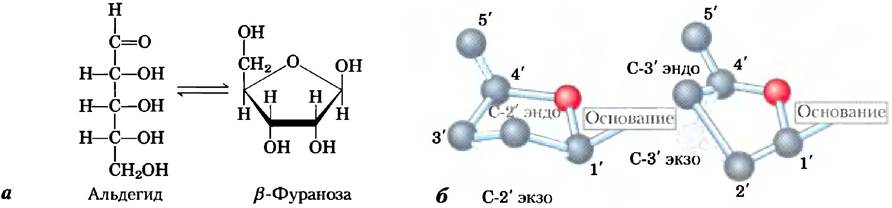
Nucleic acids contain Two Types of pentose sugars. The repeating deoxyribonucleotide units of DNA contain 2'-deoxy-D-ribose, whereas the ribonucleotide units of RNA contain D-ribose. In nucleotides, both types of pentose exist in the β-furanose form (a closed five-membered ring). As shown in Fig. 8-3, the atoms of the pentose ring are not coplanar but adopt one of several puckered Conformations.
Fig. 8-3. Conformation of ribose. a) In solution, free ribose exists in equilibrium between two forms: an open-chain (aldehyde) form and a closed-ring (β-furanose) form. In RNA, ribose occurs exclusively as the ring form, β-D-ribofuranose. Deoxyribose undergoes similar interconversions in solution, but is incorporated into DNA solely as β-2'-deoxy-D-furanose. b) The ribofuranose ring attached to a nucleotide can exist in any of four puckered conformations. In all cases, four of the five atoms are coplanar. The fifth atom (C-2' or C-3') lies either on the same side (endo) or the opposite side (exo) of the plane relative to the C-5' atom.
Key conventions.
Although There are two fundamental structural differences between DNA and RNA—their different pentoses and the presence of uracil in RNA versus thymine in DNA—it is the pentoses that define the distinct identities of these two classes of nucleic acids. If a nucleic acid contains 2'-deoxy-D-ribose, it is by definition DNA, even if it contains some uracil residues. Similarly, if a nucleic acid contains D-ribose, it is RNA, regardless of its nucleotide composition. ■
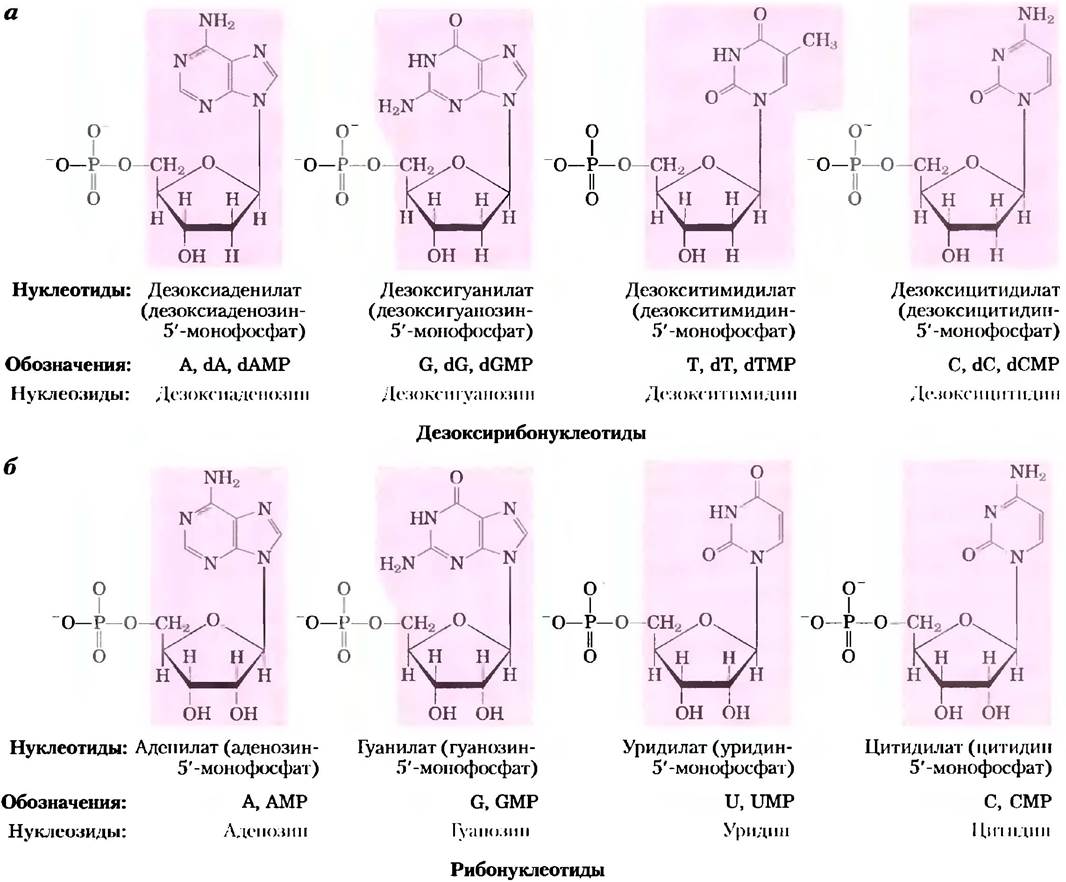
Figure 8-4 illustrates the structures and names of the four principal deoxyribonucleotides (ribonucleoside 5'-monophosphates), which serve as the structural units of DNA, and the four principal ribonucleotides (ribonucleoside 5'-monophosphates), which are the building blocks of RNA.
Figure 8-4. Deoxyribonucleotides and ribonucleotides of nucleic acids. All nucleotides are shown in their uncharged form at pH 7.0. The nucleotides of DNA (a) are commonly abbreviated as A, G, T, and C, or sometimes as dA, dG, dT, and dC; those of RNA (b) as A, G, U, and C. In their free form, the ribonucleotides are generally designated as dAMP, dGMP, dTMP, and dCMP, and the ribonucleosides as AMP, GMP, UMP, and CMP. For each nucleotide, the most commonly used trivial name appears in parentheses before the full chemical name. In all Abbreviations, the phosphate group is understood to be at the 5' position. The nucleoside moiety of each molecule is highlighted with a pink border. In this and subsequent figures, the carbon atoms of the carbohydrate ring are not explicitly labeled.
Although nucleotide compositions are dominated by the major purines and pyrimidines, both DNA and RNA also contain minor bases (Figure 8-5). In DNA, these are most commonly methylated forms of the major bases; in certain viral DNAs, specific bases may also be hydroxylated or glycosylated. These modified or uncommon bases in DNA frequently play roles in the regulation or protection of genetic information. A variety of minor bases are likewise found in RNA, particularly in tRNA (see Figures 8-25 and 26-23).
Key conventions.
The nomenclature of minor bases can be somewhat confusing. Like the major bases, most minor bases have trivial names; for example, Figure 8-5 shows the structure of hypoxanthine as a component of the nucleoside inosine. When a substituent is added to a purine or pyrimidine ring, the name of the new compound simply indicates the substituent and the number of the ring atom to which it is attached—for instance, 5-methylcytosine, 7-methylguanine, and 5-hydroxymethylcytosine (shown as nucleosides in Figure 8-5). The specific atom (N, C, or O) to which the substituent is attached is not specified. The naming convention changes if the substituted atom lies outside the ring structure; in such cases, the type of atom bearing the substituent is indicated, along with the ring position number written as a superscript. The nitrogen atom of the amino group attached to the C-6 atom of adenine is designated N6; similarly, the carbonyl oxygen and the amino nitrogen attached to the C-6 and C-2 atoms of guanine are designated O6 and N2, respectively. Examples include N6-methyladenosine and N2-methylguanosine (Figure 8-5). ■
Figure 8-5. Some minor purine and pyrimidine bases (uncommon bases found in nucleic acids), shown as nucleosides. (a) Minor bases of DNA. 5-Methylcytidine is found in animal and higher plant DNA, N6-methyladenosine in bacterial DNA, and 5-hydroxymethylcytidine in the DNA of Bacteria infected with certain Bacteriophages. (b) Selected minor bases of tRNA. Inosine contains the base hypoxanthine. Note that pseudouridine, like uridine, contains uracil; these compounds differ in the position at which the base is attached to the ribose—in uridine, the ribose is linked to the N-1 atom of uracil, as in other pyrimidine-containing nucleosides, whereas in pseudouridine, it is linked to the C-5 atom.

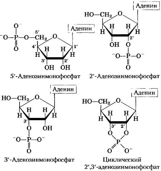
Cells also contain nucleotides in which phosphate groups are attached to positions other than the 5'-carbon (Figure 8-6). Cyclic 2',3'-ribonucleoside monophosphates are stable intermediates that can be isolated, whereas 3'-ribonucleoside monophosphates are formed as end products when RNA is hydrolyzed by certain ribonucleases. Other notable examples, such as cyclic 3',5'-adenosine monophosphate (cAMP) and cyclic 3',5'-guanosine monophosphate (cGMP), are discussed at the end of this chapter.
Figure 8-6. Several adenosine monophosphates. 2'-Adenosine monophosphate, 3'-adenosine monophosphate, and cyclic 2',3'-adenosine monophosphate are formed through the action of specific Enzymes and during the alkaline Hydrolysis of RNA.
Nucleotides in nucleic acids are joined sequentially by phosphodiester bonds
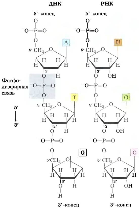
The nucleotides in DNA and RNA are covalently linked by phosphodiester bridges, in which the 5'-phosphate group of one nucleotide is joined to the 3'-hydroxyl group of the next, forming a phosphodiester bond (Figure 8-7). Thus, the covalent backbone of nucleic acids consists of alternating phosphate and pentose residues, with the nitrogenous bases serving as side groups protruding from the backbone at regular intervals. The backbones of both DNA and RNA are hydrophilic. The hydroxyl groups of the sugar residues form Hydrogen Bonds with water. The phosphate groups have a pKa near 0 and are completely ionized at pH 7; their negative charges are largely neutralized by ionic interactions with the positive charges of proteins, Metal Ions, and Polyamines.
Key conventions.
All phosphodiester bonds in DNA and RNA have the same polarity (Figure 8-7), giving each nucleic acid strand a distinct orientation with defined 3' and 5' ends. By definition, the 5' end lacks a nucleotide at the 5' position, and the 3' end lacks a nucleotide at the 3' position. Additional groups (most commonly one or more phosphates) may be present at either or both ends. The convention of writing nucleic acid sequences from 5' to 3' refers to the overall orientation of the sequence ends rather than the directional vector of each individual phosphodiester bond linking the nucleotides. ■
Figure 8-7. Phosphodiester bonds in the covalent backbone of DNA and RNA. Phosphodiester bonds (one is highlighted in the DNA molecule) link the nucleotides together in a HEAD-to-tail fashion. The backbone of alternating pentose and phosphate groups exhibits pronounced polarity in both DNA and RNA. The 5' end of the macromolecule lacks a nucleotide at the 5' position, whereas the 3' end lacks a nucleotide at the 3' position.
The phosphodiester bonds in DNA and RNA molecules undergo slow non-Enzymatic hydrolysis. In vitro, RNA is hydrolyzed quite rapidly under alkaline conditions, whereas DNA is stable; this process directly involves the 2'-hydroxyl groups of RNA (which are absent in DNA). The initial products of alkaline Treatment on RNA are cyclic 2',3'-nucleotide monophosphates, which are subsequently hydrolyzed to yield a mixture of 2'-nucleoside and 3'-nucleoside monophosphates (Figure 8-8).
Figure 8-8. Alkaline hydrolysis of RNA. The 2'-hydroxyl group acts as a nucleophile in an intramolecular displacement reaction. The cyclic monophosphate intermediate is rapidly hydrolyzed to a mixture of 2'- and 3'-monophosphates. DNA, which lacks a 2'-hydroxyl group, is stable under these conditions.

The nucleotide sequence of a nucleic acid can be represented schematically, as shown on the following page for a five-nucleotide DNA segment. Phosphate groups are depicted as  , and each deoxyribose sugar is represented by a vertical line extending from the C-1' atom at the top to the C-5' atom at the bottom (keeping in mind that the sugar in nucleic acids always adopts a closed β-furanose conformation). The bonds between nucleotides (which pass through
  ) are drawn as diagonal lines running from the midpoint (C-3') of the deoxyribose of one nucleotide to the bottom (C-5') of the next.
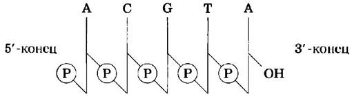
Here are examples of other simplified notations: pA-C-G-T-AOH, pApCpGpTpA, and pACGTA.
Key conventions.
Historically, The sequence of a single nucleic acid strand is always depicted starting from the 5'-end and ending at the 3'-end, oriented from left to right in the 5' —> 3' direction. ■
Short nucleic acids are called oligonucleotides. The definition of "short" is somewhat arbitrary, but polymers containing 50 or fewer nucleotides are generally referred to as oligonucleotides. Longer chains of nucleic acids are called polynucleotides.
The properties of nucleotide bases influence the three-dimensional Structure of Nucleic Acids
Free pyrimidines and purines possess weak basic properties and are therefore called bases. The purines and pyrimidines found in DNA and RNA are conjugated molecules (Fig. 8-2), which significantly affects the structure, electron distribution, and Light absorption spectrum of nucleic acids. The delocalization of ring atom electrons imparts partial double-bond character to many of the bonds. Consequently, pyrimidine molecules are flat, while purine molecules are nearly flat (with a slight bend). Free purine and pyrimidine bases can exist in two or more tautomeric forms depending on the pH. Uracil, for example, can exist in lactam, lactim, and dilactim forms (Fig. 8-9). Figure 8-2 shows the structures that predominate in solution at pH 7.0. Due to Resonance, all nitrogenous bases absorb UV light, and nucleic acids are characterized by an absorption maximum around 260 nm (Fig. 8-10).
Fig. 8-9. Tautomeric forms of uracil. The lactam form predominates at pH 7.0; other forms become more prominent as the pH decreases. Other free pyrimidines and purines can also adopt various tautomeric forms, but these occur much less frequently.

Fig. 8-10. Absorption spectrum of major nucleotides. The spectrum is shown as the molar extinction coefficient plotted against wavelength. Molar extinction coefficients at 260 nm and pH 7.0 (ε260) are given in the table. The spectra of the corresponding ribonucleotides and deoxyribonucleotides, as well as nucleosides, are virtually identical. A wavelength of 260 nm (dashed vertical line) is used to measure the absorption of a nucleotide mixture.

Purine and pyrimidine bases are hydrophobic and poorly soluble in water at near-neutral cellular pH values. In acidic and alkaline environments, the bases become charged, and their solubility in water increases. Hydrophobic interactions between adjacent bases, whose ring planes lie parallel to one another (like a stack of coins), represent one of the two most important types of noncovalent bonding between bases in nucleic acids. The bonding between parallel base planes (known as stacking interactions) is also maintained by Van der Waals forces and dipole-dipole interactions. Stacking the bases in a coin-like Column minimizes their contact with water, and the interactions between parallel bases are crucial for stabilizing the three-dimensional structure of nucleic acids, as described below.
The most important Functional groups of pyrimidines and purines are the ring nitrogen atoms, carbonyl groups, and exocyclic amino groups. Hydrogen bonding between amino and carbonyl groups serves as the second major type of interaction between bases in nucleic acids. Hydrogen bonds between bases drive the complementary association between two (and occasionally three or four) nucleic acid strands. The atoms involved in hydrogen bonding were identified by James D. Watson and Francis Crick in 1953. They discovered that adenine (A) specifically pairs with thymine (T) (or uracil U), and guanine (G) with cytosine (C) (Fig. 8-11). These two base-pairing patterns predominate in the double-Helical structures of DNA and RNA, and this specific mode of Organization is governed by the tautomers shown in Fig. 8-2. This specific base-pairing ensures The fidelity of genetic Replication, as we will see later in this chapter.

Fig. 8-11. Hydrogen bonds in Watson-Crick Base Pairs. Here, as elsewhere, Hydrogen bonds are represented by three blue lines.

Summary of Section 8.1 Basic Concepts
■ Nucleotides consist of a nitrogenous base (purine or pyrimidine), a pentose sugar, and one or more phosphate groups. Nucleic acids are polymers of nucleotides linked together by phosphodiester bonds between the 5'-hydroxyl group of one nucleotide's pentose and the 3'-hydroxyl group of the next.
■ There are two Types of Nucleic acids: RNA and DNA. Nucleotides in RNA contain ribose and the major pyrimidine bases uracil and cytosine. In DNA, nucleotides contain 2'-deoxyribose and the major pyrimidine bases thymine and cytosine. The typical purines found in both DNA and RNA are adenine and guanine.
Last update: 06/08/2026
Editorial and Educational Adaptation: This material has been compiled based on the primary/original source text. The project team performed an editorial review, corrected technical inaccuracies, structured sections, and adapted the content for an educational format.
What was processed:
- elimination of formatting defects (OCR errors, structural breaks, corrupted characters);
- editorial organization of content;
- standardization of terminology in accordance with academic sources;
- verification of factual statements against the original source text.
All mentions of the author, publication year, and origin of the primary text have been preserved in accordance with the source.