BIOTECHNOLOGY - V. H. Gerasymenko - 2006
Part I. General Biotechnology
Chapter 3. BASICS OF MOLECULAR BIOLOGY
3.1. NUCLEIC ACIDS
3.1.2. Structure of Nucleic Acids
Primary Structure. Nucleotide residues in a polynucleotide chain are linked by phosphodiester bonds between the 5'-OH group of the pentose of one nucleotide and the 3'-OH group of the pentose of another nucleotide. In abbreviated representations of nucleic acid formulas, the carbohydrate component is depicted as vertical lines with the symbols of nitrogenous bases positioned above them. Sometimes, nitrogenous bases are simply designated as Purines (Pu, Pr) or Pyrimidines (Pi, Py). Phosphodiester bonds between adjacent nucleotide residues are shown as diagonal lines with the chemical symbol for phosphorus (P) in the middle:
Class="center">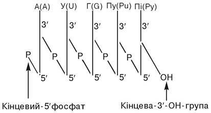
In these and even more abbreviated formulas of polynucleotides, the 5/-end of the nucleotide residue is conventionally placed on the left:

or

Hydrolysis of the polynucleotide compound will result in The formation of guanylic acid as the terminal nucleotide with a free phosphate at the 5'-end and free adenosine at the 3'-end.
The sequence or alternation order of mononucleotide residues in a polynucleotide chain determines the Introduction/19.html">Primary structure of DNA and RNA. The Formation of the primary structure involves the glycosidic bond connecting the nitrogenous bases to the pentose, the ester bond between ribose or deoxyribose and phosphoric acid, and the phosphodiester bond between NUCLEOTIDES. All these bonds are covalent and stabilize the primary structure quite firmly. It has been established that nucleotides in the polynucleotide chains of DNA and RNA are linked via 3', 5'-phosphodiester bonds. The nucleotide sequence in a DNA polynucleotide chain is presented using various schematic formats:
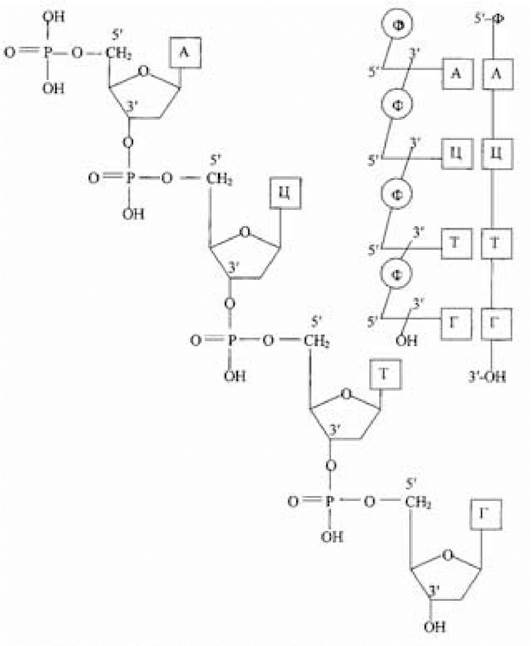
Interactions between purine-pyrimidine Base Pairs that secure the linkage of polypeptide chains in DNA are shown in Fig. 3.1. No branching has been detected in the DNA polypeptide chain.
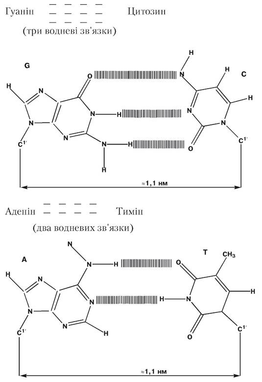
Fig. 3.1. Purine-pyrimidine base pairs in DNA
(after Bohinski R., 1986)
Elucidating the primary structure of DNA involves immense difficulties due to the fact that the Molecular Weight of even the smallest DNA molecules is calculated in millions of daltons. Consequently, initiated and supported by the USSR and the USA in 1988, the major international Human Genome Project was launched, with the ultimate goal of mapping the complete human genome and deciphering nucleotide sequences in both exonic and intronic DNA fragments. In 2001, but without the participation of the USSR, which had by then ceased to exist, data on the primary structure of human DNA were published for the first time. Thousands of scientists from more than 20 countries contributed to solving this challenge. They established that the 23 pairs of Chromosomes in The Nucleus of a human somatic Cell contain approximately 3.2 billion mononucleotide pairs, excluding the mitochondrial nucleotide composition. Interestingly, it has been reported that the total length of DNA in the nuclei of all human Cells reaches an astronomical value of ~1011 km, which is nearly 1,000 times the distance from the Earth to the Sun.
Secondary structure. Studies on the Chemical composition of Nucleic Acids revealed significant variations in the relative content of nitrogenous bases among different DNAs; however, the molar ratio of adenine to thymine, as well as cytosine to guanine, in all studied DNAs remained approximately 1:1. That is, the number of adenine residues equals the number of thymine residues, and the number of guanine residues equals the number of cytosine residues. Based on these data, E. Chargaff (1950) proposed METABOLISM/2.html">THE CONCEPT OF base pairing in DNA, or the complementarity rule, according to which nitrogenous bases interact with each other during DNA Biosynthesis in any Organism: adenine pairs with thymine, and guanine with cytosine.
R. Franklin and M. Wilkins used X-Ray Diffraction Analysis to obtain data suggesting that DNA molecules likely possess a multi-chain helical structure.
The Double Helix model of DNA was proposed in 1953 by J. Watson and F. Crick based on their analysis of X-ray diffraction patterns of DNA fibers obtained by R. Franklin and M. Wilkins. J. Watson and F. Crick established that DNA is a double helix composed of two antiparallel polynucleotide chains. The most crucial aspect of the proposed structure is the pairing of nitrogenous bases from the antiparallel chains through hydrogen bonding, which can only occur if, along the double-helical structure of DNA, adenine pairs with thymine and guanine with cytosine. Nitrogenous bases joined by Hydrogen Bonds are termed complementary, and The process of selective interaction between adenine and thymine, and guanine and cytosine, is called complementarity. The scheme of nitrogenous base pairing can now be considered experimentally proven. The nucleotide sequences in the strands of the DNA double-helical structure are complementary, but not identical. The Stability of the DNA double helix is ensured by two hydrogen bonds between thymine and adenine, and three hydrogen bonds between cytosine and guanine (Fig. 3.2). The complementarity of individual nitrogenous bases results in the DNA deoxyribonucleotide strands being complementary as a whole. The scheme demonstrates that in one strand, the linkage between nucleotides runs in the 5'→3' direction, while in the other, it runs in the opposite 3'→5' direction (Note: corrected typographical range to standard biological notation based on context). The antiparallel orientation of complementary strands has a biological basis during DNA Replication and Transcription.
In addition to hydrogen bonds between hydrogen, nitrogen, and oxygen atoms in complementary nitrogenous bases, so-called stacking interactions play a significant role in stabilizing the DNA double helix. These arise between nitrogenous bases due to hydrophobic forces acting at Van der Waals Contact distances.
X-ray diffraction analysis of fibers and crystals has shown that there are Three types of DNA Double helices (A-, B-, and Z-forms) and one type of RNA double helix. A- and B-DNAs are right-handed helices, whereas the Z-form is left-handed. The B-form of DNA is stable at a relative humidity exceeding 92%; however, when humidity drops below 76%, most deoxyribonucleotide sequences adopt the A-form. Both forms resemble flexible ladders spiraled around a central axis. Alternating deoxyribose residues and phosphate groups serve as the handrails of the "ladder," while the steps consist of complementary purine-pyrimidine base pairs.
The attachment sites of nitrogenous bases to deoxyribose residues in complementary DNA strands are not located directly opposite each other relative to the helix axis. This affects the overall conformation of DNA. On the side of the helix axis where the angle between deoxyribose rings is less than 180°, There is a groove known as the minor or glycosidic groove (toward which the glycosidic bonds connecting deoxyribose residues to nitrogenous bases are directed); on the opposite side lies the major groove, or non-glycosidic furrow. In the DNA double-helical structure, phosphate groups form the walls of the Major and minor grooves, while the "edges" of purine and pyrimidine bases form the floor. The floor of the major groove contains nitrogen and oxygen atoms capable of forming hydrogen bonds with the side chains of protein amino acid residues, playing a crucial role in recognition processes.
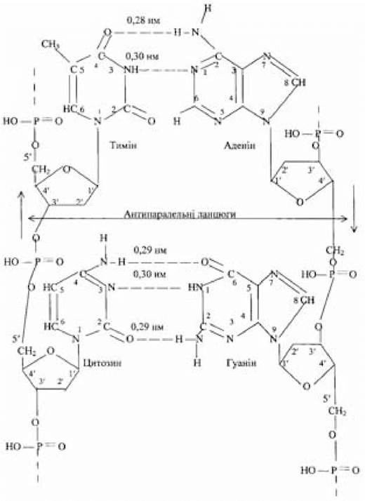
Fig. 3.2. Antiparallel complementary strands of a DNA fragment
(adapted from Berezov T.T. and Korovkin B.F., 1983)
In the purine and pyrimidine bases that form complementary pairs, the arrangement of groups capable of hydrogen bonding varies. In the A-T pair, moving from adenine to thymine, there is first a nitrogen atom acting as a hydrogen acceptor, followed by an NH group acting as a hydrogen donor, and finally an oxygen atom acting once again as an acceptor. In the complementary G-C pair, the sequence is a nitrogen acceptor atom, followed by an oxygen acceptor, and then an NH2 hydrogen donor group. Because each complementary pair can be viewed in the reverse direction (T-A and C-G), there are four possible combinations of donor and acceptor groups in the DNA strand that can be recognized by a repressor or other regulatory Proteins. Consequently, the information encoded in DNA AS A sequence of nitrogenous bases can be decoded by any other large molecule via the major groove. The minor groove is less informative, serving a different role in B-form DNA.
A- and B-forms of DNA differ primarily in the spatial arrangement and tilt angle of the base pairs relative to the helical axis. In the B-form, the planes of the bases are nearly perpendicular to the helical axis passing through the centers of the base pairs. The minor groove is narrower than the major groove due to the Asymmetry of the attachment points of the nitrogenous bases to the deoxyribose residues, while the depths of both grooves are approximately equal. In the A-form of DNA, the base-pair plane deviates from the perpendicular to the helical axis by 13-19°; the base pairs are displaced toward the outer surface, causing the helical axis to lie within the major groove without passing through the base pairs. This configuration of A-DNA results in significant differences in groove depth: the minor groove is shallow, whereas the major groove is deep. In B-DNA, there are an average of 10 base pairs per helical turn, with an axial distance of 0.34 nm between adjacent pairs; in A-form DNA, a turn comprises about 11 base pairs, reducing this distance to 0.29 nm; the distance between turns (the pitch of the helix) is 3.4 nm (for the Crick-Watson model and the B-form) and 2.8 nm for the A-form. The Z-form contains approximately 12 residues per turn. The diameter of the double helix is 1.8 nm (2 nm between phosphorus atoms) for the Crick-Watson model and the A-form, and 1.7 nm for the B-form. These parameters, derived from The Study of DNA fibers, represent averaged values.
Data obtained from the study of DNA crystals and processed mathematically indicate substantial deviations from these mean values. A comparison of the Crick-Watson model with the CHARACTERISTICS OF THE B- and A-forms of DNA shows that in terms of the number of base pairs per turn and the orientation of the base-pair plane relative to the helical axis, the B-form coincides with the Crick-Watson model; however, the displacement of base pairs relative to the axis (and the resulting central channel), as well as the positioning of the deoxyribose residues, correspond more closely to their conformation in the A-DNA helix. Parameters such as the helical twist angle (the rotation angle between two adjacent base pairs), the number of nitrogenous base pairs per turn, the axial distance between adjacent base pairs, and others determined by X-ray diffraction analysis and predicted from DNA fiber studies are in good agreement.
Of particular interest and requiring explanation are the significant deviations from average values observed in local helical twist angles, propeller twist angles (which measure the rotation of two bases of a pair in opposite directions around their connecting longitudinal axis), opening angles (in the A-form, the DNA helix wraps around the central axis, creating gaps between base pairs that open into the minor groove like accordion bellows), and other parameters. Local helical twist angles for B-DNA range from 28° to 42°, and for A-DNA from 16° to 44°. Based on these findings, it has been suggested that such conformational variations in the helix are recognized by repressors or other regulatory proteins. Furthermore, nucleotide sequence is hypothesized to influence not only Genetic information but also The regulation of its expression, where the local transition of specific regulatory regions—for example, from a right-handed B-conformation to a left-handed Z-form—can significantly affect the accessibility of genetic information and its readout process (e.g., by altering the supercoiling degree of covalently closed circular DNAs). DNA exists in the B-form during replication (DNA Synthesis on a DNA template), whereas the DNA-RNA hybrid temporarily formed during transcription is believed to adopt an A-helix structure.
The Secondary structure of RNA remains insufficiently studied. The presence of a hydroxyl group at the second carbon atom of ribose prevents RNA from forming a B-type double helix. In a hypothetical B-type double helix, the distance between the oxygen atom of the 2'-hydroxyl group of ribose and some of its surrounding atoms becomes so small that the B-form of RNA is stereochemically impossible; in the A-form, the 2'-OH group of ribose is located On the surface of the helix and is thus well-separated from adjacent atoms. Therefore, self-complementary double-stranded regions in tRNA must be Variants of the A-helix. Short self-complementary double-stranded segments in RNA (so-called hairpins) are also most likely represented by the A-form. Currently, there is a single known example of the B-form of RNA: a synthetic hybrid consisting of an adenylyl ribonucleotide strand and a thymidylyl deoxyribonucleotide strand. There is as yet no evidence for a Z-form of RNA. The secondary STRUCTURE OF THE entire tRNA molecule is most likely the cloverleaf-like planar model proposed by Holley (Fig. 3.3).
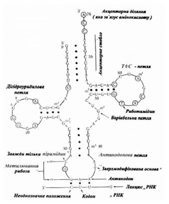
Fig. 3.3. Diagram of a phenylalanine tRNA molecule
(adapted from Metzler D., 1980)
Tertiary structure. To study the Tertiary Structure of DNA, it must be obtained in an intact (undamaged) state. To date, viral, mitochondrial, and chloroplast DNAs have been isolated in this manner. The length of the double-helical molecule in a human chromosome, if fully stretched, could reach 8 cm; in reality, its length is 5 nm. This remarkable compaction is achieved through supercoiling of the secondary structure. The degree of supercoiling (the presence of additional supercoils or superhelices) is determined by Changes in the sedimentation constant. Supercoils are frequently found in circular DNA molecules. For instance, the E. coli chromosome consists of a single closed circle. Circular DNA molecules are also common in Mitochondria, certain Viruses, and eukaryotic nuclei. Typically, circular molecules twist back on themselves, forming supercoiled molecules containing superturns. The self-twisting of the double helix results in a tightly wound right-handed structure, a phenomenon known as negative supercoiling. As a result of supercoiling, each superturn accounts for 20-25 turns of the double helix. Thanks to supercoiling, extremely long DNA molecules (1,360 µm in E. coli and 990,000 µm in humans) are packed into the tiny volume of a bacterial cell or Eukaryotic Cell nucleus. DNA topoisomerases have been isolated that catalyze supercoiling (DNA gyrases) as well as relax supercoiled structures (DNA relaxases); the existence of DNA in a supercoiled state has been confirmed using Electron Microscopy. The supercoiling of eukaryotic DNA occurs with the participation of histone proteins, which are divided into five main classes based on their Lysine and Arginine content (Table 3.2).
Table 3.2.
Lysine and arginine content in Histones, %
Histone |
Lysine |
Arginine |
H1 |
24.8 |
2.6 |
H2a |
10.9 |
9.3 |
H2b |
16.0 |
6.4 |
H3 |
9.6 |
13.3 |
H4 |
10.8 |
13.7 |
The polycationic nature of histones enables their interaction with the polyanionic pentose-phosphate backbone and, alongside hydrogen bonds, stabilizes The structure of eukaryotic DNA. In mitochondrial, chloroplast, and prokaryotic DNAs, the factor stabilizing these polyanionic macromolecules is not protein, but rather inorganic
cations. A significant contribution to the stabilization of DNA Structure is made by hydrophobic interactions among densely packed nitrogenous bases stacked in the core of the helix. The interaction of DNA with histones leads to the formation of nucleosomes, the structural units of Chromatin (Fig. 3.4). In each nucleosome particle, a DNA fragment 100-200 nucleotide pairs long is wrapped around a histone core, which is an octameric structure containing two molecules each of histones H2a, H2b, H3, and H4. Adjacent nucleosomes are connected by linker DNA regions (spacer regions) about 50 base pairs long; these spacer regions include one molecule of histone H1 as well as non-histone proteins. Histones are also thought to potentially participate in the Regulation of Gene activity. The structural heterogeneity of chromatin and its structure-related functional activity are largely driven by covalent modifications (Acetylation, phosphorylation, methylation, etc.) of the Amino Acids in histone proteins. Supercoiled DNA can be converted into an open (relaxed) circular form by breaking one or both strands of the double helix via brief enzymatic Treatment. The resulting relaxed molecular form sediments more slowly. Supercoiling affects the viscosity of DNA solutions and the electrophoretic mobility of macromolecules. In some cases, superturns can be observed using an Electron microscope. DNA is a dynamic and readily modifiable structure. The transition of supercoiled DNA into an open circular molecule is an essential step in the replication process.
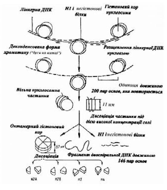
Fig. 3.4. Diagram of nucleosome structure
(adapted from Alberts B. et al., 1986)
The supercoiled structure of DNA can also be revealed by strand breakage (of one or both strands) under The Influence of intercalating agents (intercalation is the insertion of flat aromatic rings between DNA base pairs). Reagents that induce intercalation include certain drugs, Dyes, and other substances. The Use of such compounds carries a certain risk because intercalating agents are mutagenic. As the degree of intercalation increases, the turns of the DNA secondary structure unwind: each intercalating ring causes a 26° unwinding of the helix. In intact cells, intercalating agents may include the aromatic rings of amino acid side chains during protein-DNA interactions. Changes in supercoil density caused by intercalation or alterations in the ionic environment may have biological significance for Genetic regulation, particularly in maintaining sequential order
in the interaction of DNA with intracellular enzyme systems. Other modes of spatial DNA packing are also possible.
Single-stranded Ribonucleic Acids (messenger, ribosomal, and Transfer RNA) at physiological pH, Ionic strength, and Temperature possess numerous complementary regions (so-called hairpins) that determine the stability of their tertiary structure. The planar, cloverleaf-like structure of native tRNA molecules transforms into a compact structure through the folding of its various segments.
In some phages, the DNA molecule consists of a single strand; conversely, Some viruses contain double-stranded RNA whose structure resembles that of DNA.
Thus, it should be emphasized that the DNA helix contains Two Types of information, encoded and read out differently: the actual genetic information that determines Protein Structure, and information that serves as a kind of "instruction" for the selective reading of a particular record fragment. The storage and realization of both types of information are based on Physical and Chemical processes largely determined by the functional groups that make up nucleic acids. Ionized phosphate groups within nucleic acids impart a negative charge to them, which is why DNA in the organism exists in complexes with positively charged proteins (histones and protamines), Polyamines, and metals. The presence of free OH groups at the second carbon atom of the ribose residue in RNA largely dictates the conformation of these polymeric macromolecules. Polar groups 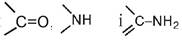 of nitrogenous bases in nucleic acids are capable of forming hydrogen bonds with proteins as well as between complementary nitrogenous bases in the double-helical DNA structure. Consequently, DNA and RNA are reactive compounds. Methylation reactions of nitrogenous bases occur relatively easily, making the view of DNA as a chain containing only four types of nucleotides somewhat simplified. Methylated nitrogenous bases undergo hydroxylation to yield hydroxymethyl derivatives. Methylation takes place following the Synthesis of the polynucleotide. Both methylation and the hydroxylation of methyl derivatives hold biological significance.
Methylation protects DNA from the action of Enzymes when viruses enter The Cell. In addition, it is hypothesized that methylated nitrogenous bases serve as markers for specific regions of genetic copies. Modifications in the RNA molecule are quite common. Stacking interactions take place between nitrogenous bases, and flat aromatic rings of amino acid side chains and other Aromatic Compounds become embedded (intercalation). Under the influence of nitrous acid, the NH2 group of nitrogenous bases is converted into an OH group:

consequently, cytosine is converted into uracil, adenine into hypoxanthine, and guanine into xanthine; formaldehyde reacts with the NH2 group. Hydroxylamine (H2N-OH) reacts even with those carbonyl  groups, especially in pyrimidines, that are part of the cyclic structure. Uracil and thymine to a greater extent, and cytosine, which are part of nucleic acids, to a lesser extent, undergo dimerization and hydrogenation under the influence of ultraviolet rays. Ultimately, this can cause a mutagenic effect.
The range of variations in the deoxyribonucleotide composition of DNA in living organisms is very wide: in prokaryotes, cytosine and guanine content ranges from 22 to 74%; in eukaryotic organisms, from 28 to 58%; and in mammals, from 35 to 45%. The content of cytosine and guanine in DNA is sometimes used to infer the phylogenetic relationship of organisms. At the same time, one must take into account the high photochemical sensitivity of thymine to ultraviolet light, which explains why a high content of guanine and cytosine is observed in Bacteria living in well-lit habitats, and a low content in those living in environments protected from the sun.
The Nucleotide Composition of DNA in organisms of the same species does not depend on age, nutritional conditions, or other factors. A constant quantitative DNA content per cell is also a distinct characteristic, regardless of the tissue from which it is isolated (Germ Cells are an exception, containing half as much DNA as somatic cells); in Cells of the same tissue across different animal species, The amount of DNA varies significantly (picograms per cell): human — 6.8; chicken — 2.3; crocodile — 5.0; carp — 3.5; Yeast — 0.05; Escherichia coli — 0.014; avian pox virus — 2.7·10-4; phage φX174 — 2.6·10-6.
DNA replication. The construction of the double-helical DNA model allowed J. Watson and F. Crick in the same year, 1953, to formulate a hypothesis regarding the replication mechanism of this macromolecule. In their hypothesis, which was experimentally confirmed in experiments using stable 14N and heavy 15N nitrogen isotopes (Meselson M. and Stahl F., 1958), J. Watson and F. Crick referred to important Structural and functional properties of DNA—base-pairing Specificity and strand complementarity. Thus, the hypothesis that DNA replication is semi-conservative was confirmed; in this mode of replication, one strand of each daughter DNA molecule is newly synthesized, while the other strand originates from the parent molecule. The double helix can be separated into its original complementary strands by heating the DNA solution (Fig. 3.5) or by ionizing the nitrogenous bases through The addition of acid or alkali. Upon heating at a well-defined temperature, the double helix unwinds almost instantaneously—this process is called melting, and the temperature at which half of the double helix separates is called the melting temperature (Tm), which depends on The ratio of G-C and A-T base pairs. The predominance of G-C pairs, connected by three hydrogen bonds, causes an increase in Tm. DNA regions where A-T pairs predominate melt earlier. Cooling the solution below Tm leads to the spontaneous reformation of the original double helix. This property of the double helix, associated with the breakdown and restoration of hydrogen bonds between complementary nitrogenous bases in complementary strands, plays a crucial biological role.
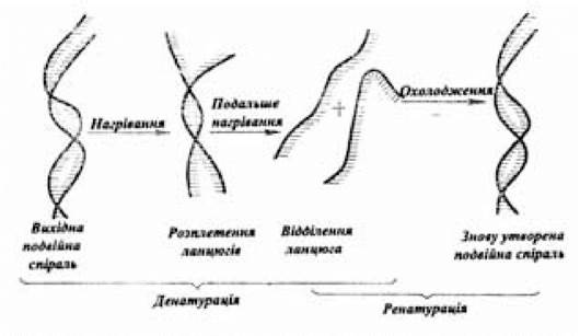
Fig. 3.5. Scheme of DNA Denaturation and renaturation
(according to Bohinski R., 1987)
Many proteins with enzymatic Functions participate in the complex process of DNA replication. As a result, A. Kornberg and his coworkers (1965) isolated, purified to a homogeneous state, and thoroughly studied DNA polymerase I, which takes part in replication. To carry out the biosynthetic function, along with DNA polymerase I, the reaction mixture must contain: a complete set of deoxyribonucleoside 5'-phosphates (dATP, dGTP, dTTP, dCTP), magnesium ions, a primer strand with a free 3'-OH end (The Role of the primer is played by a preceding DNA or RNA strand), and a template strand, which can be single- or double-stranded DNA (double-stranded DNA performs the template function provided that the integrity of its deoxyribose-phosphate backbone is disrupted). The synthesis of the polynucleotide chain is carried out as a result of a nucleophilic attack by the 3'-OH end of the template on the phosphorus atom closest to the ribose residue of only that deoxyribonucleoside triphosphate whose base is complementary to the corresponding Base of the template strand. This forms a phosphodiester bond and releases PPi, the hydrolysis of which meets the Energy Requirements of the polymerization reaction. Chain elongation proceeds in the 5'>3' direction. In one second, a DNA polymerase molecule elongates the chain by approximately 10 nucleotide residues:

DNA polymerase I also exhibits 3'→5' exonuclease and 5'→3' nuclease activity. In the first case, DNA polymerase I always removes non-complementary base residues from the 3'-OH end before attaching the next nucleotide, meaning it performs a proofreading function; the removed nucleotide must not be incorporated into the double helix (Stryer L., 1985). In the case of 5'→3' nuclease activity, DNA polymerase I hydrolyzes DNA only in Regions of the double-helical structure starting from the 5' end. In this way, pyrimidine dimers formed by UV-induced DNA damage are eliminated. Following DNA polymerase I, DNA polymerases II and III were isolated and studied. Like DNA polymerase I, they synthesize DNA starting from the 3'-OH end in the 5'→3' direction, using the same deoxyribonucleoside triphosphate precursors; in addition, DNA polymerase III, like DNA polymerase I, possesses 5'→3' nuclease activity. The role of DNA polymerases in replication is that DNA polymerase III ensures the synthesis of the bulk of the newly formed DNA, while THE CONTRIBUTION OF DNA polymerase I comes down to removing the primer and filling in the gaps. Little is yet known about the Biological Role of DNA polymerase II. Another enzyme discovered in 1967, DNA ligase, participates in the mechanisms of DNA Replication and Repair; it catalyzes the formation of a phosphodiester bond in the presence of a free OH group at the 3' end of a DNA chain and a phosphate group at the 5' end of the same chain within the backbone of the double-helical DNA structure. This reaction repairs single-strand breaks. The participation of DNA ligase is also necessary for normal DNA synthesis, the repair of damage to this macromolecule, and for joining (splicing) strands in recombinant DNA biotechnology.
The experimental hypothesis regarding the precise localization of the DNA replication origin has been confirmed. In E. coli DNA, such a site is a unique sequence near a gene. The region where DNA unwinding and replication occur simultaneously is called the Replication fork, from which The biosynthesis of daughter DNA strands proceeds simultaneously in two opposite directions. The convergence of replication forks at the point diametrically opposite to THE ORIGIN OF replication indicates that polymerization proceeds at the same speed in both directions.
DNA polymerases I, II, and III synthesize daughter DNA strands in the 5'→3' direction. Since the parental strands are antiparallel, the direction of synthesis for one of the daughter strands should be 3'→5', which contradicts the previously stated view. This contradiction was resolved when R. Okazaki determined that a portion of the daughter DNA is synthesized as fragments approximately 1,000 nucleotide residues long each. The daughter strand synthesized in the 5'→3' direction is called the leading strand and is synthesized continuously; the strand consisting of Okazaki fragments is called the lagging strand and is also synthesized in the 5'→3' direction. As synthesis proceeds, Okazaki fragments are joined together by DNA ligase, resulting in an overall chain growth direction of 5'→3'. At the same time, the synthesis process of both the leading and lagging DNA strands begins from the 3'-end of An RNA primer containing a free OH group. The synthesis of a short RNA primer chain (about 10 nucleotides) on one of the DNA template strands is catalyzed by a specialized RNA polymerase (primase) that does not require a primer. The 3'-OH terminal group of the primer is subsequently used to extend the DNA chain with the help of DNA polymerase III, whereas the RNA primer oligonucleotide fragment is hydrolyzed by DNA polymerase I; with the participation of this enzyme, the gaps formed after the removal of the RNA primer are filled with the corresponding nucleotide sequences, and DNA ligase seals the ends of the fragments. A mandatory condition for replication is the necessity of unwinding the parental DNA double helix in the region of the replication fork. During unwinding, the energy of approximately two ATP molecules is expended per base pair split with the participation of the rep enzyme (helicase). Then, single-stranded regions are stabilized by SSB proteins (single-strand binding proteins) that bind to them. Positive supercoils arising during the unwinding of circular DNA are resolved with the participation of the enzyme DNA gyrase, which acts as a molecular swivel by introducing negative supercoils into the parental DNA. The complex replication mechanism is necessary to ensure its high fidelity. According to genetic analysis data, a single error occurs per 109–1010 base pairs read (Stryer L., 1985).
RNA Synthesis. The process of transcription in Prokaryotic Cells is catalyzed by a single RNA polymerase. The transcription apparatus in Eukaryotic cells includes three RNA polymerases, one of which (RNA polymerase II) transcribes protein-coding genes; RNA polymerase I participates in the biosynthesis of high-molecular-weight ribosomal RNA, and RNA polymerase III in the synthesis of low-molecular-weight RNAs (5S ribosomal RNA, tRNA, etc.). The transcription process, the initiation of which is determined by a specific DNA sequence (promoter), proceeds in the 5'→3' direction. The termination of transcription is regulated by another specific DNA nucleotide sequence (termination signal). Both bacterial and eukaryotic RNA polymerases have approximately the same molecular weight—500 kDa; however, the Structure of Prokaryotic RNA polymerase is simpler. It consists of five polypeptide chains, whereas eukaryotic cell RNA polymerase consists of 9–11 polypeptide subunits.
The content of RNA polymerases in a cell is influenced by such an integral indicator of the functional state of a cell or organism as the growth rate. However, data (Alberts B. et al., 1986) indicate that a single higher eukaryotic cell contains approximately 40,000 molecules of RNA polymerases I and II; the number of RNA polymerase I molecules is about half as much. The average length of the nucleotide sequence synthesized with the participation of RNA polymerase II on a transcription unit (a region of DNA bounded by a specific promoter signal and a transcription termination signal for RNA polymerase II) averages 8 kb. This value exceeds by more than 6 times the Amount of Information required for the synthesis of an average-length protein molecule (400 amino acid residues).
Of the total amount of RNA contained in the Cytoplasm of mammalian cells, 95–97% is ribosomal RNA, and about 3–5%, or 360,000 molecules, is mRNA; thus, there is about one mRNA molecule per dozen Ribosomes.
The packaging of newly synthesized RNA molecules through interaction with proteins makes them appear on micrographs similar to DNA-Protein Complexes within nucleosomes. There is no convincing data on the Functional Significance of this interaction.
It is hypothesized that the formation of RNA-nuclear protein complexes is necessary to ensure the Processing of primary RNA transcripts and their subsequent transport into the cytoplasm. Polynucleotide sequences whose growth rate is 30 nucleotides per second and in the biosynthesis of which RNA polymerase II participates form the heterogeneous nuclear RNA (hnRNA) fraction. Many molecules of this fraction located in the nucleus undergo covalent modifications, thereby acquiring functional specialization. The process of covalent modifications includes capping the 5' end of the RNA synthesized by RNA polymerase II and attaching a polynucleotide fragment consisting of 100–200 adenosine monophosphate residues to the 3'-OH end of the same RNA molecule using poly(A) polymerase. These covalent modifications, leading to the formation of the primary RNA transcript, are apparently necessary for normal RNA Processing and The transport of mature mRNA molecules from the nucleus to the cytoplasm. After about 30 minutes, primary transcripts of RNA polymerase II are detected in the cytoplasm. Their total amount is about 5% of the RNA mass that made up the hnRNA fraction. The remaining 95% of RNA polymerase II primary transcripts are degraded in the nucleus within about an hour of their synthesis. At the same time, the sizes of the primary RNA transcripts decrease from 6–8 thousand nucleotide residues (hnRNA fraction) to 1.5 thousand (cytoplasmic mRNA). This is achieved during RNA processing and its conversion into mature mRNA by excising introns (non-coding sequences) ranging from 0.1 to 10 kb in size, the number of which can reach several dozen (over 50 in the procollagen α-chain gene). The RNA fragments remaining after the removal of intron sequences are spliced together and, in the form of mature mRNAs—accounting for 1–2% of The Genome's nucleotide sequences—are transported into the cytoplasm and translated. Prokaryotic RNA transcripts are translated in the form in which they are synthesized (Fig. 3.6).
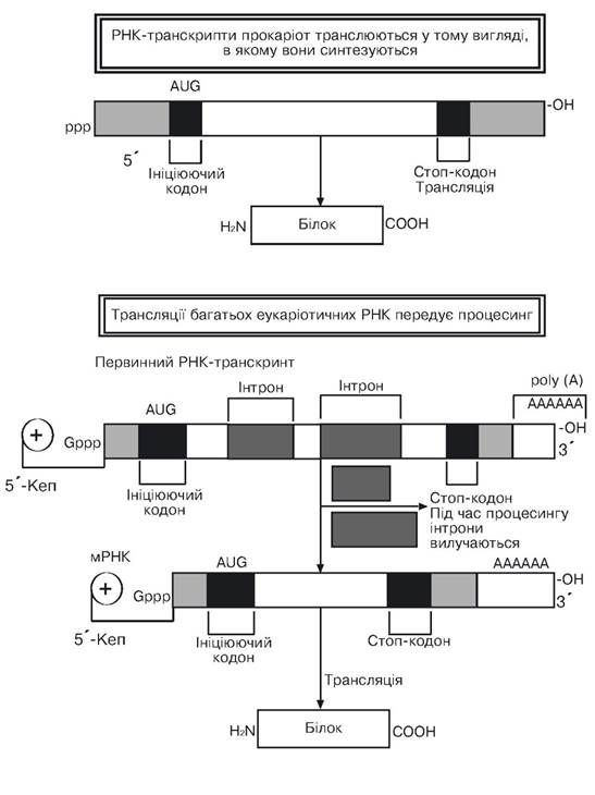
Fig. 3.6. Primary structure of prokaryotic and eukaryotic RNA transcripts
(according to Alberts B. et al., 1986)
The opinion is expressed about the possible participation of small nuclear ribonucleoprotein particles (snRNPs) in The Mechanism of intron excision and RNA splicing. It is important to maintain an optimal ratio between specific gene products to ensure a defined level of metabolic activity. Thus, certain proteins (erythrocyte Hemoglobin, Muscle cell Myoglobin) present in high amounts in the cell are encoded by genes represented in the haploid genome by single copies. However, through Translation—where about ten protein molecules are produced per minute using a single mRNA molecule, and more than 104 protein molecules during a single Cell Cycle—a high level of hemoglobin and myoglobin is achieved in a specialized cell. In another case, the final products of genes are ribosomal and transfer (rRNA and tRNA) nucleic acids, the demand for which is quite high (during one cell cycle, about 107 molecules of each of the four types of Ribosomal RNAs—28S, 18S, 5.8S, and 5S—must be synthesized, which would suffice to assemble 107 ribosomes). A solution to this problem is achieved through the gene copy number encoding the respective rRNAs and tRNAs (up to 200 copies of rRNA genes in human haploid cells, and about 600 in Xenopus).
The genes are located as tandemly repeated deoxyribonucleotide sequences separated by untranscribed regions (spacers). The primary transcripts of RNA polymerase I during the transcription of rRNA genes are 45S rRNA (13 kb), from which single copies of 28S rRNA (about 5 kb), 18S rRNA (2 kb), and 5.8S rRNA (0.16 kb) are formed and used in ribosome assembly, while a certain portion of the primary RNA polymerase I transcript mass (about 6 kb) is degraded in the nucleus. The nucleolus is the site of ribosome formation. However, after leaving the nucleus into the cytoplasm, the ribosome maturation process continues. Clusters of tandemly repeated 5S rRNA genes, as well as various tRNA genes, are transcribed by RNA polymerase III (Alberts B. et al., 1985).
Last update: 11/08/2026
Editorial and Educational Adaptation: This material has been compiled based on the primary/original source text. The project team performed an editorial review, corrected technical inaccuracies, structured sections, and adapted the content for an educational format.
What was processed:
- elimination of formatting defects (OCR errors, structural breaks, corrupted characters);
- editorial organization of content;
- standardization of terminology in accordance with academic sources;
- verification of factual statements against the original source text.
All mentions of the author, publication year, and origin of the primary text have been preserved in accordance with the source.