Biochemistry and Molecular Biology - Belyasova, N. A. 2002
Structure and Functions of Cellular Components
Cellular Polysaccharides
Structure of Prokaryotic Cell Walls
Most prokaryotes have a rigid, multilayered Cell wall On the surface of their Plasma Membrane, which provides mechanical protection and determines cell shape. The Cell wall may be separated from the membrane by a periplasmic space containing Enzymes and nutrient-binding Proteins. Unlike The Plasma Membrane, the cell wall is permeable to salts and low-molecular-weight compounds. The cell walls of most prokaryotes are based on the peptidoglycan murein. However, the Composition and Structure of cell walls vary significantly between the two major prokaryotic taxa (Gracilicutes and Firmicutes), which forms The basis of the differential Gram staining method for Bacteria.
Cell wall of Gram-positive bacteria. It consists almost entirely of murein. This heteropolymer contains alternating residues of N-acetylglucosamine and N-acetylmuramic acid linked by β(1→4)-glycosidic bonds. The murein molecule has a unique structure because it contains D-Amino Acids, diaminopimelic acid, and a y-peptide bond (Fig. 5.4).
The structural peculiarity of murein is primarily manifested in the Features of the tetrapeptide within the N-acetylmuramic acid moiety. The first position is occupied by L-Alanine, and the second by D-isoglutamic acid (or a minor amino acid). Isoglutamic acid forms a peptide bond with L-Lysine via its y-carboxyl group (y-peptide bond) rather than the usual a-carboxyl group. The third position in the tetrapeptide is occupied by L-lysine, which possesses a side-chain amino group, although it can be substituted by other amino-bearing acids (diaminopimelic or diaminobutyric) (Fig. 5.5). The final position in the tetrapeptide is always occupied by D-alanine. D-Amino acids within murein confer enhanced resistance to Proteolytic Enzymes.
Class="center">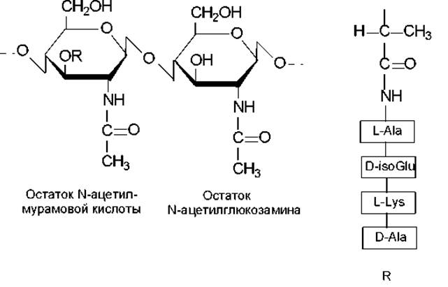
Fig. 5.4. STRUCTURE OF THE monomeric unit of the murein molecule
Linear peptidoglycan chains, consisting of alternating disaccharide-peptide units (Fig. 5.4), form the backbone of murein. Its structure is further elaborated by short Peptides that cross-link the polysaccharide chains. Numerous cross-links result in a single, giant sacculus macromolecule known as the murein sacculus (or peptidoglycan meshwork). These cross-links are most commonly pentaglycine peptides (bridges), though Other Amino Acids may also be incorporated, such as L-alanine, L-Threonine, or L-Serine. During cross-link formation, the carboxyl group of one terminal Glycine residue in the pentaglycine bridge forms a peptide bond with the side-chain amino group of lysine within the N-acetylmuramic acid tetrapeptide of another chain, while the free amino group of the other terminal residue in the pentaglycine bridge forms a peptide bond with the free carboxyl group of the terminal D-alanine residue in the N-acetylmuramic acid of the same or a different murein chain (Fig. 5.6). Thus, a single tetrapeptide can participate in forming two interchain cross-links via pentaglycine bridges.
The resulting complex murein network is reinforced by Teichoic Acids that coat or permeate the peptidoglycan layers. Teichoic acids are chains of glycerol or the pentitol ribitol molecules linked together by phosphodiester bridges (Fig. 5.5). A single teichoic acid molecule contains about 30 alcohol residues. Amino acid or sugar residues may be incorporated into this chain. The hydroxyl groups of the alcohols in teichoic acids serve to link the peptidoglycan layers together, thereby increasing the mechanical strength of the murein sacculus.

Fig. 5.5. Structure of individual components of Introduction/4.html">Prokaryotic Cell walls

Fig. 5.6. Murein structure: NAG — N-acetylglucosamine residue in the linear peptidoglycan chain; NAM — N-acetylmuramic acid residue; (Gly)5 — pentaglycine bridge cross-linking the amino acids within the N-acetylmuramic acid tetrapeptide
The cell wall of Gram-positive bacteria contains approximately 40 layers of murein, accounting for 30–70% of the dry weight of the cell wall. The wall maintains cell rigidity and shape even when the cellular contents are removed, for example, by acid dissolution.
Cell wall of Gram-negative bacteria. The cell wall of Gram-negative bacteria has a more complex Organization than that of Gram-positive bacteria. Its main distinguishing features are as follows: the murein network is single-layered and accounts for only 5–10% of the cell wall's dry weight; teichoic acids are absent; and a lipid-rich outer membrane overlies the peptidoglycan layer (Fig. 5.7). Structurally, Gram-negative murein differs from that of Gram-positive bacteria by having a lower degree of cross-linking and a lesser variety of diamino acids.
The outer membrane constitutes a thick layer of the cell wall and is composed of lipopolysaccharides, Phospholipids, and proteins. The fundamental architecture of this layer closely resembles that of the plasma membrane. A phospholipid layer lies directly above the murein layer, with the molecules oriented so that their hydrophilic heads face the murein and their hydrophobic tails face the lipopolysaccharide layer. Lipopolysaccharides serve as the major Components of the outer membrane and possess an unusual structure. Their molecules consist of three regions: lipid A, the core polysaccharide, and O-Antigens.
Lipid A—the backbone of this complex molecule—consists of disaccharide units formed by two N-acetylglucosamine residues linked by β-(1→4)-glycosidic bonds, which are repeated multiple times and interconnected by phosphodiester bridges. Fatty acid residues are attached to the N-acetylglucosamine residues, forming a lipid bilayer together with phospholipids. Protein molecules are embedded within this lipid bilayer formed by lipopolysaccharides and phospholipids. Spanning the bilayer, these transmembrane transport proteins are responsible for the Transport of substances across the outer membrane. Polysaccharide molecules are attached to the exterior of lipid A and can be divided longitudinally into two parts: the core (comprising 8–10 monosaccharide residues and having a nearly identical structure across almost all Gram-negative bacteria) and O-antigens. The latter represent the variable portion of the outer Polysaccharides and are most commonly formed by tetrasaccharide sequences that can repeat up to 50 times within a single molecule (Fig. 5.7).

Fig. 5.7. Structure of the Gram-negative Bacterial cell wall
Unlike the acyl portion of lipid A, the core Oligosaccharides and O-side chains are highly hydrophilic. Several sugars in the lipopolysaccharide are phosphorylated, giving the molecule an overall negative charge. To maintain the Stability of the lipopolysaccharide layer, Ca2+ ions are present in high concentrations. The O-side chains are located at the extreme cell surface, forming the so-called "molecular brush." They act as receptors for the adsorption of many Bacteriophages and serve as the primary antigenic determinants of the Gram-negative bacterial cell wall. Furthermore, they are referred to as endotoxins because the virulence of bacterial Cells in animals is determined by the presence and type of O-side chains. When isolated cell walls of specific bacterial species are administered to laboratory animals in experiments, the symptoms of the corresponding disease are replicated, and the animals develop Immunity against bacteria bearing the administered cell walls.
The outer membrane is linked to the murein network by Lipoproteins, which attach to the murein via diaminopimelic acid. Meanwhile, the lipophilic (hydrophobic) portions of the lipoproteins are embedded in The Lipid Bilayer, acting as an "anchor."
Various chemical agents can affect prokaryotic cell walls, notably widely used agents such as the enzyme Lysozyme and the antibiotic penicillin. The mechanisms of these actions will be discussed in subsequent sections. A common feature of these and similar agents is that, in hypertonic environments, they lead to The formation of protoplasts (forms completely devoid of a cell wall) or spheroplasts (forms partially devoid of a cell wall). These entities are characterized by osmotic fragility (undergoing lysis in hypotonic environments) and are capable of regenerating their cell walls under suitable conditions. Protoplasts and spheroplasts are widely used in Genetic Engineering for Cell Fusion and transformation experiments to generate hybrid bacteria.
Last update: 06/08/2026
Editorial and Educational Adaptation: This material has been compiled based on the primary/original source text. The project team performed an editorial review, corrected technical inaccuracies, structured sections, and adapted the content for an educational format.
What was processed:
- elimination of formatting defects (OCR errors, structural breaks, corrupted characters);
- editorial organization of content;
- standardization of terminology in accordance with academic sources;
- verification of factual statements against the original source text.
All mentions of the author, publication year, and origin of the primary text have been preserved in accordance with the source.