Antibiotics (Properties, Applications, Interactions) - M.P. Cherenko 1999
Death of Tissue (Necrosis), Ulcers, Bedsores, Fistulae, and Foreign Bodies
Necrosis (from the Greek nekros, meaning dead) refers to the localized death of living tissue or Organs within a living Organism. Small, localized areas of tissue death, typically affecting external or surface Tissues, are referred to as necrosis. The destruction of larger tissue regions, such as segments of extremities or Internal Organs, is termed gangrene (e.g., Gangrene of the FOOT, lower leg, intestine, Gallbladder, or Lungs).
Cellular death and the shedding of individual tissues—such as the epithelium of the Skin or mucous membranes, Blood Cells, and Connective Tissue—is a physiological process inherent to the natural renewal of the organism. This process occurs continuously within the body, imperceptibly or nearly so, without causing tissue defects or pathological symptoms. Physiological tissue renewal through the death of Aging cells and the generation of new ones is known as necrobiosis. Necrobiosis also encompasses pathological processes, such as dystrophies, which have a slow progression, diminish the viability of tissues or organs, and may ultimately culminate in their complete death, or necrosis. Tissue necrosis can be triggered by the action of numerous detrimental external and internal agents (mechanical, physical, chemical, and biological) upon tissues and the organism as a whole. Biological factors comprise a broad category of external and internal agents. The most frequent mechanical cause of necrosis is direct cellular and tissue injury, such as contusion, crushing, laceration, and fragmentation. Mechanical trauma damages tissue membranes and architecture, disrupts (ruptures) or compresses Blood and Lymphatic
vessels that supply nutrients and facilitate metabolic exchange, thereby leading to the death—necrosis—of tissue within the affected zone. Mechanical trauma is a frequent etiological factor in necrosis, and its pathological (necrotic) impact is predominantly complex in nature. A significant proportion of tissue necroses are caused by thermal agents, specifically high and low temperatures (Burns, frostbite). A wide range of other physical factors, such as electrical current and X-ray or radioactive radiation (gamma, alpha, beta, neutron), occupy a prominent place in the Etiology of necrosis, including cases resulting from their therapeutic application.
Active chemical substances are also frequent etiological factors in necrosis. These develop both on the body surface due to the action of chemicals on the skin and mucous membranes (chemical burns) and within internal organs following ingestion or systemic absorption (e.g., renal tubular necrosis from mercury poisoning, or necrosis of central hepatocytes resulting from mercury). Biological agents, pathogenic microbes, and Viruses cause a vast number of necrotic lesions. Such properties are characteristic of pyogenic infection pathogens, and particularly of anaerobic clostridia, non-spore-forming anaerobes, and agents of specific infections. Examples of necrosis of infectious origin include carbuncles, Hematogenous Osteomyelitis with sequestration, gas gangrene, bacteroid cellulitis, fasciitis and Phlegmon, tuberculous cavities, syphilitic gummatous necrosis, fusospirochetal lesions (such as noma, or so-called Cancer of the cheek), and fungal necroses.
Most commonly, both exogenous and particularly endogenous causes of tissue necrosis are rooted in impaired trophism associated with Circulatory Disorders (ischemia) and neurological dysfunction. These include disruptions of Blood Circulation in tissues and organs caused by the mechanical compression of Blood Vessels and the cessation of blood flow (compression of limbs and their vessels by heavy solid objects, tourniquets, or tight bandages; incarceration of the intestine along with its mesentery and vessels within a hernial ring or volvulus around its mesentery; pressure ulcers resulting from the compression of soft tissue vessels between underlying bony prominences and hard mattress surfaces), as well as the restriction or arrest of blood flow due to thrombosis, Various Forms of embolism (fat, air, thromboembolism, atherosclerotic), occlusion of other geneses, prolonged vasospasm, and other factors. Among the intravascular pathological processes that obstruct or drastically reduce vascular lumens, arterial atherosclerosis ranks first, frequently complicated by thrombosis, while isolated thrombosis or thromboembolism of vessels occurs less frequently.
Atherosclerotic lesions of the carotid artery frequently lead to ischemic Brain necrosis, whereas coronary artery disease complicated by thrombosis results in myocardial infarction.
Arterial and venous thrombosis and thromboembolism, which invariably develop acutely and are therefore particularly hazardous regarding the onset of organ necrosis, also hold a prominent position among intravascular factors that block vascular lumens, with detrimental consequences for tissue blood supply. Circulatory impairment (ischemia) serves as the primary mediator of necrosis induced by numerous other etiological factors, such as frostbite, infection, and Nervous system dysfunction. Neurotrophic necroses frequently arise following injuries to the spinal Column and brain, as well as trauma to peripheral nerve trunks.
The development, rate, and extent of tissue necrosis—particularly that associated with circulatory disorders and certain other biological and physical factors—depend on a multitude of circumstances, namely:
1) The Nature of the tissue. Brain cells are the most sensitive to ischemia, undergoing Cell death within minutes, followed by parenchymal organs (Liver, Kidneys, Spleen, Testes, and myocardium). Skin, Muscles, adipose tissue, and the intestine display somewhat higher tolerance, withstanding a cessation of blood flow for 3–5 hours. Bones, Cartilage, ligaments, and tendons exhibit the highest resistance to compromised blood supply;
2) The localization of the tissue or organ, as well as the degree of collateral circulation development within them. For instance, the distal portions of the lower extremities—the toes and feet—have the poorest Blood supply and are therefore the first to undergo necrosis upon insufficient blood inflow caused by the ligation or occlusion of major Arteries, or by other factors disrupting circulation (cooling, vasospasm). Ligation of the popliteal artery leads to Necrosis of the distal extremity in one-third of patients due to insufficient development of collaterals between the upper and lower segments, whereas ligation of the brachial artery in the elbow region is rarely complicated by hand gangrene, owing to well-developed collaterals between the arm and forearm;
3) The rate of circulatory impairment. When blood inflow to an organ decreases gradually, necrosis develops later or may not occur at all, as sufficient time is available for collateral vessels to form and develop, maintaining adequate blood supply. Conversely, the abrupt cessation of BLOOD FLOW IN major vessels precipitates necrosis much more frequently. For example, embolism or Thrombosis of the vessels of a lower extremity very commonly results in gangrene, whereas gradual arterial occlusion (in the thigh and lower leg) may fail to induce necrosis altogether. This principle applies equally to other organs;
4) The condition of the vessels themselves, including their walls and lumens. Pathological alterations in vessel walls leading to thickening, sclerosis, and a loss of elasticity and flexibility are accompanied by a narrowed vascular diameter and impaired tissue blood supply;
5) The overall state of The Cardiovascular system. Circulatory insufficiency, tissue congestion, edema, Hypoxia, and hypotension all promote The Development of necrosis;
6) Metabolic disturbances, particularly involving Proteins and CARBOHYDRATES. Hypovitaminosis, anemia, and other pathological states increase susceptibility to necrosis;
7) Disorders of the nervous and endocrine regulatory systems governing organs and tissues exert a profoundly negative impact on tissue viability. Neurotrophic disorders in patients with Spinal Cord trauma and other injuries to the central and peripheral nervous systems are widely recognized. Functional disruptions of the Pituitary Gland, Adrenal Glands, Pancreas, and Parathyroid glands (such as Diabetes Mellitus and hyperparathyroidism) provoke necrobiosis and tissue necrosis;
8) Deficient patient care and compromised sanitary-hygienic conditions occasionally play a decisive role in the etiology of necrosis (pressure ulcers, skin maceration caused by secretions of Bile, intestinal and pancreatic juices, or urine; necrosis resulting from improperly applied casts, etc.);
9) Infection, which frequently complicates tissue trauma—including surgical trauma, particularly involving interventions within the gastrointestinal tract and organ inflammation—can induce Tissue and organ necrosis both directly and indirectly (via circulatory disturbances such as vascular thrombosis). Thus, even a minor wound complicated by Putrefactive infection may, under appropriate conditions, progress to extensive tissue necrosis (necrotizing fasciitis and cellulitis). Thrombophlebitis of the mesenteric Veins in purulent Peritonitis following gastric or intestinal surgery is accompanied by necrosis (gangrene) of significant intestinal segments.
Meticulous tissue handling during surgery, minimizing operative trauma, preventing circulatory compromise during wound closure (avoiding excessive tension on wound edges and applying the justified minimum of sutures), preventing tissue compression (the tourniquet effect) during bandage application, and strictly adhering to asepsis rules during surgical interventions, dressings, and medical Procedures can minimize the risk of infection and necrosis.
The progression and rate of necrosis also depend on the Nature of the injurious agent. High temperatures—particularly flames, molten or heated metals—as well as strong acids and bases provoke rapid and overt necrosis, whereas ischemic necrosis manifests in most tissues within an average of 5–6 hours, and necrosis induced by low temperatures becomes apparent after 40 hours.
Macroscopically, dead tissues differ from living ones in color, appearing gray-pale, yellowish, or dark brown (or even black); they lack translucency (appearing dull), remain unresponsive to stimuli, and are either firm or soft and liquefied. However, the external manifestations of necrosis largely depend on the morphological type of necrosis, the affected tissue, and the nature of the causative agent. Two primary forms of necrosis are distinguished: coagulative (or dry) necrosis and colliquative (or wet) necrosis.
Dry necrosis (Fig. 92) predominantly develops As a result of either a progressively slow reduction in blood supply (ischemia of tissues with low fluid content) or the action of Physical and Chemical agents that induce rapid Water evaporation and coagulation of cellular protoplasmic proteins.
Dry necrosis can preserve tissue architecture—such as that of skin, muscles, tendons, blood vessels, nerves, and bones—for extended periods.
Wet necrosis develops more frequently than dry necrosis and arises under METABOLISM/18.html">The Influence of most injurious factors, with the exception of flames, concentrated acids, and certain other agents that rapidly dehydrate tissues. In addition to tissue characteristics, the rate of necrotic development, and the nature of the necrotizing agent, wet necrosis is promoted by secondary infection, predominantly putrefactive. Necrosis of internal organs (gangrene) invariably exhibits a wet Morphology (Fig. 93).
The macroscopic morphological feature distinguishing wet necrosis from dry necrosis is the absence of a distinct demarcation zone—a clear line of Separation between necrotic and healthy tissues—along with the blurring of this boundary. Wet necrosis, particularly when massive (gangrene), is characterized by severe systemic intoxication resulting from both tissue breakdown products and complicating infection, posing a serious threat to the organism's viability. Wet necrosis affecting tissues located either superficially or deep within the body (organ necrosis) is accompanied by tissue breakdown and conversion into an amorphous mass.
In superficial gangrenes, observed primarily in the lower extremities and originating in the distal segments (the toes and feet), the necrotic process progressively encompasses all anatomical structures of the region: the skin, subcutaneous adipose tissue, tendons, and bones.
Due to the resistance of all these structures to dissolution, their breakdown proceeds predominantly slowly (starting from the skin), even in cases of rapidly progressive necrosis of the foot in a proximal direction. Putrid ulcers form only on the toes or the foot, whereas the majority of the necrotic skin area covers the underlying necrotic and necrobiotic structures like a sleeve, along which there is almost always an accumulation of pus and detritus proximal to the visibly necrotic skin. The foot is markedly edematous, signs of inflammation (lymphangitis, phlegmon) are noticeable On the surface of still-living skin, and the edema also extends to the lower leg and sometimes the thigh. Due to the presence of a putrefactive infection, the gangrenous tissues emit a sharp odor.
Class="center">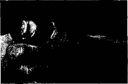
Fig. 92. Dry gangrene of the first toe

Fig. 93. Gangrene of a Small Intestine loop in an incarcerated hernia
Ischemic necrosis of brain tissue, infectious necrosis of lung tissue (abscess and gangrene), frequently tuberculous or caseous necrosis, as well as a significant number of gummatous necroses, culminate in the dissolution of the necrotic tissue with The formation of a liquid mass (tissue detritus and exudate). In the absence of infection, this mass in the brain tissue can be resorbed, forming a cavity—a cyst—or transform into an abscess if complicated by infection. When necrosis is of an infectious nature, for example in the lungs, decay products and exudate are partially or completely eliminated outward through the Bronchi, resulting in the formation of a lung abscess cavity or a cavern (in tuberculosis). In the absence of such drainage and Treatment, the process spreads through the lungs and Pleura. Massive purulent staphylococcal destructions of the lungs (widespread multiple abscesses) and pulmonary gangrene caused by mixed aerobic and anaerobic infection, accompanied by Pleural Empyema (purulent and putrid Pleurisy), frequently lead to the patient's death due to severe intoxication, poor drainage, and low resistance to infection.
Alongside moist gangrene of the extremities, There are also dry forms of gangrene. This is almost invariably ischemic gangrene that develops gradually in individuals with poorly developed soft tissues (or atrophy), lacking edema, and uncomplicated by infection.
Microscopic features of necrosis boil down to alterations in Cell Structure, primarily The Nucleus, which shrinks, becomes dense (karyopyknosis), and intensely absorbs basic Dyes. Subsequently, the nucleus breaks down into parts, fragments (karyorrhexis), loses its staining affinity, and its elements cease to be differentiated. Dead tissues tend to undergo diffuse red staining with dyes such as eosin and lose their affinity for blue and hematoxylin stains. Only dead tissues with calcium precipitates stain blue.
The Evolution of the necrotic process is determined by numerous factors: the size of the necrotic focus, its nature (dry or moist), localization and depth, etiology, complications (particularly infection), and the patient's condition.
The necrotic focus irritates adjacent healthy tissues, provoking an inflammatory response—vascular (hyperemia) and cellular (leukocytic wall)—at the boundary between healthy and dead tissues. This boundary is referred to as the demarcation line. It is distinct only in cases of dry necrosis. Proteolytic Enzymes released from dead cells and inflammatory tissues (predominantly leukocytes) dissolve the necrotic tissue along the demarcation line across its entire perimeter and base, and the scab sloughs off. The bed of necrosis (tissue defect) is cleansed and fills with granulation tissue, similar to the healing of a purulent wound by secondary intention (repair). Small foci undergo spontaneous epithelialization, whereas large ones require plastic closure. Surgical removal of dry necrosis foci and dry gangrene of the toes, lower extremities, and their segments is generally performed after the appearance of a demarcation zone, which, in cases of extensive necrosis or lower extremity gangrene, usually becomes apparent only after 1–3 weeks. Delaying the operation is associated with the absence of typical intoxication in dry necrosis and the desire to preserve healthy tissues to the maximum extent. Foci of moist necrosis, which also induce surrounding reactive tissue inflammation but without a sharply defined demarcation line, separate from healthy tissues through softening and conversion into a liquid mass composed of cellular detritus and exudate. Fat necrosis in the pancreas dissolves with the formation of soaps. Since moist necrosis is predominantly infected, the final separation of dead tissue elements occurs through purulent or putrid inflammation, leading to the cleansing of the necrotic area followed by granulation tissue ingrowth. However, this is only possible for small foci complicated by low-virulence infection, or aseptic foci, and under a sufficiently good general, particularly immunological, condition of the patient. All large foci of moist necrosis, as well as those falling under the definition of gangrene, pose a high risk to the patient's life due to intoxication, potential spread of infection—including Sepsis—and other severe complications. Therefore, all of them, especially those of significant size, require timely and intensive treatment, including surgical intervention. Among therapeutic measures, the primary ones are those that convert moist necrosis (gangrene) of external localization into dry necrosis (solux and Applications of alcohol, 3–5% potassium permanganate solution, xeroform), as well as the removal of dead tissues and the management of intoxication and infection.
If these measures fail to yield the desired results, one should not hesitate to perform radical surgical intervention (necrectomy, amputation, or organ removal).
Due to the indistinct demarcation line in moist gangrene of the lower extremities, amputation is performed much more proximally to the dead tissues within healthy, well-vascularized tissue. For instance, in gangrene of the distal foot, amputation is carried out at the border between the middle and upper thirds of the lower leg, whereas in gangrene of the entire foot, it is performed at the thigh level.
Following the elimination of moist necrosis and the cleansing of its surface, tissue defects (wounds or ulcers) are closed plastically using local tissues or free skin grafts. Only certain small foci of moist necrosis (fat necroses in the pancreas, foci of tuberculous infection in the lungs, especially the so-called primary complexes) may form tumor-like masses that become incrusted with calcium salts, vascularized, and encapsulated.
Small foci of ischemic necrosis, particularly aseptic ones in various organs—primarily the myocardium, as well as the kidneys, lungs, etc.—are resorbed and replaced by connective tissue. Although surgeons participate in the treatment and Prevention of tissue necrosis of nearly any localization and origin, necrosis and gangrene of circulatory origin affecting the extremities, Abdominal cavity organs, and body surface tissues fall exclusively within the surgical domain.
Many forms of internal organ necrosis of diverse genesis, along with their Clinical presentation and treatment, are discussed in the respective chapters of specialized surgery. This chapter focuses on necrosis in peripheral organs and tissues, caused primarily by Impaired blood supply due to vascular occlusion. Among Vascular Diseases most frequently leading to necrosis and gangrene of this localization are obliterating Atherosclerosis of the major Branches of the Abdominal Aorta supplying the lower extremities, arterial thromboembolism, obliterating endarteriosis (endarteritis or Buerger's disease), Raynaud's disease, VARICOSE VEINS OF the lower extremities, and infrequently, thrombosis of the deep pelvic and lower extremity VEINS OF THE iliofemoral segment (phlegmasia).
OBLITERATING ATHEROSCLEROSIS
Atherosclerosis (arteriosclerosis) is one of the most common human diseases, characterized by fatty degeneration (lipoidosis) and sclerotic changes (fibrosis) of the inner layer (intima) of the artery, followed by atrophy and sclerosis of the middle layer.
The term "atherosclerosis" (from Greek *athere* – gruel) was introduced to replace the term "arteriosclerosis". In its initial stage, it manifests as the embedding of fatty masses (Neutral Fats and Cholesterol) into the intima, forming flat spots or raised plaques on its surface. Such plaques appear as early as childhood and are found in the aorta or large vessels in nearly 100% of individuals by the age of 5. Lipid deposits in the arterial wall trigger a reactive splitting of fibrils, proliferation of connective tissue, and sclerosis (fibrosis) of the wall from the side of the deep endothelial layers. The process intensifies particularly between the ages of 40 and 50.
The accumulation of fatty deposits within the arterial wall leads to endothelial atrophy over the plaque, narrowing of the arterial lumen, and loss of wall elasticity and mobility. The plaque, devoid of endothelial coverage (atheroma, atheromatosis), may partially disintegrate or rupture, transforming into an atheromatous ulcer. Calcium salts are deposited in the atherosclerotic focus on the arterial wall, rendering it rigid (calcinosis). The affected artery narrows, and its lumen may become completely blocked, leading to a drastic deterioration or complete cessation of tissue blood supply in the branching zone of the affected artery if collaterals are poorly developed or absent. Furthermore, the artery loses its anticoagulant properties, causing blood cells, particularly platelets, to lose repulsion from the wall, adhere to it, and form thrombi. The altered relief and roughness of the vascular wall lead to significant Changes in the character of blood flow within the artery, manifesting as turbulent flow. All these described alterations of the arterial wall, vascular lumen, and blood flow promote the formation of thrombi within the artery—primarily in the region of atherosclerotic plaques—which very frequently completely occlude the arterial lumen, rendering it impassable. The development of connective tissue in the arterial wall is accelerated by thrombus formation and culminates in the obliteration of the vascular lumen.
The atherosclerotic process in arteries has an uneven distribution, predominantly affecting elastic and muscular type arteries (brain, Heart, lower extremities) and segments of large arteries at branching sites (bifurcation of the aorta, iliac and femoral arteries, carotid artery, etc.).
Fat deposition in arteries, even those predisposed to atherosclerosis, is more or less mosaic (map-like) and segmental, meaning specific vascular segments are affected with luminal occlusion while the lumen remains patent above and below the site of obstruction.
Atherosclerotic involvement of the aorta, or less commonly its major branches, can lead to severe thinning of their walls, the formation of pathological dilations (aneurysms), dissection, and rupture of the latter, particularly in elderly individuals.
OBLITERATING ATHEROSCLEROSIS OF THE LOWER EXTREMITY ARTERIES
Obliterating atherosclerosis of the lower extremity arteries typically occurs in individuals over 60 years of age and predominantly affects males. The latter is attributed to hormonal differences in men and their abuse of tobacco and alcohol. Diabetes mellitus is detected in 10–25% of patients, sharply increasing the risk of atherosclerosis development; among diabetic patients, the prevalence exceeds 40%. Therefore, blood glucose levels must be measured in all patients with lower extremity atherosclerosis. The manifestations of atherosclerosis, its clinical signs, and symptoms depend on the severity of the atherosclerotic process, its localization and extent, as well as the development of collateral circulation. In the early stages, patients experience pain and weakness in the lower extremities—particularly in the calf muscles and the Posterior Muscle group of the lower leg—during walking, which subsides 3–4 minutes after stopping. This symptom is known as intermittent claudication (*claudicatio intermittens*). Already in this early stage (prior to the appearance of trophic changes), the pulse in the major Arteries of the foot (in one or both) is markedly diminished, intermittent, or, more frequently, completely absent. The feet are cold and pale; pallor persists for an unusually long time after elevating the extremities and subsequently lowering them. The skin of the foot is dry, thinned, hyperkeratotic, and hairless. Increased sensitivity to cold, diminished sensation, and frequent paresthesias are observed. As atherosclerosis progresses and tissue blood perfusion worsens, accompanied by increasing ischemia and hypoxia, the walking distance patients can cover without perceptible pain progressively decreases. Following the occlusion of a main artery with inadequately developed collaterals, the pain becomes constant, even at rest. While lying in bed, patients often keep their leg hanging down toward the floor, which causes venous stasis in the foot but alleviates the pain.
The spread of the atherosclerotic process to small-caliber arteries and arterioles is accompanied by trophic and necrotic changes—dark blue or brownish-black patches (dry necrosis) starting from the toes, most frequently the big toe (loss of sensation or ulceration following even minor trauma). In the vast majority of cases, ischemic foci are rapidly complicated by infection (lymphangitis, thrombophlebitis, phlegmon), with the infection and necrosis (gangrene) spreading proximally via lymphatic and blood (venous) vessels to the foot and even the lower leg. Infection-complicated gangrene is always moist in character, manifesting as escalating pain and deterioration of the general condition. Proximal to the necrotic area, the dead tissues are edematous, dirty grey, and emit a sharp, unpleasant putrid odor; segments of the foot and lower leg are likewise edematous and increased in volume. This type of gangrene is typical in diabetic patients. Moist gangrene lacks a demarcation line. Gangrene is accompanied by systemic intoxication (fever, tachycardia, loss of appetite, lethargy, headache, etc.) and poses a threat to the patient's life. In the absence of complications in the initial necrotic focus during obliterating atherosclerosis, the dry form of gangrene develops: the dead PARTS OF THE foot and lower leg decrease in volume, become firm, and turn black due to impregnation with decay products and Hemoglobin transformation. A clear demarcation line separates the dead and living segments of the leg. Dry gangrene is not accompanied by significant systemic intoxication.
In atherosclerosis and obliteration of the iliac vessels or aortic bifurcation, patients experience pain and weakness in the legs, buttock pain, impotence, and lower extremity muscle atrophy. This localization of atherosclerosis is known as Leriche Syndrome and has a particularly severe clinical course and prognosis. This form of atherosclerosis is much less common than atherosclerosis of the main segments of the lower extremity arteries—the popliteal and femoral arteries. In the majority of patients with obliterating atherosclerosis, arterial pulsation at the level of the femoral canal is preserved.
In addition to the mechanical narrowing and occlusion of arteries caused by atherosclerosis, the condition is also accompanied by irritation and damage to the neural elements of the vessels, leading to vasospasm.
The treatment of obliterating atherosclerosis is comprehensive, combining conservative and Surgical Methods. Atherosclerosis is a systemic disease, although the arteries of specific organs or body areas are most frequently affected. Therefore, general conservative measures are of greater importance than local ones.
Early detection and prompt initiation of treatment are crucial, alongside strict adherence by the patient to medical recommendations regarding dietary hygiene, work, and rest. Patients must completely quit smoking and drinking alcohol, protect their legs from cooling and trauma, keep them in a hygienic state (clean and dry), and wear warm, non-tight footwear. Mandatory measured physical loads (walking) should be practiced—they promote the development of collaterals. Body weight control and a cholesterol-lowering diet (predominantly plant-based, dairy-carbohydrate, and protein-rich, with a high content of Vitamins, especially groups B and C) are of great significance.
Spasmolytics are prescribed (papaverine, no-shpa, and nicoshpan, along with vitamin preparations PP, B1, and C), as well as adreno-blockers and ganglion blockers; agents that increase cellular oxygen consumption (solcoseryl solution); circular novocaine blockades of the lower extremities, and intravenous and intra-arterial administration of novocaine solution, among others.
Physiotherapeutic procedures play a significant role in treatment, including warm foot baths, ultraviolet and laser (defocused beams) irradiation, and general Oxygen therapy (general oxygen chambers and local limb chambers, as well as vacuum chambers).
It is very important to reduce blood viscosity and clotting. Blood fluidity is improved by rheopolyglucukin, polyvinylpyrrolidone, anticoagulants (heparin, phenilin, syncumar, pelentan), and anti-thrombotic disaggregants (acetylsalicylic acid). Effective agents include those that stimulate metabolic and reparative processes, such as pyrimidine bases—methyluracil, nerobol, retabolin, taroschin, etc.
Surgical intervention for this disease is used in both early and late stages. Three types of interventions are applied: on the vessels, on the sympathetic paravertebral ganglia on the affected side, and amputation of the extremities at various levels. Arterial surgery is performed in four variants: balloon angioplasty of the artery (performed in X-ray suites and constituting a subject of endovascular surgery); creation of vascular bypass shunts (using autogenous veins or polymeric grafts) between the proximal and distal parts of the segmentally affected artery; thromboendarterectomy (removal of thrombi from the affected intima); and resection of the affected artery replaced by an autogenous vein or xenograft (angioplasty). Surgery on the sympathetic ganglia—predominantly the removal of the II and III lumbar ganglia (Diez operation)—is performed in pre-necrotic and, less frequently, necrotic stages. It achieves sympathetic denervation of the arteries and their dilation, which sometimes lasts for years, but more often temporarily, in an average of 50% of patients. The operation is effective in early stages. However, even in necrotic stages, especially in the stage of limited necrosis (toes), such surgery improves the condition of many patients by relieving pain and edema, and hindering the spread of the necrotic process. Naturally, necrotic tissues must be removed (necrectomy) after the necrosis stops (appearance of demarcation). With the onset of wet gangrene, antimicrobial agents, particularly Antibiotics, are used, as well as agents that facilitate its conversion into a dry form. Surgical treatment follows thereafter.
With the development of gangrene of the toes (in patients with diabetes mellitus), foot, or lower leg, surgical intervention is reduced to amputation of the extremities at various levels, determined by the nature of the gangrene and the state of blood supply at the amputation border, as well as considerations for future prosthetics. From the perspective of the latter, amputations at the lower leg level are preferable, preserving the knee joint and a stump of at least 10 cm. Unfortunately, however, such an amputation in the vast majority of patients (especially those with wet gangrene) fails to ensure wound healing due to inadequate stump vascularization caused by popliteal artery obliteration and the risk of infection spread. Therefore, amputation at the level of the lower third of the thigh (Callender operation) is more frequently performed. In cases of toe gangrene in patients with diabetes mellitus, more conservative amputations (at the foot level) are also possible.
Performing surgeries for atherosclerosis, especially vascular interventions, requires angiographic examination to clarify The Nature and level of vascular involvement by the atherosclerotic process.
In addition to angiography, other studies are used, including diagnostic-prognostic tests with nitroglycerin, lumbar novocaine blockades according to A.V. Vishnevsky (these tests determine the feasibility of sympathectomy), tissue tomography and oximetry at various levels, Doppler echography, and vascular ultrasound imaging (to determine the amputation level).
OBLITERATING ENDARTERITIS
(OBLITERATING ENDARTERIOSIS, WINIWARTER'S DISEASE, BUERGER'S DISEASE, THROMBOANGIITIS, SPONTANEOUS GANGRENE)
Obliterating endarteritis is a systemic arterial disease that primarily affects men aged 20–40 years and very rarely women (3.4% — O.M. Shabanov, V.P. Kotelnikov, 1983). Patients are typically heavy smokers. Unlike obliterating atherosclerosis, which predominantly affects large arteries, this condition begins in the arterioles (arteries up to 100 µm in diameter) and small-caliber arteries, mainly of the legs, primarily their distal parts—the toes and foot.
Pathomorphological changes in the arteries consist of intimal edema, histolymphocytic infiltration, endothelial proliferation, Hypertrophy of the muscular layer, proliferation of connective tissue in the vessel walls and perivascular tissue. Changes are also observed in the vasa vasorum. These changes and arterial spasm lead to a reduction in the lumen of the latter, their thrombosis, and ultimately complete occlusion and obliteration. Tissue ischemia and hypoxia cause the development of necrosis, which usually begins in the toes and often extends to the foot. As the disease progresses, pathological changes spread to the main leg arteries (tibial, popliteal, femoral). Along with the leg arteries, arteries in other body areas and organs are also affected, most frequently those of The Heart, brain, and arms. The disease is often accompanied by venous involvement in the form of migratory superficial thrombophlebitis of the lower leg and thigh (inflammation of varicose-unaltered veins occurs alternately in different areas of the lower extremities).
The etiology of the disease remains unestablished.
Early researchers associated it with arterial inflammation, which was reflected in the name of the disease, "obliterating endarteritis," proposed by Friedländer in 1876. This view was maintained by some scientists later on (A.A. Vvedensky, 1892; L. Buerger, 1911). Nowadays, the most important etiological factors are considered to be cold injury to the legs (vessels) and nicotine intoxication. Nicotine acts as a ganglionic and vascular poison that operates both directly and through irritation of the Sympathetic division of the Autonomic nervous system, causing vasospasm (D.F. Skrypnichenko, M.F. Mazurik, 1972; O.M. Shabanov, V.P. Kotelnikov, 1983). Less significance is attributed to mechanical trauma (D.I. Panchenko, 1954).
The Pathogenesis of obliterating endarteritis is also not yet fully elucidated. Several theories have been proposed to explain the pathogenesis of the disease. The Theory of vascular inflammation as a manifestation of systemic, particularly rheumatic, vascular involvement was not confirmed. The endocrine theory (hyperadrenalinemia theory), put forward by V.A. Oppel (1928), gained significant popularity in the 1930s. This theory was based on the detection of elevated blood adrenaline levels in patients, which the author considered a manifestation of adrenal hyperfunction. Based on this concept, V.A. Oppel proposed treating the disease by adrenalectomy (epinephrectomy). However, treating obliterating endarteritis by this method yielded no positive results. Furthermore, it is known that catecholamines (adrenaline and noradrenaline) are produced not only by The adrenal medulla but also at all levels (central and peripheral) of the sympathetic division of the autonomic nervous system, and hyperadrenalinemia in numerous pathological states and various irritations, including this disease, is a manifestation of sympathoadrenal system activation. The adrenal medulla is an element of it, a derivative Formation of the peripheral sympathetic division of the autonomic nervous system. Nevertheless, V.A. Oppel viewed this pathology as a general disorder and established that vasospasm plays an important role in the formation of the disease.
In the 1950s, the corticovisceral or corticoorgan theory of the pathogenesis of obliterating endarteritis was put forward (A.V. Vishnevsky, A.A. Vishnevsky, N.A. Elansky, et al.). It was based on the assumption of the formation and consolidation of a focus of pathological excitation in the Cerebral Cortex under the influence of various external harmful factors on the organism, particularly on the lower extremities and their vessels. This focus subsequently maintains the spasm of arterioles and arteries and their obliteration due to the development of connective tissue as a reaction to ischemia and hypoxia (the latter being inherent to the vessels themselves via vasospasm of the vasa vasorum). Although higher brain centers, particularly the cerebral cortex, serve as the supreme integrating mechanism in the intact organism (I.P. Pavlov), and unconditioned and conditioned Reflexes from the cortex play a major role in many forms of pathology, this theory—like similar pathogenetic theories of other diseases (peptic ulcer, Hypertension, etc.)—proved theoretically declarated (ignoring vascular regulation by lower levels of The Nervous System and especially local self-regulation) and practically ineffective.
The nature of pathomorphological changes in vessels and perivascular tissues in obliterating endarteritis, alongside immunological research data—specifically the detection in patients' blood of Antibodies against arterial wall intimal proteins (G.N. Zakharova, 1972, etc.)—formed the basis for the theory of an immunogenic (autoimmune) genesis of this disease. Autoimmunization probably plays a prominent role in disease progression, but its role in the initiation of the disease remains insufficiently studied.
The most well-argued and widespread theory is the neurohumoral theory of the pathogenesis of obliterating endarteritis. It proceeds from the multi-etiological nature of the disease. All nociceptive factors (cold and mechanical trauma, toxins such as nicotine), acting on the reactive endothelium of arterioles, venules, small arteries, veins, and neural (sympathetic) elements, cause functional (spasm) and histostructural changes in the vascular walls (edema, histolymphocytic infiltration of the intima, perivascular inflammation, sclerosis). Changes in the vascular wall are mediated by the action of catecholamines, biogenic amines (histamine, serotonin), and kinins generated in ischemic cells. Injury to the intima of arterioles and arteries can lead to delayed-type autoimmune reactions and deepening histostructural changes within them. Disorganization of local microcirculatory regulation mechanisms and metabolic disturbances in the affected limb area (hypoxia, acidosis) lead to connective tissue development in the arteriolar and arterial walls, luminal narrowing, hypercoagulation, vascular thrombosis, obliteration, and necrosis of the distal lower extremity tissues (toe(s) or foot).
Local changes, acting as a source of irritation to the nervous and endocrine systems, sustain the hyperactivity of the sympathetic division of the autonomic nervous system (both higher and ganglionic levels), as well as adrenal and cortical secretion, creating a peculiar vicious circle. The pathological process spreads to larger-caliber arteries. Gangrene and toxemia exacerbate local and general bodily disorders, primarily cardiovascular and neurological, which frequently become the cause of patient mortality.
The disease has a chronic course with periods of stabilization—especially with treatment and smoking cessation—and acceleration or exacerbation when patients fail to follow medical recommendations. The course of the disease is divided into 4 stages (O.M. Shabanov, V.P. Kotelnikov, 1983). Initial symptoms are vague, potentially limited to leg fatigue after walking and sometimes increased sensitivity to cold. Subsequently, the intensity of these disturbances increases, and calf muscle pain appears during walking, forcing patients to stop (so-called intermittent claudication). Patients predominantly seek medical attention at this stage of impairment. Objective Examination at this stage reveals pallor of the toes and foot, dry skin, sometimes increased sweating, reduced Temperature; pulses in the foot arteries are either absent, significantly diminished, or palpable only periodically. This stage (I) is called spastic or ischemic. With disease progression, trophic disorders appear on the toes and feet—hyperkeratosis, sometimes skin fissures on the toes, brittle or absent nail growth, decreased toe sensitivity, paresthesia, reactive skin hyperemia of the foot, and edema, occasionally hyperhidrosis, which frequently migrates into lower leg thrombophlebitis. Foot arterial pulses are absent. The pain-free walking distance becomes very short, and pain often troubles the patient even at rest. Patients frequently Sleep with their leg dangling off the bed, which relieves pain through venous congestion but causes increased edema. This is the stage of trophic disorders (II). Even minor trauma (bruise, scratch, abrasion) causes a non-healing ulcer complicated by infection, marking the onset of the necrotic stage (III). In this stage, pain becomes constant, and the patient walks very little. Insomnia sharply worsens their psycho-emotional and physical state. Local disorders—inflammatory-necrotic phenomena (edema and hyperemia, toe ulcers)—increase. Infection easily spreads in edematous tissues, leading to the Development of the gangrenous stage (IV) accompanied by fever and intoxication.
Diagnosis of the disease in most cases is straightforward and achievable using general clinical (physical) Research Methods. Particular attention is paid to age, sex, smoking history, and past illnesses or injuries. Special Methods (capillaroscopy, thermometry, oximetry, Doppler echography) can also be informative, especially in the early stage. However, patient examination must be comprehensive to identify concomitant diseases (diabetes mellitus, rheumatism, neurological disorders, and connective tissue diseases—collagenoses).
Treatment consists of smoking cessation, avoiding leg cooling, maintaining proper foot hygiene, and wearing clean, dry socks and non-tight footwear. Patients must avoid excessive leg strain, specifically taking rest breaks while walking even before calf muscle pain arises, and protecting their toes from trauma. These simple measures, especially smoking cessation, can delay disease progression.
Physiotherapeutic modalities — such as warm foot baths, suberythematous ultraviolet and UHF irradiation of the lower extremities, foot self-massage, vacuum therapy in a low-pressure chamber, and dry heat procedures — help reduce arterial vasospasm and promote collateral vessel development. These methods are indicated in the early (pre-necrotic) stages. Among the numerous drugs proposed for treatment, antispasmodics and metabolic enhancers (such as papaverine, No-shpa, solcoseryl solution, nicotinic and ascorbic acids, thiamine, and pyridoxine) are of particular importance. The intravenous administration of rheologically active solutions (e.g., polyvinylpyrrolidone, rheopolyglucukin) and The Use of antiplatelet agents (acetylsalicylic acid) have proven effective. The administration of prednisolone and other anti-inflammatory drugs is also appropriate.
Surgical interventions on the Sympathetic trunk, primarily its lumbar segment (removal of the 2nd and 3rd lumbar sympathetic ganglia), have become widely adopted. They are most effective in stages I–II, although they can occasionally be beneficial even in the necrotic stage. While sympathectomy relieves vasospasm, stimulates blood flow, and eliminates edema in the majority of patients, a lasting effect is achieved in no more than half of those operated on during the spastic stage and the stage of trophic disorders.
When necrosis develops, therapeutic measures should aim both at halting its spread and, crucially, at preventing it from progressing to a wet form, as well as converting wet necrosis into dry necrosis (using alcohol compresses, antiseptics, solux lamps, etc.).
The removal of dry necrosis on the fingers (necrectomy and amputation of a digit or digits) should only be performed once the necrosis has stabilized. If the necrotic process spreads and gangrene develops, amputation is performed at various levels of the foot, lower leg, or thigh. In most cases, amputation is limited to the foot and lower leg. Vascular surgery is rarely used in these patients because, unlike obliterating atherosclerosis (atheromatosis), the process has a diffuse rather than segmental distribution, although localized lesions of the main lower-extremity arteries do occasionally occur.
Disease prevention involves eliminating adverse environmental factors, specifically quitting smoking, protecting the feet from frostbite and chilling, avoiding injuries, and preventing physical and neuroendocrine overload.
RAYNAUD'S DISEASE
Raynaud's disease (syndrome) is sometimes an underlying cause of ischemic tissue necrosis. Its pathophysiological basis is a severe, prolonged spasm of small arteries and arterioles, leading to impaired capillary circulation in the vascular bed of peripheral organs, primarily the fingers, and more rarely the lower extremities, Nose, or ears. The etiology of the disease remains unknown. It predominantly affects young women, and less frequently those of middle age. Vasospasm is triggered by hypothermia—especially of the hands or other body parts—and negative emotions. There is good reason to consider the disease an angioneurosis caused by dysfunction of the higher autonomic centers in the brain.
We have long monitored a female patient with diencephalitis, in whom Raynaud's syndrome was a persistent component. Substantial evidence points to a crucial role of altered adrenoceptor activity in the pathogenesis of the disease, specifically abnormalities in both a2-adrenoceptors and presynaptic ß-receptors. An attack typically begins with a sudden pallor of the skin on one or all fingers (or toes), accompanied by pain, paresthesia, and numbness. It lasts from 10 to 30 minutes, sometimes longer, after which the vasospasm is replaced by vasodilation and the development of reactive skin hyperemia. The early, spastic stage of the disease eventually progresses to the obstructive stage, which manifests as cyanosis, decreased blood pressure in the arteries and arterioles (during the spastic stage), and frequently subsequent necrosis.
The latter is invariably symmetrical (affecting both hands or feet and other organs), which is why the condition is also referred to as symmetrical gangrene.
A characteristic feature of circulatory impairment in Raynaud's syndrome is the preservation of patency in the main arteries of the upper and lower extremities, along with a palpable pulse, even when digital necrosis is present.
The disease is treated conservatively, with surgical interventions reserved exclusively for cases where necrosis has developed.
Acute attacks are managed with warm hand or foot baths, antispasmodics, and analgesics.
Mild forms of the disease often resolve after the patient quits smoking and avoids exposure to cold.
The mainstay of conservative management is the administration of calcium channel blockers, specifically nifedipine and its analogues. This approach is particularly effective during the spastic stage of the condition. Sedatives, procaine fascial blockade, and physical therapy modalities (such as Sollux lamps, paraffin and ozokerite applications, vacuum chambers, etc.) also yield good results.
Surgical interventions on the sympathetic division of the autonomic nervous system (thoracic or lumbar sympathetic trunk)—which were previously used, sometimes with significant but short-lived benefits—are no longer performed. In cases resulting from arterial embolism of the palm or fingers, surgery can be highly effective.
THROMBOSIS AND EMBOLISM
Vascular causes of necrosis frequently include arterial and venous thrombosis and embolism. Thrombosis is the intravascular coagulation of blood resulting in the formation of a soft, elastic mass (a thrombus) within a specific vascular segment—an artery or a vein (rarely within the broader vascular network)—leading to the complete or partial occlusion of its lumen, or within the heart chambers. According to the classical doctrine of R. Virchow, thrombus formation in blood vessels requires three conditions: slowing of the blood flow, morphological alteration of the inner vessel surface, and changes in the Chemical Composition and coagulability of the blood.
Causes of endothelial and intimal injury include trauma, inflammation, or degenerative changes of the intima, most commonly atherosclerosis (atheromatous lesions of the vessel or endarteriosis).
Thrombi frequently form in the heart during myocardial infarction on The surface of necrotic and inflammatory-altered ventricular walls.
Sluggish blood flow, turbulence, or blood stasis are critical factors in thrombosis. Under conditions of rectilinear and rapid blood flow, as typically seen in arteries, throm-
bosis is rare. Conversely, under conditions that lead to a marked deceleration of blood flow—such as in dilated leg veins, hemorrhoidal varices, aneurysmal arteries and veins, or in the atrial appendages during heart failure caused by valvular heart disease (e.g., mitral stenosis)—thrombosis occurs very frequently.
The coagulability of blood is often altered under the influence of various factors: toxic substances (various poisons such as arsenides, mercury compounds, potassium chloride, and snake venom); foreign proteins and tissue extracts introduced into the bloodstream; physiological agents such as adrenaline released during stress and defense reactions; elevated blood lipid concentrations (hyperlipidemia); and systemic conditions such as polycythemia, malignant tumors, diabetes mellitus, and Eclampsia.
Arterial thrombosis, particularly in major or trunk arteries of organs, develops either as a complication of their atherosclerotic involvement (as a terminal stage) or, less commonly, as a result of trauma. In atherosclerosis, other arterial diseases, and trauma, all conditions conducive to thrombosis are present: morphological changes in the arterial wall, turbulent blood flow, sluggish circulation, and blood hypercoagulability.
Most commonly, arterial thrombosis—primarily against the Background of underlying atherosclerosis—is observed in the femoral and popliteal arteries, and less frequently in the aortoiliac segment and the tibial arteries of the lower extremities. Among visceral arterial thromboses, thrombosis of the Internal Carotid Artery and coronary arteries of the heart is most prevalent, while thrombosis of the superior mesenteric, renal, and splenic arteries occurs less frequently.
Thrombosis of the femoral artery within the femoral canal or of the popliteal artery causes acute pain in the leg, particularly in the calf and toes, a drastic reduction in sensation, limb weakness, and a feeling of coldness. In the distal leg (foot and toes), thrombosis manifests as skin marbling—paleness combined with a mosaic cyanotic pattern. These circulatory disturbances in the extremity are especially pronounced when thrombosis affects an artery whose prior narrowing had not yet produced Clinical symptoms of ischemia (the latter typically emerge when the vessel diameter is reduced by more than 50% on average). Thrombosis of an atherosclerotic artery in a patient with pre-existing clinical signs of circulatory insufficiency (such as intermittent claudication or cold hypersensitivity) is accompanied by somewhat milder pain and ischemic manifestations and follows a more favorable clinical course, although a sudden intensification of pain and exacerbation of other symptoms may still occur.
The impact of arterial thrombosis on the viability of a limb and the organism as a whole depends on the specific artery involved, the site of the occlusion, and the condition and extent of collateral circulation in the affected limb. In thrombosis of the superficial femoral artery, due to the presence of robust collaterals between the profunda femoris and popliteal arteries, necrosis typically occurs only in elderly patients with pre-existing atherosclerotic lesions in both the collateral vessels (such as the crural, deep femoral, or iliac arteries) and the main pathways. Conversely, thrombosis of the iliocemorl-femoral segment and the popliteal artery is significantly more likely to result in lower extremity gangrene.
Venous thrombosis occurs much more frequently than arterial thrombosis, particularly in the lower extremities, yet it rarely culminates in gangrene.
Thrombosis (thrombophlebitis) of visceral veins, which is predominantly infectious in etiology, frequently leads to organ infarction (e.g., mesenteric vein thrombosis causes intestinal gangrene, splenic vein thrombosis leads to infarction often complicated by a large abscess, and so forth).
Most commonly, however, thrombosis affects the superficial veins of the lower extremities. This is primarily attributed to the exceptionally high incidence of varicose veins, especially in the calf and less frequently in the thigh, which creates a predisposing environment for thrombus formation. Deep vein thrombosis (DVT) of the lower limbs is observed mainly in the calf veins and popliteal segment, and less frequently in the iliofemoral region. Deep vein thrombosis in any segment of the lower extremities carries a significant risk of Pulmonary Embolism or detachment of emboli lodging in its branches, potentially causing infarction, Pneumonia, or even sudden death (in cases involving occlusion of the main trunk or a major branch). Nevertheless, limb gangrene is observed exclusively in thrombosis of the iliofemoral deep vein segment and never occurs with isolated thrombosis of the popliteal or crural veins. Thrombosis of the latter and the popliteal vein presents with calf pain that intensifies upon passive dorsiflexion of the foot (Homans' sign), accompanied by marked edema of the foot and partially the lower leg. Iliofemoral thrombosis—known as phlegmasia coerulea dolens, which is frequently associated with malignant tumors or pelvic infections—causes severe pain along the entire limb and massive Swelling extending above the inguinal ligament, accompanied by a cyanotic-purple skin discoloration. Because the thrombotic process in this localization often extends into the microvasculature (venules), phlegmasia frequently leads to lower limb gangrene (in 20–40% of patients), which typically begins in the toes and foot before progressing to the calf.
For the diagnosis of deep vein thrombosis of the lower extremities, alongside physical examination, clinicians utilize non-Invasive Methods such as Doppler Ultrasonography, as well as phlebography, which remains the diagnostic gold standard for this condition. Phlebography not only defines the extent (level) of thrombosis but also detects potential embolization, namely the detachment of thrombus fragments and their proximal migration into the vena cava. The presence of embolization is an indication for surgical prophylaxis of pulmonary embolism via the placement of an INFERIOR VENA CAVA filter or external caval plication.
Thrombosis of other major systemic veins is considerably less common than lower extremity DVT and is observed primarily in the subclavian and axillary veins. Thrombosis in these vessels typically arises from trauma to the proximal segment within the costoclavicular space (Saveliev & Yablokov, 1982) and is clinically recognized as Paget–Schroetter syndrome.
In recent years, the incidence of subclavian vein thrombosis has risen due to the widespread use of therapeutic catheterization, particularly for intensive care and resuscitation management.
Among other superficial veins, pathologically dilated hemorrhoidal veins are particularly susceptible to infectious thrombosis. These can sometimes undergo necrosis when internal thrombosed hemorrhoids prolapse through the anal canal and become incarcerated.
Thrombosis (phlebothrombosis) of anatomically unaltered veins—predominantly superficial and less frequently deep—is frequently observed in patients with visceral malignancies.
Aside from anatomical venous alterations, predisposing factors for thrombosis include infection, malignant tumors, advanced age, obesity, polycythemia, and, in younger individuals, the uncontrolled use of estrogen-containing contraceptives.
ULCERS
An ulcer is a defect of the skin or mucous membrane, and occasionally of underlying tissues, resulting from localized necrosis. It is characterized by a chronic course and impaired healing. The etiology of ulcers is identical to that of Other forms of necrosis and may stem from mechanical, thermal, chemical, or radiation trauma, among other causes. In cases of mechanical, chemical, or Thermal Injury, the ulcer is typically preceded by a wound that fails to heal within the expected timeframe due to various local, systemic, or sometimes iatrogenic factors.
In radiation injury, tissue destruction does not occur immediately at the time of exposure, but rather after a latency period, as radiation induces dystrophic and necrobiotic changes in the functional structures of cells that ultimately lead to cell death (Fig. 94).
Many ulcers are of infectious origin. Specific infections such as tuberculosis, Syphilis, and actinomycosis are accompanied by the formation of unstable, necrotizing granulomas that eventuate in ulceration.
Ulcers may also develop as a result of The breakdown of superficial malignant tumors or impaired blood supply within benign neoplasms, such as on the apex of a pendulous Lipoma.
Ulcers frequently have a neurotrophic origin, meaning they are caused by severe dysfunction of the nervous system—both central, particularly the spinal cord (Fig. 95), and peripheral (involving somatic and autonomic nerves). For instance, injury or transection of the sciatic nerve frequently results in perforating ulcers of the foot (mal perforans pedis). Following TRAUMATIC SPINAL CORD transection, patients rapidly develop ulcers on the lower extremities and lower trunk (such as the sacral region), which manifest as pressure ulcers within the first few days post-injury.
Nevertheless, the vast majority of ulcers arise from impairments in either arterial or Venous Circulation (atherosclerosis, endarteritis, thrombosis, embolism, or arterial compression/transection). Necrosis resulting from arterial trauma that persists as a chronic ulcer has already been discussed in the context of obliterating atherosclerosis and endarteritis.
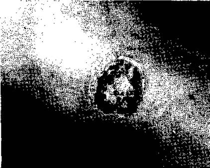
Fig. 94. Radiation skin ulcer

Fig. 95. Perforating-type neurotrophic heel ulcer

Fig. 96. Varicose leg ulcer
Among ulcers caused by venous circulatory disorders, the most prevalent are varicose (Fig. 96) or trophic ulcers, which develop in patients with varicose veins and chronic lower extremity venous insufficiency. These ulcers are the consequence of severe venous outflow obstruction and elevated pressure within superficial varicose veins of the calf belonging to the greater and lesser saphenous systems (v. saphena magna and v. saphena parva). This engorgement leads to tissue edema, profound hypoxia, and the local accumulation of metabolic byproducts and acidic metabolites. Driven by tissue hypoxia and the extravasation of fluid, Plasma Proteins, and blood cells (leukocytes and erythrocytes) through congested venules and capillaries, connective tissue proliferates within the skin and subcutaneous tissue of the lower leg. This process culminates in dermatosclerosis (dermatoscleroz), most pronounced in the lower third of the medial calf, typically posterior and slightly superior to the malleolus. Within the zone of dermatosclerosis, characteristic trophic changes emerge: skin thinning and brownish hyperpigmentation (resulting from hemoglobin degradation and its conversion into hemosiderin). Often, these trophic disturbances are exacerbated and accelerated by secondary complications of varicose veins, such as thrombophlebitis or minor trauma (e.g., excoriations due to varicose-associated pruritus) complicated by secondary infection (infiltrate, dermatitis, lymphangitis, Erysipelas, etc.). The cascade of trophic disorders ultimately culminates in the formation of a roundish ulcer of variable dimensions, typically 2–3 cm in diameter, situated at the center of the dermatosclerotic focus. Its characteristic Location posterior to the medial malleolus is explained by the maximal venous pressure at this site. This elevated pressure is due to the convergence of the great saphenous vein (formed by the confluence of plantar veins) and the presence of perforating veins connecting the superficial and deep venous systems, where valvular incompetence—secondary to varicosities—causes blood to flow in a retrograde pathological direction from the deep to the superficial veins during calf Muscle contraction.
The ulcer and its associated infection (characterized by the discharge of inflammatory exudate and necrotic debris onto the skin surface) induce skin maceration, contact dermatitis, and the spread of the necrotic process. Such ulcers are frequently complicated by erysipelas, which, by obstructing lymphatic vessels, further impairs tissue fluid drainage from the distal lower extremity, intensifying edema and compounding tissue ischemia and hypoxia. Over time, these ulcers enlarge, occasionally encompassing nearly the entire circumference of the lower third of the leg (circumferential ulcers). They become covered with dirty-gray necrotic slough, pale granulation tissue (which may sometimes be entirely absent), and frequently extend deeply into the underlying sclerotic tissues.
Ulcers primarily occur on the skin, but many also develop on the mucous membranes of both the digestive tract—starting from the outer mucosal layer of the Lips—and the urogenital tract.
Among the mucosal ulcers of the digestive tract, chronic ulcers of The Stomach and duodenum are the most common (with acute or stress ulcers occurring less frequently, indicating their neurotrophic origin), along with ulcers of the colon and rectum. In the urogenital tract, the most frequent lesions are ulcers (erosions) of the cervix, Vagina, and Urinary Bladder.
Ulcers of the mucous membranes of internal organs are predominantly of inflammatory and neurotrophic origin, and less commonly tumor-related. Esophageal ulcers arise mainly in reflux esophagitis—a pathological syndrome whose pathophysiological basis involves the regurgitation of acidic gastric contents into the Esophagus due to cardiac sphincter incompetence. This occurs as a result of functional or morphological changes in the esophagus itself or in adjacent organs (cardiospasm, PEPTIC ULCER DISEASE, hiatal hernia, etc.). However, any chronic esophageal ulcer should be regarded not only as a potential source of cancer due to malignant transformation (precancerous condition), but must also be differentiated from malignant tumors, which can present as ulcerated forms in the esophagus and other parts of the digestive tract.
A Chronic Gastric Ulcer (Fig. 97) and duodenal ulcer serve as the morphological substrate of peptic ulcer disease. The latter is one of the most widespread and severe human pathologies; although its etiology is not yet fully elucidated, it is well established that both local aggressive factors (hypersecretion of gastric acid, bile reflux, an increased number of Hydrochloric acid-producing cells in the stomach, infection, etc.) and general neuroendocrine disorders play a significant role in its pathogenesis.
Ulcers in the small intestine are rare and are predominantly of infectious origin.
In the Large Intestine, ulcers may be solitary (rare) or multiple (predominant), forming the morphological basis of several severe systemic conditions, notably Ulcerative Colitis, in the pathogenesis of which immunopathological, vascular, and infectious mechanisms play a role. Solitary ulcers of the large intestine (particularly the rectum) can arise as a result of inflammatory processes, predominantly infectious, including specific infections (tuberculosis, actinomycosis, syphilis).
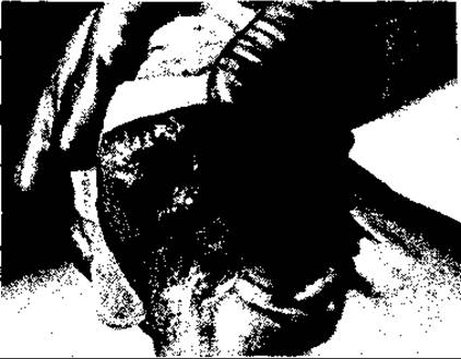
Fig. 97. Chronic gastric ulcer (peptic)
Ulcers of the outer mucosal layer of the lips may stem from vascular (arteriosclerosis), inflammatory-infectious, dystrophic, or tumoral origins. The formation of ulcers is facilitated by both local Anatomical and physiological Features of the affected sites and a range of systemic disorders and diseases (anemia, atherosclerosis, hypoproteinemia, disturbances in protein, carbohydrate, or vitamin metabolism, endocrine disorders, etc.).
The localization, clinical appearance, shape, base, edge characteristics, and course of ulcers depend on numerous factors, primarily their etiology. Determining the appropriate treatment strategy relies on medical history data alongside physical, microbiological, endoscopic, vascular, and other diagnostic examinations.
The most common varicose ulcers on the lower extremities differ markedly in appearance and localization from ischemic ulcers. Varicose ulcers are typically shallow, round, and initially develop posterior and superior to the medial malleolus. They present with a dirty-gray appearance and fragile, easily traumatized, bleeding granulations, surrounded by sclerotic and pigmented tissues (skin and subcutaneous fat). Ulcers on the foot usually have an arterial (ischemic) origin. In most cases, ulcers associated with insufficient arterial blood supply appear on the toes—primarily the first toe—are irregular in shape, dark-brown or dark-gray in color, and lack granulation tissue. The surrounding skin is pinkish-blue or pale-blue, cold to the Touch, and foot pulses are absent. Such ulcers do not bleed.
Tuberculous ulcers (which can occur on the neck, chest—particularly the mammary gland—face, etc.) typically have an irregular shape, flat or papillary growths, and a pinkish-gray color. The surrounding tissues are frequently puckered due to reactive sclerosis. Lupus vulgaris ulcers exhibit a typical localization (a butterfly-shaped distribution symmetrically across the tip of the nose and Cheeks).
Syphilitic ulcers in the primary stage have an acute course, a rounded shape, distinct contours and margins, a lardaceous base, and are localized on the genitalia, in the rectum, and occasionally on the lips and in the Oral Cavity.
Tertiary syphilitic ulcers (gummatous ulcers) are located predominantly on the face, cranium, and shins (anterior surface). They are irregularly shaped, deep, frequently invading the bone, with uneven, firm edges and a gray-pink or brownish hue.
Ulcers as a form of malignant tumor growth have an irregular shape, firm edges raised above the skin surface, and a base filled with gray-pale-pink cauliflower-like proliferations of tumor tissue. Their localization varies, but they are most commonly found on the face and hands (basal cell carcinoma). Many malignant ulcers develop on the background of long-standing non-healing wounds, simple ulcers, or nevi—especially pigmented ones—as a result of malignant transformation (scar cancer on the legs, malignant degeneration of varicose ulcers, gastric and rectal ulcers, and trauma-induced malignant transformation of Papillomas and nevi, etc.).
Mycotic ulcers are localized mainly in the interdigital spaces (on the feet), the Perineum, and rarely on the thighs. They are superficial, cover a significant area, and are preceded by the formation of fluid-filled blisters and accompanied by pruritus.
Treatment of ulcers depends on their origin. Specific ulcers are managed with targeted conservative therapies, while surgical intervention is employed to close defects resulting from tissue breakdown. Neurotrophic ulcers caused by spinal cord or nerve damage and diseases respond very poorly to treatment; therefore, the priority in these cases is to prevent the spread of infection, particularly sepsis, which frequently causes patient mortality. Varicose ulcers also present a complex therapeutic challenge. Because they invariably develop against the backdrop of chronic venous insufficiency of the lower extremities and are complicated by infection, primary measures include improving venous circulation in the limbs and administering local antiseptics, sorbents, and agents that promote tissue regeneration (such as solcoseryl gel). Treatment of concomitant dermatitis is mandatory. Patients must keep their legs elevated while in bed and wear compression stockings or elastic bandages when walking. Varicose veins complicated by ulceration are surgically removed. Such measures improve venous and capillary circulation and often facilitate ulcer healing if the dimensions are small and surrounding tissues are not severely sclerosed. In rare cases, a well-cleansed, regenerating ulcer can even be closed with a split-thickness skin graft. However, in most instances, simple skin grafting fails to achieve the desired outcome. First and foremost, all necrotic tissue must be removed, and the blood and lymphatic capillaries in the base and surrounding tissues must be unblocked using vacuum debridement according to A. A. Safonov's method. Once treated in this manner (leaving a healthy base covered with capillary blood resembling dew), a split-thickness skin graft can be successfully applied, although permanent results are not always guaranteed in every case.
Ulcers resulting from arterial circulation disorders are treated primarily through surgery (necrectomy, amputation of toes or the foot) combined with measures aimed at improving arterial blood supply via collateral vessel development.
Benign ulcers of the mucous membranes of the digestive tract and other pathways are treated conservatively; if conservative measures fail or complications arise, surgical intervention of varying types—predominantly organ resection—is utilized.
Complications of ulcers may include Hemorrhage (which is particularly hazardous in intestinal mucosal ulcers such as those of the stomach, duodenum, and colon), infectious diseases including sepsis, malignant transformation, luminal stenosis (e.g., pyloric stenosis in gastric ulceration, small bowel stenosis in tuberculous ulceration), as well as severe infections resulting from gastrointestinal perforation (peritonitis due to gastric or intestinal perforation, mediastinitis from esophageal perforation, etc.).
Effective ulcer treatment requires timely diagnosis, a combination of conservative and surgical therapies, high clinical expertise, immense patience, and proper patient care.
BEDSORES (PRESSURE ULCERS)
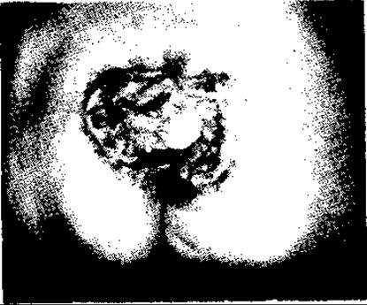
Fig. 98. Pressure sore in the sacral region
Bedsores (pressure ulcers) represent necrosis of the skin and underlying soft tissues in areas where they are compressed between a hard bed frame or bandage and the bony prominences of the patient's body. The pathogenesis of bedsores is related to impaired blood circulation in tissues resulting from the compression of these structures and their blood vessels. Bedsores most frequently develop over superficial skeletal landmarks—the sacrum (Fig. 98), scapulae, heels, iliac crests, spinous processes of the vertebrae, and other muscle-free bony prominences—in patients who are unable to independently change position in bed or turn over, and who lack adequate Sanitary and hygienic care. Their development is promoted by underdeveloped subcutaneous fat, anemia, hypoproteinemia, tissue edema, cardiovascular insufficiency, and various systemic diseases (diabetes mellitus, atherosclerosis, sepsis, nervous system dysfunction). For instance, bedsores develop rapidly in patients with spinal cord injuries (with spinal Shock), who may form pressure sores within just 24 hours over the sacrum, vertebral spinous processes, greater trochanters, and even the heels. In patients without impaired neurotrophic regulation, the primary cause of bedsores is inadequate overall patient care, particularly poor skin hygiene. Bedsores begin with reactive hyperemia and skin inflammation in the compressed zone, followed by moist necrosis due to secondary infection or (less frequently) dry necrosis resulting from ischemia. Typically, necrosis is limited to the skin and subcutaneous tissue, but it frequently extends through the entire thickness of the skin and fascia down to the bone.
The treatment of pressure ulcers (bedsores) primarily consists of eliminating the causes of necrosis and its progression. This includes proper patient care, placing a soft, air-filled rubber ring (covered with fabric) beneath the patient in the necrotic area (predominantly the sacrum), wiping the skin with alcohol and keeping it clean, preventing the bedding from becoming damp, and changing it frequently. For pressure ulcers caused by the pressure of a rigid bandage, the bandage must first be removed and replaced with another.
Necrotic tissue (skin and subcutaneous tissue) in the area of the pressure ulcer is removed, and the tissue defect is treated according to the standard principles of managing a purulent wound. In cases of dry necrosis, the scab is removed only after it has completely separated from the adjacent tissues (provided there is no pus underneath); until then, it serves as a biological "dressing" that protects the wound from irritation and infection.
The prevention of pressure ulcers relies on meticulous patient care. Patients must be turned onto their side in a timely manner, their skin wiped with alcohol or cologne, and the bedding smoothed out carefully. For patients with severe conditions, particularly the elderly, foam or inflatable rubber cushions should be placed under the sacrum from day one, along with gentle back massages and other measures to improve skin circulation. In recent years, specialized anti-decubitus beds have also been widely utilized.
When rigid bandages, especially plaster casts, are applied, bony prominences must be padded with a layer of cotton wool, and the condition of the skin underneath the bandage must be regularly monitored. Clinicians should also pay close attention to any patient Complaints of discomfort in the area of the bandage. Naturally, management cannot be limited to local measures alone. Eliminating or reducing systemic disorders (such as heart failure, hypertension, anemia, or Carbohydrate Metabolism disorders) both pre- and postoperatively, as well as controlling infection, is crucial for preventing pressure ulcers associated with improperly applied rigid dressings. Furthermore, the skill and qualifications of both physicians and nursing staff applying the bandages play a major role.
FISTULAE
A fistula is an abnormal tract connecting deep tissue layers or hollow organs to the external body surface (skin or mucous membrane), or connecting hollow organs and their chambers to one another. Based on this, fistulae are classified into external and internal. Depending on the location of its inner tissue terminus, a fistula may be blind (incomplete) or patent (complete), penetrating into the cavity of an internal organ (such as the Pharynx, intestine, biliary, or urinary tracts).
Fistulae can be congenital or acquired. Congenital fistulae are always lined with epithelium (or endothelium), whereas acquired fistulae are predominantly lined with granulation tissue, and more rarely with epithelium.
Congenital fistulae frequently arise from disruptions in Organogenesis during Embryogenesis, meaning they are developmental anomalies or defects. The most common among these are fistulae of the digestive tract opening externally or into neighboring hollow organs, as well as cardiovascular anomalies. Examples include lateral cervical fistulae connecting the pharynx to the skin (Fig. 99), abnormal communications or fenestrations between various heart chambers, persistent connections or ducts between the aorta and pulmonary artery known as the patent ductus arteriosus (Fig. 100), esophagotracheal or esophagobronchial fistulae (Fig. 101), fistulae extending from the rectum to the perineal skin, urinary bladder, or vagina, and fistulae extending from the small intestine or bladder to the umbilical region due to the failure of the vitelline duct (d. omphalomesentericus) and urachus to close, respectively.
Acquired fistulae generally share the same etiology as necrosis. They can result from any type of trauma (including surgical), infection, necrotizing-dystrophic processes, or tumors. Acquired fistulae are mostly accidental, though they can also be intentional or artificial, created for therapeutic purposes. Examples of the latter include gastric fistulae created in cases of esophageal obstruction of various origins. These fistulae may be temporary or permanent. Even more common are artificial fistulae of the large intestine, predominantly the sigmoid colon, which are established temporarily or permanently for rectal malignancies (known as an artificial anus, or anus praeternaturalis; Fig. 102, a).
Acquired accidental fistulae of various origins pose a significant threat to the body. Traumatic fistulae result from injuries—particularly stab or gunshot wounds—to the face, neck, or abdomen (e.g., the formation of a salivary fistula due to damage to the parotid duct, or an esophageal or intestinal fistula resulting from penetrating trauma). Fistulae of the biliary and urinary tracts are also frequently observed as complications of surgeries on the gallbladder, Urinary Tract, female internal genitalia, rectum, and other structures.
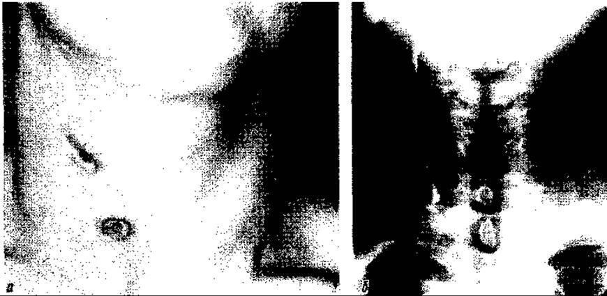
Fig. 99. Lateral cervical fistulae: a — general view; b — fistulogram
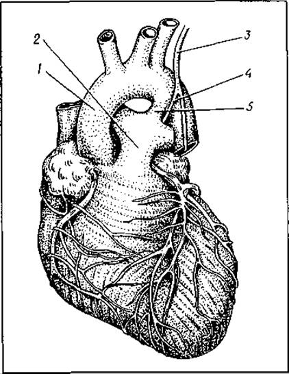
Fig. 100. Patent ductus arteriosus:
1 — aorta; 2 — pulmonary artery; 3 — n. vagus; 4 — n. reccurens; 5 — ductus arteriosus
Fistulae as complications of surgical infections can occur in various parts of the body or organs. External fistulae most commonly develop as a result of wound suppuration (both accidental and surgical), osteomyelitis, infectious inflammation of the perirectal tissue (anal fistulae or chronic paraproctitis), and certain other infections. The formation of postoperative fistulae due to wound infection is facilitated by the presence of foreign bodies deep within the tissues, notably non-absorbable sutures (Fig. 102, b), tissue fragments (especially bone sequestra), vascular and joint prostheses, and similar Materials. Such fistulae persist until the foreign bodies are surgically removed or spontaneously expelled from the depths of the tissues.
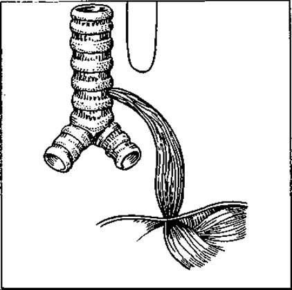
Fig. 101. Tracheoesophageal fistula
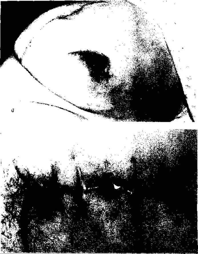
Fig. 102. Artificial intestinal fistula — anus praeternaturalis (a) and infectious fistulae — ligature fistulae (b)
Following surgical interventions on abdominal organs (such as appendectomy, bowel resection, gallbladder, pancreatic, or gastric surgeries), an external intestinal fistula may develop—without generalized peritonitis—as a consequence of infection within the operated organ's wound and localized limited peritonitis, infection of the abdominal wall wound, or (more frequently) concurrent infection of both. If such a fistula of the intestine, biliary, or other tracts opens directly into the free peritoneal cavity due to anastomotic leakage, it causes generalized peritonitis.
Accidental intestinal fistulae are morphologically classified into tubular and labial (lip-like) forms.
The former are characterized by the intestinal mucosa not everting outward through the margins of the tract, whereas in labial fistulae, the mucosa rolls outward onto the external surface of the intestine. The shape of an intestinal fistula depends primarily on its size (diameter) and the dimensions of the opening in the intestinal wall. Tubular fistulae have small openings, while labial fistulae have large ones (due to the redundancy of the mucosal surface relative to the muscular layer and its high mobility).
These Anatomical Features largely determine the prognosis of intestinal fistulae. Tubular fistulae tend to close spontaneously, whereas labial fistulae lack this propensity (as the mucosal edges do not fuse with one another).

Fig. 103. Median cervical fistula following purulent inflammation of a median cyst
Fistulas of the middle region of the neck also have an infectious etiology. They typically result from the suppuration of a median cervical cyst (Fig. 103; a congenital anomaly), coccygeal fistulas, etc.
Acquired fistulas (both internal and external) of hollow organs develop as a result of severe chronic diseases of various origins—neurotrophic, infectious-inflammatory, or tumoral. Examples include internal fistulas between the stomach and the transverse colon caused by the penetration of a gastric ulcer into the transverse colon; fistulas between the gallbladder and the duodenum in Chronic Calculous Cholecystitis (where a large gallstone frequently enters the intestine, causing intestinal obstruction); and external biliary fistulas on the skin in the right hypochondrium resulting from pressure sores and infectious destruction of the anterior abdominal wall in chronic calculous cholecystitis. Other instances include external purulent fistulas on the lateral abdominal wall, which sometimes develop in malignant tumors of the large intestine, as well as purulent rectovaginal fistulas.
Congenital external fistulas most commonly occur in complex anatomical regions, primarily the neck and the perineum. These include lateral cervical, rectoperineal, rectovesical, and rectovaginal fistulas.
The localization of lateral cervical fistulas depends on the branchial cleft (arch) from which they originate: if from the first cleft, the fistula opens at the earlobe; if from the second, in the middle third of the neck (anterior to the sternocleidomastoid muscle); if from the third, above the clavicle anterior to the aforementioned muscle (Fig. 99). They can be single or multiple, and rarely bilateral (with a maximum of three on each side). However, they are most frequently solitary and originate from the second, or less commonly, the third cleft. Most of these fistulas are complete, meaning they penetrate into the pharynx. Pharyngeal fistulas originating from the second cleft open above the tonsil, while those from the third cleft open below it. Rarely, these fistulas are blind, lacking a pharyngeal opening.
Fistulas connecting the rectum with the perineal skin and/or the cavities of the bladder, Urethra, or vagina are invariably associated with embryonic maldevelopment of the rectum—specifically, atresia of its distal end (Fig. 104) and the anus.
Clinical manifestations of fistulas depend on their etiology, nature, location, and other factors. Their impact on the patient's body varies widely—ranging from localized inconveniences (such as certain congenital lateral neck fistulas) to severe, life-threatening conditions (gastrointestinal, cardiovascular, tracheoesophageal, bronchopleural, rectovesical, and perirectal fistulas, etc.). Congenital fistulas adversely affect children's development and may also act as a source of tumorigenesis.
Diagnosis is based on Anamnesis, fistulous discharge, and the LOCATION OF THE fistula. Investigation involves physical examination, probing, and staining of the tract to determine its nature (blind or complete), supplemented by fistulography. To assess the patient's general condition, physical, radiological, endoscopic, ultrasonic, and other diagnostic modalities are employed.
The nature of a fistula, particularly an external one, can in most cases be determined by its discharge (pus, intestinal contents, bile, mucus, saliva, urine, gas, etc.) and its anatomical location. Nevertheless, even with overtly specific discharges, it is sometimes challenging to establish either the Origin of the fistula or its exact level within the digestive tract, bronchial tree, or other organs and pathways (esophagus, stomach, intestines, lungs, bile ducts, etc.). Diagnosis is generally more straightforward for postoperative purulent (suture) fistulas, perirectal fistulas, fistulas of osteomyelitic origin, as well as certain other external fistulas, notably congenital lateral mucous fistulas of the neck, mucopurulent median neck fistulas, and congenital fistulas connecting an underdeveloped rectum to surrounding pelvic organs and tissues.
Purulent discharge from one or more openings on the surface of a fresh or mature scar, frequently accompanied by hypergranulation around the fistulous openings, indicates a suture-related (ligature) etiology. The drainage of pus, occasionally accompanied by small bone fragments through a small wound against the background of old scars in the lower extremities (leg, foot, or less frequently other segments), points to an osteomyelitic fistula. Perirectal fistulas are characterized by their location around the anus on the buttocks or perineum (following prior acute tissue inflammation in this area) and a foul-smelling discharge.
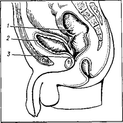
Fig. 104. Fistula of the atretic rectum into the Ureter:
1 — atretic rectum; 2 — urinary bladder; 3 — fistula
In addition to specific discharges, the Clinical presentation of fistulas originating from abdominal hollow organs is characterized by acute dermatitis and skin maceration around the opening, which are particularly pronounced in the presence of gastric, small intestinal, or Pancreatic juice and bile.
A significant depth of an external purulent fistula and its spontaneous formation (following the rupture of an abscess) with a tendency toward a chronic course may indicate that the fistula is connected to an inflammatory or tumoral process in adjacent organs—such as the large intestine, Kidney, or Skeletal System (BONES OF THE lower extremities, Ribs, etc.).
Both the Diagnosis and treatment of fistulas can range from simple to highly complex. Spontaneous closure or conservative healing of fistulas is possible (typically over a prolonged period) only in select cases—provided that foreign bodies are removed and that the intestinal fistula is tubular in nature, meaning its mucous membrane does not evert onto the bowel wall or skin surface. In most cases, however, fistulas require surgical intervention.
Many postoperative purulent wound fistulas caused by implantational infection (ligature fistulas) can be resolved using simple procedures, such as removing the ligatures with a hemostats introduced through the fistulous opening, or (less commonly) by laying open the tract and removing foreign bodies (threads, granulation tissue, etc.). Conversely, the majority of external fistulas—both acquired and especially congenital—as well as all internal fistulas, regardless of origin, are treated using surgical techniques of varying complexity. These range from simple longitudinal incision (syringotomy) or excision to complex reconstructive procedures aimed at eliminating the fistula (resection of an intestinal segment, pulmonary lobectomy, thoracoplasty, separation of organs at the fistula site with closure of their defects, ligation and division of the tract, etc.).
The timing of surgery is determined by the nature of the fistula and its impact on the patient's general condition and vital Functions. Congenital mucinous fistulas (e.g., lateral cervical) can be operated on during preschool age, provided they are not complicated by infection. Conversely, small intestinal fistulas—particularly high-output ones, as well as pancreatic or biliary fistulas—should be treated surgically as early as possible, once intra-abdominal and perilesional tissue infections have been resolved. Such fistulas lead to severe Metabolic Disorders that cannot be fully compensated by parenteral or even
enteral tube feeding. Only certain postoperative fistulas of traumatic and infectious origin—such as cecal fistulas following appendectomy or duodenal fistulas following gastric resection, which frequently present as tubular tracts—tend to close spontaneously within 2–4 weeks. Consequently, they may initially be managed with intensive conservative treatment (aspiration of secretions, administration of sandostatin, etc.). Spontaneous closure of labial (lip-like) intestinal fistulas, where the intestinal mucosa everts onto the external surface of the bowel or skin, is impossible.
Congenital tracheoesophageal and bronchoesophageal fistulas are treated surgically immediately after birth, as without intervention the infant dies within the first few days due to pneumonia and respiratory failure.
Congenital fistulas and defects in The Heart and major vascular trunks are predominantly treated surgically at various ages, depending on the severity of hemodynamic and respiratory impairment, but primarily during infancy and preschool childhood.
The prevention of fistulas (typically acquired ones) involves avoiding infectious diseases, strictly adhering to asepsis rules during surgical procedures, and performing operations in an atraumatic, technically and biologically sound manner.
As for congenital fistulas, preventive measures are practically nonexistent at present, as these anomalies form predominantly During the first trimester of Pregnancy.
Foreign bodies in THE ORGANISM AND THEIR REMOVAL
Foreign bodies are defined as any objects, parts thereof, or formations of organic or inorganic origin that enter the body and should not normally be present there.
Foreign bodies can enter the body under various circumstances and through different pathways, namely: during mechanical trauma (firearm and non-firearm, surgical) via wounds in the skin and mucous membranes; during certain physiological acts (swallowing and breathing, during which foreign bodies enter the digestive tract and respiratory pathways); and through mischief, hooliganism, or sexual deviance, whereby foreign bodies may enter through natural orifices and bodily channels (into the rectum or urinary bladder).
The structure, size, and shape of foreign bodies vary widely. Most commonly, these are metallic, wooden, Glass, and stone objects, though they can also include organic matter (fish and animal bones, fruit stones and pits; tricho- and phytobezoars, i.e., masses formed by Hair and plant residues in the stomach, as well as cereal awns, etc.).
Parasites residing in tissues and organs, calculi forming in the urinary and biliary tracts, as well as xenoprostheses and xenomaterials introduced into the body for therapeutic purposes, are not classified as foreign bodies.
The pathological impact of a foreign body on The Human Body depends on numerous factors, including its nature (structure), size, shape, mode of entry, localization, degree of contamination, type of microflora, etc. Identifying early manifestations of pathology caused by foreign bodies that enter tissues and organs through the skin and mucous membranes—predominantly during blunt or closed trauma—is nearly impossible, as they are masked by local and general symptoms of trauma (pain, hemorrhage, inflammation with or without suppuration, tissue edema, and organ dysfunction). Only after a wound heals without surgical debridement or, conversely, when healing is delayed even after debridement due to an unremoved foreign body, can a patient's complaints and dysfunctions be attributed entirely or partially to the presence of the foreign body in the tissues or organs. If a foreign body penetrates the body through its natural orifices and channels, it causes either partial or total obstruction of the channel or cavity, impairing their patency. This is accompanied by a corresponding clinical syndrome, such as asphyxia or Pulmonary Atelectasis, esophageal or intestinal obstruction, etc. However, foreign bodies entering through bodily orifices and channels provoke acute symptoms only under specific conditions (dimensions significant and sufficient to cause channel obstruction, or a sharp shape that traumatizes the mucosal lining of the cavity).
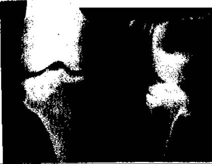
Fig. 105. Foreign body in the tibial metaphysis
In other instances, a foreign body may reside within a cavity for a considerable period until it eventually triggers a complication from the affected organ (inflammation, pressure sores leading to hemorrhage or severe infection, or the development of a fistula).
Foreign bodies in soft tissues resulting from trauma are observed most frequently. During wartime, these are typically fragments of shells, mines, and aerial bombs, and less commonly, bullets. In addition to superficial soft-tissue foreign bodies of ballistic origin, wartime conditions often present numerous foreign bodies within parenchymal organs (lungs, liver) and deep tissues, particularly within bones (Fig. 105). Such foreign bodies are very frequently multiple, especially in cases of injuries sustained from mine explosions. Foreign bodies in soft tissues resulting from non-firearm injuries include needles, metal shards, or stones detached from processed materials under the impact of a hammer or other tool, as well as wooden splinters and glass particles that penetrate soft tissues through the skin.
Soft-tissue foreign bodies, particularly sharp and small ones, are accompanied by pain and an inflammatory process in the early stages. The latter is typically serous in nature, subsides after a few days, and the wound heals by primary intention. Connective tissue proliferates around the foreign body (stimulated by its presence), forming a dense capsule (the body becomes encapsulated).
Foreign bodies in superficial soft tissues (especially blunt, heavily contaminated ones such as stone and metal fragments, wood splinters, etc.) are frequently accompanied by the development of purulent infection around them and within the wound (abscess, phlegmon), which may recur if the foreign body is not removed.
Soft-tissue foreign bodies penetrating via the skin (a wound), regardless of whether they cause suppuration of the wound and surrounding tissues, can be complicated by acute specific infections, such as tetanus or anaerobic clostridial infection. These types of acute infection are particularly dangerous when foreign bodies are located in the soft Tissues of the lower extremities and buttocks. Alongside infection and inflammatory complications, soft-tissue foreign bodies can cause pressure necrosis (bedsores) of vessel walls leading to erosion hemorrhage, and when localized near a nerve trunk, they can cause compression and traumatic neuritis.
Occasionally, connective tissue proliferates so intensively around a soft-tissue foreign body that a tumor-like granuloma—a foreign body granuloma—is formed. Despite the encapsulation of the foreign body in soft tissues, purulent infection in the area of its localization can very often reactivate, even after many years, causing an abscess or phlegmon and the spontaneous discharge of the foreign body through an opening. If the foreign body is not expelled, the infection relapses.
Foreign bodies in internal parenchymal organs (such as the lungs and liver), which are almost invariably of firearm origin, are more frequently complicated by infection, particularly shrapnel fragments. They tend to encapsulate primarily in penetrating bullet wounds where the bullet itself serves as the foreign body. This is explained by the prior contact of shell and mine fragments with the ground and their greater microbial contamination compared to bullets. Encapsulated foreign bodies in internal organs are far more likely than those in superficial soft tissues to eventually become complicated by infection or hemorrhage from an eroded blood vessel. It should be noted that foreign bodies of ballistic origin, especially shrapnel, can be either primary (shrapnel, bullet) or secondary (particles of soil, clothing, buttons, etc.). A combination of such foreign bodies is typically complicated by severe forms of purulent infection, and frequently by anaerobic and specific infections.
A significant number of varied foreign bodies enter the body through natural orifices and channels. This is due to the heavy functional load placed on these channels, the anatomical and physiological features of the digestive and respiratory tracts—specifically the lack of anatomical isolation between them at the oropharyngeal level—as well as other factors (children's curiosity, psychiatric disorders, etc.).
The clinical characteristics of foreign bodies entering human anatomical channels include potential acute disruption of respiratory function (asphyxia), swallowing difficulties, psychological shock, and Technical Challenges during medical intervention to restore the patency of these channels.
Foreign bodies enter the digestive tract most frequently. This typically occurs in children under the age of 5, who tend to taste everything, and in psychiatric patients who frequently swallow various objects (Fig. 106).

Fig. 106. Foreign bodies in the stomach (a). Foreign objects removed from the stomach (b)
Swallowed objects predominantly enter the stomach, and less frequently become impacted in the esophagus. The latter occurs either when sharp, large, irregularly shaped objects are swallowed (needles, Nails, dentures, etc.) or in the presence of esophageal narrowing or impaired propulsive function. The presence of foreign bodies in the esophagus is accompanied by esophageal pain and impaired patency, or in other cases, by inflammation or perforation of the esophageal walls and acute mediastinitis.
Foreign bodies in the respiratory tract are also observed predominantly in children under 5 years of age who, due to inadequate supervision, put various small items into their mouths (coins, buttons, beads, pebbles, etc.); less frequently in adults in a state of alcohol intoxication and in elderly individuals (dentures, loose Teeth, etc.) or people with impaired superior laryngeal nerve function. Foreign bodies can also enter the respiratory tract in individuals with a professional habit of holding various objects in their teeth (needles, nails), as well as in those who talk and laugh extensively while eating. Impaction of objects in the respiratory tract that occludes the lumen of the Trachea or Larynx causes asphyxia and can lead to patient death within minutes. Small foreign bodies that do not occlude the larynx or trachea pass into the bronchi, causing their obstruction and the development of aspiration lung abscesses.
Foreign bodies in the urinary tract (urinary bladder), which enter the body during childhood play or masturbation, are observed rarely.
A patient may be unaware of the presence of foreign bodies in their tissues. Therefore, suppuration in a poorly healing wound, the presence of excessive granulation tissue within it, persistent pain in a healed wound, or suppuration in the scar area following trauma should raise suspicion of a retained foreign body in the tissues.
Foreign bodies entering soft tissues and parenchymal organs typically do not migrate. In contracting muscles, they may shift slightly, particularly sharp objects. When entering major blood vessels, sharp foreign bodies (needles or their fragments) can be transported by the bloodstream over varying distances. Blunt foreign bodies, both in the subcutaneous tissue and in organs, when accompanied by suppuration (abscess or phlegmon, especially recurrent ones), can shift over some distance under the influence of gravity within the purulent cavity in a caudal or dorsal direction.
Foreign bodies in hollow organs, particularly in the digestive tract, move during the contraction of the smooth Muscles of the stomach and intestine until they exit naturally, become stuck in a narrow section, or catch on the organ wall with a sharp end (needles, fish or chicken bones, etc.). This usually culminates in intestinal wall perforation and the exit of the foreign body into the peritoneal cavity, leading to peritonitis, or into an adjacent organ that has adhered to this intestinal site as a result of inflammation. The greater omentum is most frequently the organ involved.
The clinical manifestations of foreign bodies residing in tissues or cavities depend on their structure and localization, size, shape, and the nature of the complications they provoke. These may include superficial tissue abscesses, lung abscesses, asphyxia, dysphagia, intestinal obstruction, peritonitis, mediastinitis, cardiac dysfunction, fistulas, and the presence of hypergranulation tissue around them.
The diagnosis of foreign bodies is based on medical history, physical examination of the tissue penetration site, and Palpation of superficial tissues and organs. Metallic, stony, bone, and certain glass (radiopaque) foreign bodies are detected using two-view radiography. Fistulography (injecting a radiopaque agent into the affected organ followed by imaging) plays a crucial role in diagnosing bodies with an external fistulous tract. Endoscopic examinations—such as bronchoscopy, esophagoscopy, gastroscopy, rectosigmoidoscopy, and cystoscopy—are highly effective for diagnosing foreign bodies in the respiratory tract, esophagus, stomach, rectum, urinary bladder, and other hollow organs. Ultrasonography and computed tomography have also become widely used in diagnosing foreign bodies in cavities and parenchymal organs. Laparoscopy and thoracoscopy are relevant only in cases of complications caused by foreign bodies in the abdominal and pleural cavities (e.g., peritonitis, pleuritis).
Treatment involves the removal of foreign bodies, except for those that, due to their quantity, small size, and the absence of pain or functional organ impairment, do not require extraction. Likewise, certain small, solitary foreign bodies in organs that do not cause functional disorders and whose removal would pose a high risk to the patient's life (such as metal fragments deep within the brain or liver) should not be removed.
The majority of foreign bodies in soft tissues and organs, as well as all foreign bodies in the respiratory tract, esophagus, digestive tract, urinary tract, and Body Cavities, are indications for removal.
Patients with foreign bodies in the stomach and intestines (except for small ones with smooth surfaces) should be hospitalized in a surgical unit for observation (monitoring the object's movement via fluoroscopy and radiography, and inspecting stool). During this period, patients are given a fiber-rich diet (porridge, bread, potatoes). If a foreign body moving through the intestine is retained for a prolonged period (1–2 days), even in the absence of acute symptoms, it must be removed surgically (via laparotomy and enterotomy). If the foreign body causes acute symptoms, the patient must be operated on immediately, regardless of the object's location.
Certain foreign bodies—where one end penetrates deeply into tissues while the other protrudes through the skin (such as a knife, chisel, large nails, or wooden ski poles piercing a skier's body completely)—should not be removed during first aid (only the protruding ends of a pole may be cut off to facilitate patient transportation). Such objects may act as "plugs" preventing hemorrhage from a heart chamber or a blood vessel. They should only be removed in a specially equipped operating room.
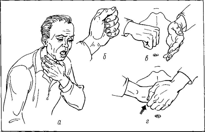
Fig. 107. The Heimlich maneuver for foreign body airway clearance (a – d)
To remove a foreign body, particularly from soft tissues, an incision of the skin and underlying tissues is made directly over the projected location of the object.
Sharp foreign bodies, such as needles, located in certain parts of the body (e.g., the mammary gland) can sometimes be removed by compressing the gland between the fingers: this causes the needle point to pierce the skin outwardly, allowing it to be grasped with a clamp.
Foreign bodies encapsulated within tissues must be removed along with their capsule, followed by tetanus immunization. This is because the tetanus bacillus may persist on the foreign body and its capsule.
Many foreign bodies, particularly those in the trachea, bronchi, esophagus, stomach, and rectum, are removed endoscopically. Certain objects, such as metal fragments in soft tissues, the eyes, and other areas, can be extracted using magnetic probes.
Foreign bodies in the respiratory tract are particularly dangerous as they frequently lead to asphyxiation. Small objects incapable of causing mechanical obstruction of the larynx or trachea provoke a sharp initial reaction consisting of coughing fits, dyspnea, and often laryngospasm and asphyxia. If such bodies migrate into the bronchi, pulmonary atelectasis, abscesses, and pneumonia develop. Similar complications occur when food particles and gastric juice enter the respiratory tract (Mendelson's syndrome). Large foreign bodies—such as teeth, dentures, coins, and buttons—entering the larynx or trachea cause sudden asphyxia, which often results in the victim's death within minutes. In such cases, only a tracheostomy can save a life. This Procedure can be performed solely by a physician under specific conditions. In the absence of such conditions, conservative foreign body expulsion techniques must be employed. One of these is the technique proposed by American physician N. Heimlich, known as the Heimlich maneuver (Fig. 107). It is performed as follows: if the standing patient is upright, the rescuer stands behind them and wraps their arms around the patient's waist (Fig. 108). The fingers of the right hand are clenched into a fist, with the thumb placed over the curved four fingers and aligned along the axis of the forearm. The clenched fist is placed on the patient's abdomen on the midline, slightly above the navel and somewhat below the costal margin of the xiphoid process. The palm of the left hand is placed on top of the fist, and both hands deliver a rapid inward and upward thrust into the abdomen. As a result of this abrupt movement, the Diaphragm is elevated and the lungs are compressed, forcing the air out to expel the foreign body from the respiratory tract. If necessary, this maneuver is repeated several times. If the patient is lying down, the maneuver can be performed by kneeling astride the patient's hips. For children, it is performed using the index fingers while standing over them from the front. A patient familiar with this technique may perform it on themselves.
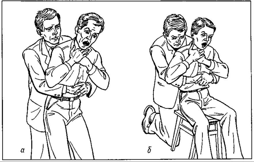
Fig. 108. Performing the Heimlich maneuver in the standing (a) and sitting (b) positions
A distinct category comprises foreign bodies accidentally introduced into tissues or body cavities (forgotten) during surgical operations, i.e., iatrogenic foreign bodies. These include gauze Sponges and balls, drains, and instruments such as hemostatic clamps, scissors, and even ring forceps. Foreign bodies left in a wound or cavity provoke acute or chronic infectious inflammation, including abscesses, phlegmon, peritonitis, pleuritis, acute or chronic intestinal obstruction, intestinal perforation, pressure ulcers on major blood vessels, and massive hemorrhage.
The difficulty in the timely diagnosis of retained foreign bodies lies in the fact that their clinical presentation in the first few days is masked by general postoperative systemic changes. This leads to delayed surgical intervention and severe consequences. Whenever complications arise following a laparotomy, the possibility of their association with a foreign body must always be considered. Fluoroscopy, radiography, and ultrasonography should be utilized for diagnosis, while prompt relaparotomy is required for treating complications. Every surgeon must know how to avoid these occasionally fatal errors. Among preventive measures, the most crucial ones include:
1) maximum attentiveness by the surgeon and assistants during surgery, particularly on abdominal organs and especially in cases of intra-abdominal hemorrhage and peritonitis;
2) avoiding the use of small gauze balls deep within a wound, even if secured with ring forceps;
3) securing all large sponges and abdominal packs introduced into a cavity to the skin;
4) ensuring the operating nurse accurately tracks the number of large sponges used and performs instrument counts before and at the end of the surgery;
5) performing a mandatory final revision of the wound and cavity prior to closure;
6) securely anchoring all drains placed in the wound and ABDOMINAL CAVITY AND exteriorizing their long ends.
The prevention of foreign body penetration into the organism remains insufficiently developed due to its complexity and the multitude of diverse causes involved. Nevertheless, several preventive directions can be outlined. We have already addressed the prevention of iatrogenic foreign bodies. To prevent occupational injuries, industrial safety regulations must be strictly observed. Reducing the incidence of foreign bodies in the respiratory tract and esophagus in children can be achieved through proper childcare. Strict supervision of psychiatric patients can likewise prevent them from swallowing various foreign bodies. A multifaceted approach to combating various types of trauma—including domestic injuries—simultaneously serves as prevention against the penetration of foreign bodies into tissues. Combating alcoholism, rowdy behavior, and proper sex education are all measures with the potential to prevent foreign bodies from entering the body.
Last update: 08/08/2026
Editorial and Educational Adaptation: This material has been compiled based on the primary/original source text. The project team performed an editorial review, corrected technical inaccuracies, structured sections, and adapted the content for an educational format.
What was processed:
- elimination of formatting defects (OCR errors, structural breaks, corrupted characters);
- editorial organization of content;
- standardization of terminology in accordance with academic sources;
- verification of factual statements against the original source text.
All mentions of the author, publication year, and origin of the primary text have been preserved in accordance with the source.