Antibiotics (Properties, Application, Interactions) - M.P. Cherenko 1999
Chronic Specific Infection
The group of chronic specific infections includes several common forms (tuberculosis, Syphilis, actinomycosis) as well as A number of less prevalent conditions (tularemia, brucellosis, leprosy, leishmaniasis).
Characteristic Features of all these infections include a chronic clinical course and similar morphological tissue reactions to the pathogen, a key element of which is The formation of a so-called unstable granuloma.
Most of these diseases (such as tuberculosis and syphilis) are highly contagious and can assume epidemic proportions, particularly under unfavorable social living conditions. These infections are of significant interest to surgeons because Surgical Methods are integrated into their Treatment regimens at specific stages and for particular localizations. Furthermore, surgeons must avoid critical and potentially devastating diagnostic errors when differentiating these conditions—most of which are managed conservatively—from Various Forms of common (nonspecific) infections and pathologies of other etiologies.
TUBERCULOSIS
Tuberculosis is caused by the tubercle bacillus (Mycobacterium tuberculosis), discovered by R. Koch in 1882. There are three main strains of the pathogen: human (typus humanus), bovine (typus bovinus), and avian (typus avium), alongside several atypical variants. The human and bovine types are pathogenic to humans, while atypical strains rarely cause disease (typically in immunocompromised individuals). In addition to infected humans, sources of transmission to humans include infected cattle, as well as their meat and milk.
The tuberculosis pathogen appears as a slender, straight or slightly curved rod measuring 1.5–4 µm in length and up to 0.5 µm in width. It lacks spores, capsules, and flagella. It is an obligate aerobe, Gram-positive, and stains bright red using the Ziehl-Neelsen method. Fresh cultures isolated from patients grow very slowly (3–4 weeks) on various media (such as potato, egg, and broth) enriched with glycerin. The bovine tubercle bacillus exhibits certain morphological and cultural differences: it is thicker and shorter, and does not require glycerin for growth. Mycobacteria tuberculosis do not produce exotoxins; their virulence is associated with a complex of Lipids, lipoids, and Other Compounds (virulence factors). These mycobacteria produce Enzymes such as lecithinase, catalase, peroxidase, and urease.
The metabolic product of mycobacteria was discovered by R. Koch and named tuberculin. It is utilized in the Diagnosis of tuberculosis, predominantly in a purified form derived from old tuberculin, and consists of pure mycobacterial Proteins mixed with polysaccharide fractions.
Tuberculosis is a frequent disease, particularly in economically disadvantaged countries with poor living conditions, inadequate healthcare systems, and limited medical resources.
However, in recent years, the incidence of tuberculosis has risen even in regions where it was previously uncommon, driven in part by a decline in preventative measures.
Improving socio-economic living conditions plays a paramount role in the Prevention and eradication of tuberculosis.
Transmission of tuberculosis—namely, The entry of mycobacteria into the body—occurs primarily via the respiratory tract (airborne droplets). It is rarely transmitted through the gastrointestinal tract (via deposition on the mucous membrane), in which case it affects either the initial segment (oropharynx) or, more frequently, the middle segment (cecum), and uncommonly the terminal segment (rectum).
Host resistance to tuberculosis is lowest during childhood and after the age of 65.
Different Tissues and Organs exhibit varying degrees of susceptibility to mycobacteria. The most susceptible are Lymph Nodes (affected in 100% of infected children), the Meninges, and the Spleen (particularly in children). The Kidneys, Adrenal Glands, fallopian tubes, and Pituitary Gland are also susceptible. Conversely, The Stomach, Pancreas, Thyroid Gland, and Testes exhibit low susceptibility and are rarely affected.
Infection (Primary tuberculosis) occurs in the vast majority of individuals during childhood via the tracheobronchial route, typically associated with contact with active Pulmonary Tuberculosis patients. Such transmission is facilitated by crowded environments, poor living and sanitary conditions, and public transportation.
The primary tissue reaction in human Lungs following infection with tubercle bacilli manifests as the formation of a so-called primary complex. This complex consists of a small focus of specific Pneumonia and involvement of the regional mediastinal lymph node, presenting as specific lymphadenitis. The morphological manifestations of tissue infection include a nonspecific reaction (hyperemia, edema), leukocyte infiltration, Connective Tissue Cell proliferation, and the Formation of the specific element of tuberculosis under the Influence of the mycobacteria and their metabolic products—the so-called tubercle (tuberculum, Fig. 79) or granuloma (a conglomerate of tubercles). A defining characteristic of this granuloma (ranging from the size of a millet seed upwards) is its propensity for necrosis and caseous breakdown.
Class="center">
Fig. 79. Tubercle (tuberculum)
The fundamental unit of a tubercular lesion and the center of the tubercle consists of so-called epithelioid Cells. Constant cellular elements of the tubercle also include Langhans giant cells and lymphocytes (distributed along the periphery of the tubercle), along with connective tissue.
Epithelioid cells are large mononuclear cells packed with disintegrated products of dead leukocytes and mycobacteria, featuring a pale, foamy Cytoplasm resulting from a high lipid content.
The accumulation of mononuclear cells resembles tightly packed epithelial cells, which is why they were termed epithelioid cells. The fusion of the cytoplasm of mononuclear cells into multinucleated structures produces giant cells, which are located both in the center and at the periphery of the tubercle. The center of the tubercle contains caseous (cheese-like) necrotic masses (a form of dry necrosis). The tuberculosis granuloma, like other granulomas, is of immunogenic origin (I.V. Davydovsky, 1969). Its development proceeds through four phases: primary tissue alteration, Development of the cellular granuloma, destruction of granuloma cells, and terminal sclerosis or encapsulation of the lesion.
The primary complex (tuberculosis) lacks specific and distinct clinical manifestations. Patients may experience mild fatigue, allergic phenomena—such as erythema nodosum or other Skin rashes on the shins—frequent enlargement of cervical lymph nodes, low-grade fever, and a cough. Due to its mild symptom profile, primary infection often goes undiagnosed. After 2–3 weeks, the primary complex begins to subside in the majority of infected individuals, becoming restricted (encapsulated) by a Fibrous connective tissue capsule and infiltrated by calcium salts (via phosphates released from lysed cells) to form so-called calcifications. In some individuals, encapsulation is incomplete, and the mycobacteria within it (especially within the lymph node) do not completely perish, causing the process to smolder. This is evidenced by cases of primary complex reactivation accompanied by mycobacterial dissemination and disease development many years later, despite increased Immunity to the Bacteria.
In 1–2% of infected individuals, the primary complex fails to encapsulate, leading to The Development of pulmonary tuberculosis and, less frequently, Extrapulmonary tuberculosis.
Both primary infection and active tuberculosis were previously observed predominantly in children and adolescents. Today, in economically developed countries—including Ukraine—adults and the elderly contract tuberculosis, many of whom experience primary infection at an older age. Disease progression is promoted by numerous factors, including anemia, hypoproteinemia, hypovitaminosis, Diabetes Mellitus, other Metabolic Disorders, and genetic predisposition.
Heightened sensitivity and bodily sensitization to Mycobacterium tuberculosis are of critical importance. Among local factors, silicosis and Other forms of pulmonary anthracosis facilitate the development of tuberculosis.
Tuberculosis, like any inflammatory process, is accompanied by exudation, alteration, and cell proliferation. The relative proportions of these phenomena vary widely depending on the virulence of the pathogen, host reactivity, and treatment modalities, ultimately determining the development of predominantly exudative-infiltrative or necrotizing-productive (proliferative) forms of tuberculosis.
The tuberculous process spreads through the lungs via contact, bronchial, lymphogenous, and hematogenous routes, causing various forms of pulmonary and pleural disease, and frequently affecting other organs as well. Bronchial destruction leads to the discharge of tissue necrosis products and mycobacteria into its lumen, propagating the infection throughout the lung and resulting in cavitary forms. Meanwhile, vascular destruction with the breakthrough of mycobacteria into the bloodstream causes the so-called miliary form of tuberculosis.
Alongside specific pathological changes, tuberculosis is often accompanied by non-specific reactions and complications, such as Hemorrhage, Atelectasis of a lung lobe (caused by bronchial compression from lymphadenitis—most commonly the middle lobe bronchus of the right lung—or due to intrinsic bronchial occlusion), pulmonary emphysema, and others.
Primary infection may also involve other organs. Mycobacteria from the primary pulmonary complex can enter the bloodstream (via vascular destruction or through macrophage transport laden with mycobacteria), spreading to various organs—particularly bones, kidneys, and lymph nodes—where they settle and cause disease. All such forms of extrapulmonary tuberculosis resulting from the dissemination of mycobacteria from the primary pulmonary complex are classified as secondary tuberculosis, which until recently formed The basis of what was known as surgical tuberculosis.
The term "surgical tuberculosis" is somewhat artificial, given that virtually all forms of tuberculosis—including its most common pulmonary manifestations (fibrocavitary forms, Tuberculoma, Pleural Empyema, and bronchopleural fistulas)—are also amenable to Surgical treatment. Pulmonary tuberculosis, which accounts for over 75% of all localizations, can be successfully treated conservatively when diagnosed early and managed with highly effective therapeutic agents, particularly Chemotherapy (Antibiotics, etc.), leading to a progressive decline in surgical interventions for these forms. Nevertheless, surgery plays a vital role in the comprehensive treatment of extrapulmonary tuberculosis, which is primarily secondary—especially peripheral forms (Bone and joint Tuberculosis, lymph node tuberculosis, and visceral involvement such as Urogenital tuberculosis).
Gastrointestinal tuberculosis is predominantly observed in the ileocecal region, accompanied by lesions in the intestinal wall and mesenteric lymph nodes. Caseous breakdown of the latter leads to tuberculous Peritonitis, characterized by the formation of numerous specific granulomas, adhesions, and exudation into the peritoneal cavity. Gastrointestinal tuberculosis can be either primary or secondary. In pulmonary tuberculosis, lesions of the gastrointestinal tract, Larynx, and often the cervical lymph nodes contain Mycobacterium tuberculosis As a result of swallowing infected sputum.
Oropharyngeal tuberculosis, particularly affecting the Tonsils, can present as a primary form in the absence of pulmonary disease.
Very rarely, a primary focus of tuberculosis may develop on damaged skin (such as on the hands).
This occurs most frequently among meat handlers, physicians, and veterinarians.
All other localizations of tuberculosis—particularly so-called surgical tuberculosis (affecting bones, kidneys, cervical lymph nodes, and certain other organs)—are almost invariably secondary, regardless of the presence or absence of pulmonary involvement; that is, they result from the dissemination of mycobacteria from the primary pulmonary complex.
Surgical interventions for tuberculosis can be radical, involving the removal of the affected organ or its part (such as a lung lobe or entire lung, Kidney, Epididymis, or lymph nodes); auxiliary, which do not eliminate the focus but facilitate healing (such as the division of adhesions or thoracoplasty for pulmonary tuberculosis, and osteoplastic spinal fixation); or corrective, designed to correct and eliminate the sequelae of tuberculosis, such as spinal deformities or limb shortening.
Pulmonary tuberculosis. Primary pulmonary tuberculosis, which was previously observed predominantly in children and adolescents, is now also encountered in adults. Due to its propensity for pleural and lymph node generalization, it is very rarely treated surgically. Surgical management is generally reserved for Secondary forms of pulmonary tuberculosis (arising from primary complex foci due to disease reactivation) complicated by cavities, bronchopleural fistulas, and empyema, or large caseous foci (caseomas or tuberculomas), which predominantly develop in patients with infiltrative and disseminated forms of tuberculosis. Fibrocavitary forms, caseomas, and tuberculomas (Fig. 80) do not tend to generalize and are tuberculin-insensitive; consequently, they generally present as localized lesions, which favors successful surgical outcomes (N.M. Amosov, 1958; M.S. Pylypchuk, 1973).
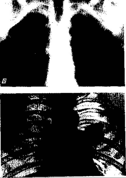
Fig. 80. Tuberculoma in the right (a) and left (b) lungs
Bone and joint tuberculosis is the most common form of extrapulmonary (peripheral) tuberculosis, accounting for up to 10% of all tuberculosis cases. It is believed that Osteoarticular Tuberculosis develops as a result of the entrapment and proliferation of mycobacteria within the bones during hematogenous dissemination from the primary pulmonary complex, which explains why it predominantly affects children. This predisposition is facilitated by the sensitization of Cytology/practical/86.html">Red Bone Marrow to tuberculosis. Both tubular and flat bones are affected (spine, scapula, clavicle, etc.). The process is most frequently localized in the spinal Column, Femur, and Tibia, and less commonly in the BONES OF THE upper extremities. Among joints, the hip and knee are the most frequently involved.
In 90% of patients with tubular bone involvement, the process begins in the epimetaphysis, as the vascular architecture there traps mycobacteria. A small, spherical focus of destruction—a primary ostitis (tuberculous Osteomyelitis)—is formed here. P.G. Kornev (1971) refers to this stage as the prearthritic stage. The process then extends toward the joint, eventually involving it (the arthritic stage), and only occasionally progresses along the bone outside the joint, forming specific cold migratory abscesses in the soft tissues that eventually rupture and create cutaneous fistulas.
Diaphyses of tubular bones are rarely affected, typically occurring only in young children. In adults, tuberculosis of this localization is observed in the Phalanges of the hands.
When the tuberculous process extends from the epimetaphysis into the joint, a specific Arthritis develops—most commonly exudative synovitis, or less frequently fungous osteoarthritis, which is characterized by a predominance of proliferative-alternative inflammation, replacement of the joint cavity with mushroom-like granulations, and destruction of the Cartilage and articular bone ends.
Primary (extraosseous) involvement of the synovial membrane resulting from hematogenous dissemination is rare.
Primary ostitis manifests as a focus of caseous necrosis (the coalescence of tuberculous tubercles) which, following purulent melting, transforms into a bone cavity. The process expands actively, primarily toward the joint, and in only 5–10% of cases extends outward, involving the cortical bone layer, periosteum, soft tissues, and skin.
Cutaneous destruction leads to the formation of fistulas discharging thin, white-gray pus, occasionally containing small fragments of necrotic bone (sequesters). The bone surrounding the cavity is osteoporotic, and periosteal reaction is virtually absent. Significant periosteal proliferation is observed only in cases of diaphyseal localization, causing the bone (or finger) to enlarge in its middle section and acquire a fusiform shape.
When the process extends to the joint in its exudative form, numerous tubercles form on the synovial membrane—particularly abundant in the articular recesses (miliary form). Swelling and hyperemia of the synovium, edema of the villi, and accumulation of serous exudate with fibrin within the joint cavity are observed, while the cartilage becomes turbid and friable. The joint swells markedly, and ballotment of the Patella can be elicited.
At this stage, the anatomical changes are still reversible; under treatment, the exudate may be reabsorbed, and the tubercles may resolve either completely or partially or undergo encapsulation. If the process extends from the synovium to the cartilage, ligaments, Joint Capsule, and bone, The breakdown of specific tubercle conglomerates leads to Purulent Arthritis or joint empyema. Edematous synovial villi within the joint may detach, forming so-called rice bodies.
In the proliferative-alternative or fungous form of the lesion (arthritis), the entire joint cavity is filled with soft specific granulations that frequently invade the cartilage and bones, causing a marked enlargement of the joint. Exudate within the joint cavity is practically absent.
Due to dystrophic-sclerotic Changes in the soft tissues surrounding the joint and the compression of Blood Vessels, the skin overlying the joint is pale and stretched, while the joint itself appears tumor-like against the backdrop of limb Muscle atrophy—hence this form of arthritis was historically termed "white swelling" (tumor albus).
During the course of development, necrotic masses are absorbed and sequestered, and the foci become scarred (postarthritic stage). However, complete resolution of the process is extremely rare, and relapses and exacerbations are frequent. Once the process subsides, fibrous contracture or ankylosis develops in the joint due to the proliferation of spongy Bone tissue, and the cartilage does not regenerate. Without adequate treatment, bone and joint tuberculosis results in structural destruction and patient disability.
In Tuberculous Spondylitis, the vertebral bodies are predominantly affected, particularly their anterior section, where a primary osteitis forms, from which the pathological process extends to the intervertebral discs.
Considering this progression, P.G. Kornev (1971) distinguishes the pre-discal, discal, and post-discal phases of tuberculous spondylitis.
The general clinical and morphological features of osteoarticular tuberculosis include significant (and, in joint involvement, severe) pain—even with minimal morphological changes—along with pronounced functional impairments of the joints, extremities, and spine. Neurotrophic (atrophic and dystrophic) changes in bones and Muscles manifest very early, such as Osteoporosis, muscle atrophy, and decreased muscle tone, alongside suppressed reparative processes (absent or poorly expressed periosteal reaction), immune impairment, and systemic allergy.
The diagnosis of bone and joint tuberculosis is based on patient history (taking into account the patient's age), physical, serological, and instrumental (such as radiological) examination methods, as well as the results of blood, urine, joint exudate, fistula discharge, or punctate analyses. A significant role in tuberculosis diagnosis belongs to the detection of tuberculin hypersensitivity. Tuberculin skin testing was proposed by R. Koch in 1880, who administered small subcutaneous doses of a tuberculoprotein—tuberculin ("old tuberculin"). This preparation was poorly purified, contained numerous impurities, and was therefore difficult to dose. Nowadays, a purified protein derivative recommended by the WHO is used. Its standard preparation contains 50,000 IU per 1 mg. It is administered in diluted form intradermally using the Mantoux method at a dose of 0.1 ml of the tuberculoprotein derivative (protein solutions are applied).
The reaction is evaluated after 24–72 hours by measuring the size of the erythema and skin edema at the injection site (volar surface of the forearm).
Most researchers consider an erythema spot diameter of 10 mm or more to indicate a positive reaction—that is, tuberculous allergy resulting from infection or disease, especially when other clinical signs of the illness are present. It should be kept in mind that a positive reaction may also occur in individuals previously immunized with the BCG vaccine.
The Treatment of tuberculosis, particularly osteoarticular tuberculosis, involves a comprehensive combination of general and local measures.
Among general measures, a vitamin-rich diet with a balanced ratio of proteins, CARBOHYDRATES, and fats is highly important, alongside antibacterial (ftivazide, isoniazid preparations, PAS) and antibiotic (streptomycin, rifampicin, kanamycin, etc.) therapy aimed at combating both specific and banal microflora, particularly gram-positive bacteria.
A major part of the general measures involves creating favorable Sanitary and hygienic conditions in hospitals (wards with adequate oxygen and sunlight, patients spending time outdoors in bed) and providing sanatorium-climatotherapy.
Local measures primarily entail the immobilization of the spine or extremities, mostly using plaster casts, skeletal or skin traction, and an appropriate bed posture. Other orthopedic and surgical interventions may also be performed if necessary. For instance, in the arthritic phase, joint or abscess puncture is performed; in the post-arthritic phase (corresponding to the discal and extra-discal phases, respectively), joint resection and arthrodesis, spinal correction, plastic leg lengthening, etc., are carried out.
Tuberculous spondylitis. Tuberculosis of the spinal column is the most frequent form of bone tuberculosis, accounting for approximately 40% of cases. The disease typically begins in childhood (ages 3–15, predominantly at 3–5 years) and results from the hematogenous seeding of mycobacteria from a primary focus in the lungs. The discal form is the primary presentation, while the extra-discal form is rare. The process starts as osteitis in the body of a thoracic or lumbar vertebra (more frequently), and rarely in the cervical spine. It spreads toward the disc, destroys it, and then extends to the adjacent vertebra—upper or lower—causing subluxation with the formation of a sharp gibbus. When spreading along the anterior surface of the vertebra, cold abscesses develop anteriorly and laterally to the spine. In adults, tuberculous osteitis spreads along the anterior surface of the bodies of adjacent vertebrae, which is why vertebral subluxation and humps are rare in adult patients.
Destruction of 2–3 or more vertebrae leads to severe impairment of spinal mobility, shortening of the spine (while limb length remains normal), and deformity (Kyphosis or kyphoscoliosis), and occasionally to compression and damage of the Spinal Cord. Most patients develop cold abscesses. In upper thoracic tuberculosis, these abscesses are located anteriorly or bilaterally in a "swallow's nest" pattern or slightly higher. In lower thoracic or lumbar involvement, the abscesses track downward along the fascial planes of the psoas muscles following the nerve and vascular trunks into the inguinal region or the sacral area. In cases of cervical tuberculous spondylitis, cold abscesses are located either in the supra- or subclavian region or in the retropharyngeal space.
Clinical Features of spinal tuberculosis include intoxication, weakness, decreased appetite, anemia, frequent low-grade fever, tachycardia, sharp spinal pain during movement (due to irritation and compression of spinal nerve roots) radiating to the upper limbs (if the cervical region is affected), the back, or the lower limbs (in thoracic or lumbar spondylitis), and frequently paresthesia or even paresis or paralysis of the legs. Blood tests reveal lymphocytosis in addition to low Hemoglobin levels.

Fig. 81. Tuberculous spondylitis:
a — impaired mobility of THE Vertebral Column (normally, flexion forms a smooth curve; a patient with a spinal lesion spares the back and squats by bending the legs and supporting themselves with hands on the thighs); b — formation of varying shapes of deformity (gibbus)
Local signs of spondylitis include deformity of the vertebral column (ranging from a gibbus to unusual spinal curvature) and the torso, particularly the rib cage (Fig. 81, b); restricted mobility of the vertebral column, especially during forward bending: patients bend at the hip joints rather than the spine to lean forward (Fig. 81, a); radicular symptoms and other neurological disorders.
During the active phase, patients with spondylitis exhibit hypertonicity of the back muscles, which, like reins forming two ridges or bundles, extend upward toward the affected vertebra (P.G. Kornev's sign, or the rein sign). Over time, muscle hypertonicity is replaced by sclerotic hardening.
The main diagnostic method for spondylitis is radiography—imaging in two projections (anteroposterior and lateral). It reveals destruction and flattening of the vertebrae, with subluxation in the majority of cases.
Treatment primarily reduces to immobilization of the spine using a plaster shell bed that encompasses the HEAD (occiput), neck, chest, and pelvis (Fig. 82). In the quiescent stage, in cases of significant spinal deformities and subluxations, corrective orthopedic surgeries are performed.
General treatment is identical to that for other forms of bone tuberculosis, including sanatorium-resort care. Since the treatment course is prolonged, school-aged children continue their education during treatment.
Tuberculous coxitis. Tuberculosis of the hip joint, or coxitis, accounts for up to 20% of all forms of osteoarticular tuberculosis. The disease almost always affects a single side. Coxitis, much like spondylitis, is observed in childhood, predominantly at ages 5–10, and ranks among the most severe forms of bone tuberculosis. Primary osteitis (a bone cavity) develops mainly in the cancellous bone forming the acetabulum (in 2/3 of patients), and less frequently in the femoral head or neck (P.G. Kornev, 1971). The primary focus (osteitis) varies in size from a pea to a plum or larger. Its shape is predominantly spherical or oval. The Contents of the primary focus most often resemble a cheesy (caseous) mass with small sequestra (which look like melting sugar cubes), and less frequently consist of granulation tissue or pus.
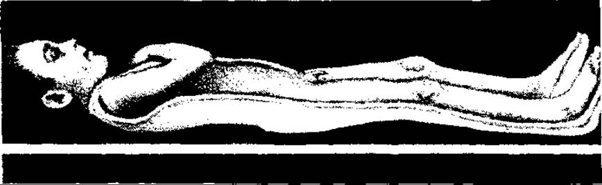
Fig. 82. Plaster shell bed for treating a patient with tuberculous spondylitis
In the majority of patients, the primary osteitis process progresses and spreads toward the joint, eventually involving it. In contrast, in a small number of patients—particularly when the osteitis is localized in the peripheral PARTS OF THE acetabulum or the femoral neck region—it extends via an extra-articular pathway into the bone tissue, with necrotic products breaking through the surrounding tissues to form cold abscesses and fistulae.
The transition of a tuberculous process from the bone to the joint typically occurs gradually, via contact involvement of the synovial membrane, and less frequently through the rupture of a primary focus (ostitis) into the joint through destroyed cartilage. Synovial involvement manifests as hyperemia, edema, accumulation of serous-fibrinous exudate (nonspecific changes), the appearance of specific tubercles On the surface, and the development of tuberculous granulations.
Caseous breakdown of the granulations transforms the serous exudate into purulent fluid. The exudate may breach the capsule, resulting in the formation of cold abscesses.
The proliferation of granulations from the synovial membrane extends to the articular cartilage, destroying it (sometimes completely when granulations grow beneath it, between the cartilage and the bone) and reaching the joint-
forming surface of the bones, causing their destruction (secondary ostitis). Destruction of joint elements leads to subluxations and dislocations of the hip. Over time, the inflammatory process in the joints subsides, and tuberculosis transitions into the post-arthritic phase (metatuberculous osteoarthritis). Connective tissue forms within and around the joint, and adhesions develop that restrict and demarcate caseous necrotic masses and granulations, which often trigger exacerbations and relapses of the process. Occasionally, the articular surfaces of the bones fuse either through fibrous adhesions or directly, resulting in ankylosis. Bone destruction ceases, their ends thicken, and manifestations of osteoporosis diminish. However, bone tissue does not regenerate. The cavities of the primary ostitis (bone cavities) with sequestered bone fragments and granulations persist for a particularly long time (posing a risk of disease reactivation in the coming months or years). The degree of morphological changes in the joint ranges from deforming osteoarthrosis-arthritis to complete joint destruction and the development of pseudoarthrosis (intra-articular or, less commonly, extra-articular — in cases of complete hip dislocation).
The clinical manifestations of coxitis depend on numerous factors, primarily the stage of the process, the localization of the primary ostitis, The Nature and intensity of its spread, as well as the patient's age and immunological and nonspecific reactivity.
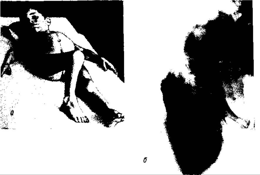
Fig. 83. Tuberculous coxitis: a — general view; b — radiograph of a hip joint affected by tuberculosis (post-arthritic stage)
In the first, pre-arthritic stage, symptoms are mild and inconsistent. General signs include a feeling of fatigue and lethargy, especially with movement (play or other physical loads), and periodic pain in the leg (frequently referred to the knee joint), particularly at night. Sometimes patients protect the leg from weight-bearing and develop a limp. Since intoxication signs are subtle, specific tests assessing the body's sensitization to Mycobacterium tuberculosis (such as the Mantoux tuberculin test) hold significant diagnostic value during this period. The mainstay in diagnosing tuberculous coxitis at the primary ostitis stage is radiographic examination (anteroposterior and lateral radiographs) of the hip joint, which reveals characteristic changes in adjacent bones: a destructive focus in the acetabulum or femur, often containing minute sequestra, alongside osteoporosis of the bones forming the joint.
In the arthritic stage of tuberculous coxitis, clinical signs are usually pronounced, particularly during the stage of tissue breakdown. These include symptoms of systemic intoxication, restricted joint mobility, nearly constant joint pain exacerbated by movement (both active and passive), impaired gait and flexion, occasional overgrowth of the limb due to irritation of the growth plate and epiphyseal hyperemia (if epiphyseal plates have not yet closed), as well as muscle atrophy and contracture in the affected leg (Fig. 83, a).
With contracture, the leg assumes a position of flexion in the Hip and knee joints, combined with internal rotation (adduction and inward rotation). Local symptoms of coxitis also include an increase in joint volume, especially in the exudative form of arthritis and upon the formation of cold abscesses, which are predominantly observed anterior to the greater trochanter, beneath the inguinal ligament, and in the adductor muscle region of the thigh. In addition to the aforementioned trophic disorders in the Tissues of the leg—such as muscle Atrophy of the affected limb—an important early sign of coxitis is thickening of the skin fold over the joint (Alexandrov's sign).

Fig. 84. Tuberculous gonitis. Exudative form (joint effusion)
Radiographic signs in the early stage of the arthritic phase (synovitis) include widening of the joint space and bone osteoporosis, whereas later stages reveal narrowing of the joint space, irregularity of articular surface contours, and destruction of the bones comprising the joint.
In the post-arthritic stage (the stage of sequelae of tuberculous coxitis), severe impairments of joint function (gait, mobility) and body posture occur, caused by the destruction of joint structures (Fig. 83, b), hip subluxations and dislocations, limb length discrepancy, Scoliosis, and other degenerative-dystrophic processes. Radiologically, this manifests both as signs of joint destruction, subluxations, and dislocations, and as reparative-adaptive features (terminal bone overgrowth, sclerotic-atrophic phenomena, and pseudoneoarthrosis).
Treatment for tuberculous coxitis, like other forms of osteoarticular tuberculosis, is comprehensive: it includes orthopedic methods (immobilization with plaster casts and, less frequently, traction), chemotherapy, surgical interventions, and sanatorium-resort therapy.
Tuberculosis of the knee joint (gonitis) ranks third in frequency among forms of osteoarticular tuberculosis. Similar to coxitis, it is observed in children and adolescents and develops through the hematogenous dissemination of mycobacteria from a primary focus.
Primary gonitis, like coxitis, occurs in exudative and productive (or fungous) forms of arthritis. Exudative arthritis is accompanied by hyperemia and edema of the synovial membrane (synovitis) with an accumulation of serous exudate rich in fibrin within the joint cavity. The formation of tuberculous tubercles on the synovial membrane is minimal. At this stage, changes in the joint are reversible. However, frequent precipitation of fibrin, particularly in the joint recesses (pouches), can compartmentalize the cavity, while its subsequent Organization alongside the tubercles (coupled with their resorption) may lead to restricted joint mobility (contracture). Involvement of the synovial villi and their detachment into the joint cavity, as well as the precipitation of fibrin clots, leads to the formation of so-called rice bodies—a white granular mass. In cases of protracted exudative tuberculous synovitis (due to delayed or inadequate treatment), the condition may progress to the second, fungous form.
Tuberculous synovitis is characterized by pain in the joint, especially during movement, impaired function (limited range of motion and reduced weight-bearing capacity), occasional low-grade fever, headache, sweating (particularly at night), and mild lethargy. The joint increases in volume due to fluid accumulation and periarticular soft tissue edema, its contours become smoothed out (Fig. 84), and visible muscle atrophy of the leg occurs alongside a thickening of the skin fold over the affected limb (Alexandrov's sign).
The so-called patellar tapotement (ballotment) is observed, which is a pathognomonic sign of fluid within the joint. This sign is elicited by compressing the sides of the joint with both palms while balloting the patella downward and backward with the index fingers. Upon release, the patella immediately rebounds upward, striking the femoral bone surface.
The fungous form of gonitis is accompanied by the development within the joint (in addition to infiltration of the synovial membrane by exudate and leukocytes) of mushroom-like granulations sprinkled with tuberculous tubercles. These granulations typically invade the cartilage, leading to its resorption and detachment. With inadequate treatment, the process spreads to the articular ends of the bones, the joint capsule, and ligaments, resulting in subluxations and dislocations. Furthermore, the caseous breakdown of granulations leads to the accumulation of purulent exudate within the joint, which extends beyond the capsule, forming cold abscesses and fistulae due to skin breakdown. Fistulae frequently cause secondary conventional bacterial infections (superinfections) within the tissues, significantly worsening the patient's condition and potentially leading to Sepsis.
The fungous form of tuberculous arthritis is characterized by severe alterations in the joint and surrounding tissues. The joint markedly increases in size, becoming dense and tumor-like; soft tissues swell, blood vessels are compressed, and the overlying skin becomes pale («white swelling»). Due to the enlargement of the joint against the Background of muscle atrophy, the limb acquires a fusiform (spindle-like) shape (Fig. 85).
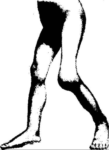
Fig. 85. Tuberculous gonitis. Productive or granulation (fungous) form (tumor albus)
Joint function is severely impaired—movements are restricted (contracture) and painful, and unassisted ambulation becomes impossible.
In the majority of patients, tuberculous gonitis results from the extension of a tuberculous process (primary tuberculous ostitis in the epimetaphysis of the tibia and femur) into the joint; thus, it represents a secondary process, corresponding to the arthritic stage of osteoarticular tuberculosis. During the ostitis phase (pre-arthritic stage), patient Complaints are vague. They experience mild, intermittent joint pain and subtle functional impairments of both the joint and the entire limb (rapid fatigue and protective unloading). As arthritis develops, the clinical picture becomes distinct. The disease course depends on its morphological form—exudative or fungous—and the patient's overall reactivity.

Fig. 86. Tuberculosis of the tubular bone — spino ventosa (a) and cervical lymph nodes (b)
The treatment of tuberculous gonitis, much like coxitis, involves conservative measures (chemotherapeutic drugs, physical therapy, sanatorium-resort treatment, vitamin-rich Nutrition, etc.), as well as orthopedic and surgical methods (plaster casts with immobilization of the hip, knee, and ankle joints; following the subsiding of the inflammatory process, various surgeries ranging from drainage and necrotomy to joint resection and plastic-corrective interventions, including limb lengthening via segmental osteotomy, etc.).
Tuberculous involvement of other Bones and joints is observed much less frequently than that of the spine, hip, and knee joints. In descending order of frequency, other joints are affected as follows: the ankle (5–7%), shoulder, elbow, wrist, and others. These arthritides develop through the same mechanism as coxitis and gonitis, i.e., primarily due to the spread of the process from the bones to the joint. Lesions of the diaphyses of tubular bones, particularly short ones (metacarpals, forearm bones, phalanges), are rare.
A distinctive feature of tuberculosis in rare locations (arthritis and ostitis) is its frequent onset in adulthood and a less severe degree of patient disability. Tuberculosis of the diaphyses of long and short tubular bones causes their fusiform deformation — spino ventosa (Fig. 86, a).
Lymph node tuberculosis is a frequent form of extrapulmonary tuberculosis, both primary and, especially, secondary. It is predominantly observed in children and adolescents, and is rare in young adults.
The most commonly affected lymph nodes are the cervical, bronchial, tracheal (mediastinal), and mesenteric lymph nodes. Superficial (external) lymphadenitis occurs mainly in the neck — submandibular, submental, and lateral jugular lymph nodes along the vascular bundle of the neck (including supraclavicular ones).
The cervical lymph nodes become infected through the mucous membrane of the Oral Cavity and Nose, where mycobacteria enter either during primary infection of the body via the digestive tract or with sputum during coughing in patients with pulmonary tuberculosis. Mediastinal and mesenteric lymph nodes are affected when mycobacteria penetrate through the respiratory tract and digestive canal from infection foci in the lungs and intestines, respectively.
A characteristic feature of lymph node tuberculosis is the multiplicity of affected nodes with the formation of conglomerates. Mediastinal, mesenteric, and retroperitoneal tuberculous lymphadenitis (acting as lymphogenic metastases of tuberculous infection of the lungs, intestines, and other organs, or more rarely as a consequence of hematogenous dissemination of mycobacteria from the primary complex in the lungs) are discussed in the respective sections of special surgery. Tuberculosis of external (peripheral) lymph nodes, primarily of the neck, is traditionally studied in the general surgery course.
Tuberculosis of the cervical lymph nodes has two clinical and morphological forms. In the first form, a productive process prevails in the lymph nodes, accompanied by phenomena of connective tissue proliferation and the formation of specific granulomas; consequently, the lymph nodes are dense, painless, mobile, and not fused with the skin or with each other. In the second form, characterized by the predominance of an exudative-necrotic process, they are fused with each other and the skin (as a consequence of periadenitis), painful, dense-elastic or soft, and form large matted clusters.
Necrosis within the lymph nodes, accompanied by the liquefaction of necrotic masses and pus formation, leads to the appearance of multiple fistulae on the skin of the neck, through which a liquid grayish-white purulent exudate and crumbs of necrotic masses are discharged.
Involvement of the cervical lymph nodes is almost always bilateral (Fig. 86, b). The lymph nodes vary in size; often, the clusters consist of one large lymph node surrounded by a chain of smaller ones. Patients frequently exhibit general pathological reactions — subfebrile body Temperature, lethargy, decreased appetite, anemia, lymphocytosis, and an elevated ESR. Patients are pale and emaciated. These general disorders are particularly characteristic of cervical lymph node tuberculosis complicated by the formation of purulent abscesses and fistulae, the so-called scrofuloderma. Edema of the face and Lips, significant intoxication, and secondary purulent infection frequently supervene.
The diagnosis of tuberculous lymphadenitis is challenging. Advanced forms are virtually non-existent today, particularly scrofuloderma — lymphadenitis accompanied by facial edema, subcutaneous abscesses, and fistulae. Tuberculous lymphadenitis must be differentiated from banal lymphadenitis, Hodgkin's disease, leukemic lymphadenopathies, and metastatic Cancer in the cervical lymph nodes. Specific tuberculosis tests and, above all, aspiration biopsy play a vital diagnostic role.
Treatment is comprehensive: specific chemotherapeutic agents (streptomycin, phthorafur/ftivazide, PAS, rifampicin, isoniazid, etc.), general strengthening agents (Vitamins, proteins, a nutritious diet), ultraviolet and solar irradiation, and—in cases of secondary banal infection—other antimicrobial agents, particularly antibiotics, administered after determining the sensitivity of the microflora. Well-circumscribed lymph node conglomerates can be radically excised, followed by specific conservative treatment.
SYPHILIS

Fig. 87. Spirochete (Treponema pallidum)
Syphilis is a sexually transmitted disease caused by the pale spirochete, or treponeme (Treponema pallidum). The spirochete was discovered in 1905 by F. Schaudinn and E. Hoffmann. It is believed that syphilis was an endemic disease of the Western Hemisphere and was brought to Europe in the Middle Ages by Columbus's sailors. The name "syphilis" originates from the shepherd Sypylus, the hero of Fracastoro's poem "Syphilis, sive morbus gallicus" ("Syphilis, or The French Disease"). Treponema pallidum is a thin, motile, spiral-shaped spirochete with sharp coils, measuring 5–15 µm in length; it is a facultative anaerobe that stains pale pink with Giemsa stain and cultivates well in tissues. Transmission occurs primarily sexually from an infected person to a healthy one, both heterosexually and homosexually, although direct transmission via skin wounds is also possible. Transmission can also occur indirectly through everyday objects contaminated with treponemes (spoons, cups, toothbrushes, mouthpieces, towels, etc.).
Treponemes penetrate the body predominantly through lesions in the mucous membrane or skin. However, they are capable of penetrating even intact mucous membranes. Within The Human Body, treponemes spread very rapidly — as early as 2 days after infection, they can be detected in regional lymph nodes. The incubation period typically ranges from 3 weeks from the moment of infection to the development of the disease.
Laboratory diagnosis is performed microscopically. Dark-field Microscopy reveals characteristically shaped, motile spirochetes (Fig. 87). A serological method, the Wassermann Complement fixation test, is also used. For this test, the patient's blood serum or CEREBROSPINAL FLUID (in cases of progressive paralysis) is examined. As an antigen, Wassermann originally used an extract of the Liver of a patient with syphilis; currently, the source of the antigen is beef Heart. Spirochetes are very easily detected in the Early stages of syphilis and very difficult to detect in the late stages.
Syphilis has a staged course. Primary, secondary, and tertiary periods of the disease are distinguished. Primary syphilis manifests on the 5th to 8th day after the penetration of spirochetes into the mucous membrane or skin as the so-called hard chancre (primary sore). This is an inflammatory reaction of the mucosa or skin to the inoculation of spirochetes, characterized by exudation, infiltration by lymphocytes and macrophages, proliferation of connective tissue elements, and degeneration and necrosis of epithelial cells. The clinical and morphological manifestation of this reaction is a small, dense, firm, painless ulcer on the mucous membrane, predominantly of the genitalia — the Penis or labia (in cases of sexual transmission) — or, more rarely, on the mucosa or skin in other locations (rectal mucosa, lips, Tongue, perineal skin, etc.) resulting from sexual or domestic transmission.
The ulcer (chancre) has a rounded shape, a bright pink color, clear ridge-like edges, and protrudes slightly above The surface of the mucosa or skin. The development of a hard chancre is always accompanied by regional lymphadenitis, specifically inguinal lymphadenitis in cases of sexual infection. The regional lymph nodes are painless, small, mobile, firm, and not fused with the skin.
The hard chancre, or primary syphilis, typically disappears spontaneously 2 weeks after its appearance, leaving no scar. A month or later, the lymphadenitis also resolves. After 5–8 weeks, the second period of the disease begins — secondary syphilis, which is a manifestation of generalized bodily involvement. It is characterized by general symptoms (lethargy, chills, headache, generalized lymphadenopathy, body aches, tonsillar enlargement) as well as localized syphilids — maculopapular, or more rarely pustular, rashes on the skin, mucous membranes of the Mouth, Vagina, and infrequently the conjunctiva. Around the genitalia and the anal canal, flat condylomas develop due to skin irritation by secretions, followed later by indurative keratosis.
Papules on the hands and soles disappear spontaneously. In untreated patients, they may persist for up to 3 years. Secondary syphilis is highly contagious. During this period, the blood and secretions contain A large number of treponemes.
The tertiary period, or tertiary syphilis, usually develops after the secondary stage, or less frequently several years (sometimes decades) later. It is observed in untreated or inadequately treated patients.
Its morphological substrate is the formation of a so-called gumma, usually single, less frequently multiple, located in various organs and tissues. A gumma is a proliferative-necrotic form of localized inflammation, an unstable granuloma caused by primary spirochetemia. It predominantly develops in Internal Organs, particularly The Heart and large Arteries, the Central Nervous system (CNS), and the liver, and less commonly in the lungs, testes, and kidneys. Even rarer are gummas developing in muscles, bones, joints, the skin, and mucous membranes. They can form in any area of the trunk. Microscopically, The Structure of a gumma includes perivascular proliferation of the organ stroma, lymphocytic and plasmacytic infiltration, fibroblasts, atrophy of specialized tissue, and vascular obliteration, primarily affecting arteries. The latter causes a degenerative-necrotic process within the gumma and its subsequent breakdown, resulting in large defects in organs or ulcers in superficial tissues.
Epithelioid cells and Langhans cells are rarely found in a gumma and are practically absent, which distinguishes gummas from tuberculous granulomas. Macroscopically, a gumma has a rubbery (guttapercha-like) consistency, a grayish-yellow color, and is opaque.
Syphilitic gummas rarely resolve without breakdown and ulceration. They leave behind coarse, retracted, and deforming scars.
Necrosis and breakdown of gummas, which are observed in the majority of patients, lead to irreversible changes in the cardiovascular, central nervous, and musculoskeletal systems. In one out of 13 untreated patients, this causes cardiovascular disease; in one out of 25, disability (paralysis); in one out of 44, irreversible CNS damage; and in one out of 200, blindness (N.M. Sommers, 1980).
The breakdown of superficial gummas leads to the formation of deep ulcers that frequently penetrate through the entire thickness of soft tissues and bones (Fig. 88), creating apertures in the latter. This disfigures the patient's appearance—such as a gaping nose resulting from the destruction of nasal bones—impairs the Functions of organs and tissues, and poses a threat of Acute Purulent Infection complications.


Fig. 88. Syphilis of the bones of the lower leg (a) and bone eburnation—sclerosis and disappearance of the medullary cavity (b)
Syphilitic ulcers have sharply cut edges, as if punched out, with a yellowish-gray floor resembling old lard.
In the tertiary stage of syphilis, both flat bones (bones of the facial Skeleton, Skull, clavicles) and long tubular bones (tibia, humerus, etc.) are affected, manifesting as periostitis, osteitis, and osteomyelitis. Syphilitic periostitis is accompanied by a significant osteoblastic periosteal reaction of a locally limited type, resulting in painful periosteal nodes and bone deformation. Osteitis manifests as focal bone destruction with the formation of small sequestra and marked proliferation of the periosteum.
Syphilitic osteomyelitis, along with focal necrosis and bone sequestration, is accompanied by a sharp osteoblastic periosteal reaction that frequently leads to the obliteration of the medullary cavity, its ossification, and calcification. As a result, the bone (e.g., the tibia) acquires the appearance of a dense, deformed structure known as bone eburnation, or ivory bone (Fig. 88, b).
Painless syphilitic nodes around large joints—such as the knee and elbow—are formed due to fibrosis of the subcutaneous adipose tissue.
Clinically, tertiary bone syphilis manifests as nocturnal bone pain and minor functional impairments of the limbs despite substantial morphological changes in the bones. The disproportion between functional impairment and structural bone alterations is explained by decreased pain sensitivity in patients due to nervous system involvement.
In congenital syphilis, bone lesions present with deformation and eburnation. They are most commonly localized in the facial skeleton and the tibia, which acquire a saber-like shape.
Syphilis treatment is specific: antibiotics, particularly penicillin-group drugs. Early forms of syphilis (Primary and secondary) must be differentiated from banal ulcers, lymphadenitis, perianal condylomata lata, and other superficial inflammatory and dystrophic processes. Destructive bone changes are treated surgically and orthopedically.
To prevent complications of late (tertiary) syphilis, timely diagnosis and adequate Treatment of the early forms of the disease (primary and secondary) are essential.
In gummatous syphilis, the restoration of the morphological state and function of organs and tissues is impossible. Disease prevention, avoidance of casual sexual relations, and the promotion of a higher culture of Sexual Behavior play a colossal role in combating syphilis.
ACTINOMYCOSIS
Among numerous fungal diseases, actinomycosis is of the greatest interest to surgeons. Actinomycosis (from *aktis* – ray, *mykes* – fungus) is a chronic specific inflammatory disease caused by ray Fungi, or actinomycetes, particularly *Actinomyces israelii*. An actinomycete is a thin, filamentous microorganism that is more closely related to bacteria than to fungi (its branching filaments easily fragment into bacillary forms). It is a normal commensal Organism that vegetates in the oral cavity and intestines of both humans and animals, especially domestic livestock, and is an anaerobic bacterium. A characteristic feature of this microorganism is the formation of consolidated bodily masses known as yellowish sulfur granules. Under certain conditions, the microorganism invades tissue and, in Symbiosis with vulgar microflora, causes an inflammatory suppurating process. In the oral cavity, it is found in dental plaque, gingival pockets, cavities of carious Teeth, tonsillar crypts, etc. The disease occurs in both humans and animals—cows, and less frequently horses and dogs. The main source of actinomycetes for humans is the oropharynx. Human infection occurs via endogenous microorganisms (autoinfection), although exogenous transmission is also possible (through the ingestion of actinomycete-contaminated cereals or Water). Microorganisms spread via Lymphatic vessels and by direct contact. All tissues can be affected, but connective tissue, particularly adipose tissue, is especially vulnerable.
The disease occurs in three forms: cervicofacial, bronchopulmonary, and abdominal. Actinomycosis of the central nervous system, kidneys, and Ovaries, as well as its generalized form, are rare. The cervicofacial localization is the most common, accounting for 60% of all lesions. Bronchopulmonary and abdominal forms each account for approximately 20%.
The cervicofacial form typically develops following a tooth extraction or another oral cavity Procedure that creates a wound, facilitating the entry of pathogens into the tissues. The disease progresses slowly. A dense, almost painless infiltrate (swelling) appears in the region of the angle of the lower jaw, with the overlying skin acquiring a pinkish-bluish hue. The face becomes asymmetric (involvement is almost always unilateral). Subsequently, softening occurs in the center of the infiltrate due to necrosis and tissue lysis, leading to the formation of an abscess (Fig. 89). The abscess typically ruptures externally through multiple openings, or fistulae. These discharge fluid pus containing yellowish crumbs or grains (granules) known as sulfur granules (Fig. 90), which harbor accumulations of microorganisms.
These granules are specific elements of discharge in superficial actinomycosis and aid in diagnosing the disease. Around the inflammatory lesion in the tissues, particularly along the fistulae, a pronounced fibroplastic reaction develops due to the sensitizing effect of actinomycetes, resulting in fibrous tissue formation and sclerosis. This leads to puckering of the skin in the area of the infiltrate and the formation of skin folds along tension lines (Langer's lines). Due to the involvement of the masticatory muscles in the inflammatory process, mouth opening is restricted (trismus), and chewing and digestive functions are impaired.

Fig. 89. Cervicofacial form of actinomycosis
The inflammatory process frequently spreads to the periosteum and the bone of the lower jaw, causing actinomycotic periostitis and osteomyelitis. Even before jaw involvement develops in cervicofacial actinomycosis, metabolic disturbances occur in the jaw bones in the form of osteoporosis. With the formation of skin fistulae, the disease becomes complicated by secondary pyogenic infection, which increases pain and intoxication. In most cases, the General condition of patients remains largely unaffected.

Fig. 90. Actinomycete micelles (so-called sulfur plugs in the center of an actinomycotic abscess)
Aside from the maxillofacial form of actinomycosis, other external localizations of the disease occur, albeit very rarely, notably actinomycosis of the buttocks (Fig. 91) and the Perineum. These manifest as a multi-nodular sclerosis of the subcutaneous tissue accompanied by abscesses, bluish skin discoloration, and the presence of fistulas in the affected area.
Diagnosis of the disease is based on typical clinical and morphological manifestations, as well as the results of MICROBIOLOGICAL EXAMINATION OF fistula discharges—the detection of actinomycetes, and particularly their clusters, i.e., sulfur granules or micelles, indicates actinomycosis.
The bronchopulmonary form develops when actinomycetes enter the respiratory tract of patients with inflammatory and other underlying conditions, coupled with decreased bodily resistance. The pathogens invade the lung parenchyma, forming massive foci of interstitial-type pneumonia accompanied by numerous small abscesses (with a scant amount of bloody sputum) and pulmonary parenchymal fibrosis. The process may spread to the Pleura and even the Ribs, resulting in fistula formation on the chest wall, as well as extending through the Diaphragm into the Abdominal cavity. This form of actinomycosis severely compromises the patient's general condition, presenting with burning chest pain, especially when the pleura is involved (Spasokukotsky's sign), weight loss, fever, a cough with scant sputum containing traces of blood, intoxication, weakness, sweating (particularly at night), and dyspnea. The latter is especially characteristic when the process spreads to the Mediastinum. X-ray Examination reveals broad areas of pneumonia with frequent consolidation of the basal lung segments. Microbiological examination of sputum (presence of drusen—sulfur granules) is of paramount importance, along with histological examination of biopsy specimens and resected tissues.
The intestinal form of actinomycosis develops when the microflora penetrates through the mucous membranes of the digestive tract, most commonly occurring in the ileocecal region (Appendix, cecum, and the terminal ileum). The lesion originates in the Cellulose tissue surrounding these organs and clinically mimics acute appendicitis, terminal ileitis, a cecal tumor, or intestinal obstruction. The process spreads beyond the mucous membrane (which remains intact) through the adipose tissue, muscles, and serosa of the organs, eventually extending to the pelvic cellular tissue, Urinary Bladder, and other structures.

Fig. 91. Actinomycosis of the buttock
This condition is accompanied by pain (sometimes colicky in nature), elevated body temperature (occasionally reaching high levels), chills, vomiting, and weight loss. Abdominal examination frequently reveals localized tenderness and rigidity of the abdominal wall, often allowing the Palpation of tumor-like masses.
Treatment of actinomycosis is comprehensive, combining conservative and surgical approaches. Purulent superficial foci are incised and drained, while internal ones are excised (segmental or partial lung resection, removal of the affected intestinal loop and involved connective tissue, or evacuation of pus and subsequent drainage). Conservative treatment involves The Use of actinolysate and antibiotics. Among the latter, benzylpenicillin sodium salt in high doses (20,000,000 IU every 6 h) is the mainstay. It can be combined with streptomycin sulfate or other sulfonamides. Broad-spectrum antibiotics are prescribed if the indigenous microflora exhibits resistance to penicillin-group drugs. Iodine preparations, such as iodonate and povidone-iodine, are used primarily topically for cutaneous forms of the disease. In such cases, radiotherapy of the infection foci is also prescribed.
Actinolysate therapy is performed According to the methods of G.R. Sutyayev (intramuscularly) and D.I. Asnin (intradermally).
In intramuscular therapy, actinolysate is injected into the gluteal muscle at 3 ml twice a week, whereas in intradermal therapy, it is administered into the skin of the anterior forearm. Doses for intradermal administration are increased gradually from 0.5 ml to 2 ml. Each successive dose is increased by 0.2 ml, and no more than 0.5 ml is injected into a single site; thus, upon reaching a 2 ml dose, the drug is administered across 4 sites. As with intramuscular therapy, the preparation is administered twice a week. The course of treatment lasts 3 months. The intradermal method is somewhat more effective, albeit more complex. With timely diagnosis and adequate treatment, actinomycosis relatively rarely leads to fatal outcomes (which occur primarily in bronchopulmonary and abdominal actinomycosis).
Acquired Immunodeficiency Syndrome (AIDS)
Acquired immunodeficiency syndrome (AIDS), which became known in 1981, can be provisionally classified as a chronic specific infection. It is a viral disease targeting immunogenic cells, resulting in the development of opportunistic infections and specific tumors within the body. The disease is caused by a retrovirus—HUMAN IMMUNODEFICIENCY VIRUS type 1 (HIV-1). This virus exhibits an exceptional tropism for CD4 molecules found on T-helper/inducer cells (lymphocytes) and monocyte-macrophages. The entry of the virus into these cells and its Replication therein lead to The production of progenes and the destruction or functional Impairment of the cells.
The immunodeficiency virus is transmitted sexually, via blood transfusions and blood components from HIV-1-infected Donors, and through the use of virus-contaminated needles, syringes, and other instruments. AIDS transmission also occurs from an infected mother to her child during the perinatal period. Furthermore, HIV infection can be contracted by surgeons as a result of puncture injuries (from needles, scalpels, etc.) sustained while operating on patients infected with the immunodeficiency virus.
HIV infection is diagnosed using serological testing—the identification of anti-HIV-specific Antibodies in blood serum. It may remain clinically silent for a long time; however, it is estimated that virtually all HIV-infected individuals develop the disease within 7–10 years. The condition may present acutely as a mononucleosis-like syndrome, followed by a prolonged asymptomatic period lasting years before the appearance of various localized manifestations.
HIV infection is classified as follows (C. Edward and I. Boucher, 1994):
I. Acute HIV infection — mononucleosis syndrome.
II. Asymptomatic infection.
III. Persistent generalized lymphadenopathy.
IV. Other HIV-related conditions:
A. Constitutional diseases — AIDS-related complex (ARC);
B. ARC with neurological disorders;
C. With symptomatic or invasive secondary infectious diseases;
D. Secondary malignancies;
E. Other conditions (thrombocytopenia).
Common general manifestations include fatigue, weight loss, fever, and night sweats. Local (systemic) disorders vary widely on an individual basis. These are predominantly infections or diseases developing as a result of lowered host resistance. They include pneumonia, generalized lymphadenopathy, fungal esophagitis or prolonged enterocolitis, acalculous cholecystitis, proctosigmoiditis with fistulas and perianal abscesses (especially in homosexuals), mucocutaneous herpes simplex lasting over 5 weeks, histoplasmosis and Toxoplasmosis, non-Hodgkin's lymphoma, and Kaposi's Sarcoma (a vascular sarcoma) in individuals under 60 years of age, among others.
Lymphomas are classified as high-grade B-cell neoplasms.
In the majority of patients, the syndrome develops within the first 2 years following infection (although the incubation period ranges from 1 to 10 years), and the average duration of the disease prior to a fatal outcome is also approximately 2 years. AIDS caused by HIV-1 is spreading rapidly worldwide, particularly in the USA and Europe. In Africa, HIV-2 is more prevalent. AIDS caused by this type is endemic in nature.
AIDS presenting with the course of a conventional infection has posed a Differential diagnosis challenge for surgeons, distinguishing it from standard surgical infections, and has complicated patient management.
Surgical conditions in patients with AIDS often exhibit an atypical clinical course. The goal of differential diagnosis is to avoid erroneous surgical interventions in AIDS patients presenting with certain symptoms of surgical diseases, notably pain (internal, especially abdominal, manifestations of AIDS).
Thorough serological blood testing in patients with suspected AIDS eliminates the need to diagnose lymphomas via needle biopsy.
The AIDS virus (HIV-1) is easily and rapidly inactivated by heat and disinfectants (such as glutaraldehyde and hydrogen peroxide). AIDS prevention involves modifying sexual behavior, avoiding casual contacts with unknown partners, and performing serological screening of donor blood and blood products.
Preventing infection among medical personnel during surgical Procedures and manipulations requires, in addition to vigilance and strict adherence to asepsis rules, that surgeons and nurses performing various procedures—especially invasive ones—wear masks, water-impermeable gowns, and two pairs of gloves. All instruments and used Materials must be thoroughly sterilized and disinfected, and stored in separate rooms or designated areas. In the event of an accidental injury (such as a needle stick), the affected area should be immediately treated with an alcoholic iodine solution (after expressing a few drops of blood), followed by the administration of immunoglobulin.
Last update: 08/08/2026
Editorial and Educational Adaptation: This material has been compiled based on the primary/original source text. The project team performed an editorial review, corrected technical inaccuracies, structured sections, and adapted the content for an educational format.
What was processed:
- elimination of formatting defects (OCR errors, structural breaks, corrupted characters);
- editorial organization of content;
- standardization of terminology in accordance with academic sources;
- verification of factual statements against the original source text.
All mentions of the author, publication year, and origin of the primary text have been preserved in accordance with the source.