Antibiotics (Properties, Application, Interactions) - M.P. Cherenko 1999
Acute purulent infection
Purulent finger diseases
The hand and fingers are the most essential PARTS OF THE upper extremity. The Role of the hand and fingers in The Human Body is multifaceted; their movements are characterized by a wide range of motion, coordination, precision, and diversity. They perform static, dynamic, and sensory Functions. When opened with straight fingers, the hand serves as a shovel or scoop; with curved fingers, it acts as a hook or forceps; and the opposition of the thumb to the other fingers provides the hand's grasping function.
In addition, each finger performs a distinct function. Special attention should be given to the first digit, namely the thumb. It acts independently of the others, making the human hand an organ of labor. Thanks to the thumb, a person can grasp objects and manipulate them according to their intentions. The index (second) finger possesses fine tactile sensitivity and is the first to initiate a grasp. Its role is particularly crucial in pinch and sculptural grips. The middle (third) finger is more massive and longer, providing strength and stability to the grasp. The ring (fourth) finger, thanks to its well-developed sense of Touch, regulates Muscle movement, which is essential during manual work. The little finger (fifth digit) secures the grasp, closes the palmar cup, and gives stability to the hand.
Nail plates are of great importance for the function of the hand, as they ensure the reliability of the pinch grip.
The hand and fingers perform more than just a grasping function. They serve as a delicate Organ of Touch, providing The connection between a person and the outside world.
Consequently, the loss of any part of the hand or its functional inadequacy affects a person's working capacity and their interaction with the environment.
Various pathological conditions can lead to the loss of hand and finger function: trauma, inflammatory, degenerative-dystrophic processes, tumors, and congenital anomalies.
This chapter focuses on inflammatory processes of the fingers (felon or panaritium), as they are the most frequently observed.
Felon (panaritium) is a collective term for all acute inflammatory processes affecting various structures of the fingers, predominantly on the hands (specifically their palmar surface).
The clinical course of a felon on the hands largely depends on the anatomical and functional Features of the fingers.
The Skin of the PALMAR AND DORSAL surfaces of the fingers differs. The skin of the palmar surface lacks pigment apparatus, Hair, and Sebaceous Glands—ruling out The Development of furuncles and carbuncles—yet it is rich in Sweat Glands. The skin of the palmar surface is densely innervated, which accounts for its pronounced tactile sensitivity and severe pain when an inflammatory process develops in the fingers.
The skin on the dorsal surface of the fingers is soft, elastic, and mobile. It features hair and sebaceous glands (except for the terminal phalanx). Therefore, furuncles and carbuncles can develop only on the proximal and middle Phalanges.
On the palmar surface, the adipose base is more abundant than on the dorsal side. There is no subcutaneous fascia here; instead, Cytology/practical/45.html">Dense Connective Tissue bands extend almost perpendicularly from the papillary layer, weaving into the periosteum on the distal phalanx, and into the wall of the tendon sheath on the middle and proximal phalanges. These cords are densely intertwined with connective tissue fibers running parallel to the skin surface, forming numerous chambers within the subcutaneous tissue. This Structure limits skin mobility and hinders the spread of edema; the accumulation of edema quickly leads to the compression of Blood Vessels, Impaired blood supply to fat lobules, and a sharp increase in pain due to elevated interstitial pressure. The vertical walls of these chambers prevent the inflammatory process from spreading across the surface while facilitating the penetration of infection toward the bone.
On the dorsal surface of the fingers, the connective tissue fibers lie parallel to the body surface and do not impede the spread of edema.
The posterior surface of the terminal phalanx is protected by the nail. The nail plate is quadrilateral in shape. Its proximal and lateral edges are recessed into the nail groove. A skin fold—the eponychium, or nail fold—hangs over the proximal edge of the plate (the matrix). Connective tissue fibers extend from the inner surface of the nail plate to the periosteum of the phalanx, fixing the nail to the bone, which facilitates the spread of a purulent-inflammatory process to the nail phalanx. Nail regeneration after removal takes 4–6 months.
On the palmar surface, each finger except the thumb has two tendons: the superficial and deep flexors. The thumb has a single flexor tendon (the long flexor of the first digit).
The tendon sheaths of the first and fifth digits are longer than the others. They pass through Pirogov's space into the distal part of the forearm, forming a common synovial bursa there and in the forearm. The tendon sheaths of the second through fourth digits are shorter, terminating at the level of the metacarpophalangeal joints (Fig. 71). This is of great practical significance in clinical pathology. Due to the long sheaths of the first and fifth fingers and their fusion in the wrist region, inflammatory processes in these tendon sheaths have a more severe course and are complicated by deep phlegmons of the hand. Inflammation of the flexor tendons of the second through fourth fingers remains localized to those specific fingers.
Digits II–V have 3 phalanges each, while digit I has 2. The phalanges are interconnected by hinge joints.
Each finger is supplied with blood by 4 Arteries: 2 palmar and 2 dorsal. The Blood supply to the terminal phalanx has its own specific features. While the middle and proximal phalanges have a dedicated feeding artery, the terminal phalanx is supplied by a branching (dispersed) type of vascular network. Furthermore, the blood supply to the proximal and distal epiphyses is better than that of the remaining parts of the phalanx because they receive separate arterial branches of varying calibers that penetrate the bone primarily from the palmar surface. This plays a significant role in the Pathogenesis of bone felon of the distal phalanx.
Innervation of the fingers is provided by sensory and motor branches of 3 nerves: the ulnar, radial, and median, which divide into terminal branches to innervate their respective areas. The pulp of the terminal phalanges has the densest innervation, which explains the intense pain when a pathological process develops there.
Class="center">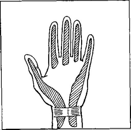
Fig. 71. Tendon sheaths of the fingers
Lymph drainage from the fingers occurs via superficial and deep Lymphatic vessels. A prominent lymphatic network is present on the dorsal surface of the terminal phalanx, the palmar surface of the fingers, and the palm. From here, lymphatic vessels run along the lateral surfaces of the fingers into the interdigital folds and through them penetrate the dorsum of the hand. Consequently, when an inflammatory process occurs on the palmar surface, hand edema is more pronounced on the dorsal side.
The lymphatic Vessels of the fourth and fifth fingers drain into the ulnar and partially into the axillary Lymph Nodes. Lymphatic vessels from the thumb, index, and middle fingers primarily drain lymph into the subclavian, supraclavicular, and deltopectoral groups of glands. This explains why felon of digits I–III has a much more severe course and can be complicated by subpectoral Phlegmon.
Etiology. Purulent inflammation of the fingers is observed in 20–25% of patients visiting outpatient surgical clinics. Various types of minor trauma play a crucial role in its onset, most commonly puncture wounds and splinters. Accordingly, felon occurs predominantly in individuals whose professions involve a risk of finger injury (fish Processing workers, butchers, chefs, medical personnel—especially surgeons and obstetricians, etc.).
Major injuries less frequently cause felon because they receive prompt medical attention. More often, the causative agents of felon are streptococci and staphylococci. An inflammatory process in the fingers is more likely to develop when their skin is dry and calloused.
In most patients, felon (panaritium) is localized on the right hand due to its increased functional load. The distal phalanx is most frequently affected.
Classification. Depending on the depth of microbial penetration and the development of infection, the following types of felon are distinguished (Fig. 72):
1) superficial — cutaneous (panaritium cutaneum); subcutaneous (panaritium subcutaneum); nail (periungual, or paronychia — panaritium parunquale s. paronychia); subungual (panaritium subunquale);
2) deep — tendinous (panaritium tendinosum s. tendovaginitis purulenta); osseous (panaritium osseum); articular (panaritium articulare).
If the pathological process involves all Tissues of the finger, pandactylitis develops.
Deep felon is usually the consequence of untimely and inadequate Treatment of superficial felon.
Clinical Features and surgical tactics for Various Forms of felon have their own specific characteristics.
Cutaneous felon occurs when the infection penetrates superficially. In this case, the inflammatory process develops beneath the epidermis. The latter detaches, forming a blister filled with pus. Most frequently, this type of felon is localized on the palmar surface of the finger. The appearance of the blister is accompanied by mild pain that gradually increases. The blister can sometimes reach sizes that involve the entire phalanx. Signs of lymphangitis appear on the dorsal surface of the hand and forearm, accompanied by fever. The General condition of the patient with this type of felon is usually not impaired.
Treatment of cutaneous felon is surgical: after preparing the surgical field, the edges of the blister are trimmed with small scissors. Pus is removed, and the necrotic epidermis is excised. Afterwards, one must ensure that the subcutaneous felon is not connected to deep tissues. To do this, the affected area is pressed with a finger. If the infection has penetrated beneath the skin from the depth, pus will ooze out. In such cases, using an appropriate anesthesia method, the subcutaneous abscess is incised. The operation is concluded by applying a dressing soaked in an antiseptic. If the cutaneous felon is complicated by lymphangitis, immobilization of the finger and hand, as well as UHF therapy, is performed.
On the dorsal surface, cutaneous felon is less common and is usually localized on the middle phalanx. The average duration of a cutaneous felon is 4–5 days.
Subcutaneous felon is the most common form. The entry of infection into the subcutaneous tissue is caused by puncture microtraumas. In 80–90% of cases, it is localized on the palmar surface of digits I–III of the right hand. Subcutaneous felon occurs more frequently in women than in men (due to needle pricks).
Subcutaneous felon is accompanied by severe pain in the finger that prevents the patient from sleeping. Sometimes the pain has a throbbing character synchronous with the pulse. This is due to the fact that connective tissue septa extending vertically from the skin to the periosteum prevent the edema fluid from spreading, thereby increasing its pressure in the tissues and compressing nerve receptors.
The finger swells, especially its dorsal surface. When the process is localized exclusively in the subcutaneous tissue, skin redness may initially be absent. In cases of pronounced edema, the skin even becomes pale due to the compression of blood vessels.
By pressing on the affected area with a probe, the zone of maximum tenderness is identified. The patient spares the finger and keeps it slightly flexed. In subcutaneous felon, the patient's general condition is hardly disturbed. Along the course of connective tissue fibers, the process can spread to the bone, tendon sheaths, joint, as well as lymphatic and blood vessels.
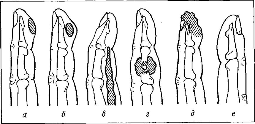
Fig. 72. Types of felon:
a — intraepidermal; b — subcutaneous; c — tendinous; d — articular; e — osseous; f — sub- and periungual
Patients with subcutaneous felon rarely seek medical help initially. Usually, they do so after the first sleepless night, which is a grave mistake, because by that time, deep destructive and necrotic processes have already developed in the finger tissues in most cases.
During the first 2–3 days (in the serous or serous-infiltrative stage), conservative therapy may be administered under a physician's supervision. It is necessary to immobilize the hand, apply local and systemic antibiotic therapy, and prescribe sulfonamides. Local antibiotic therapy consists of administering the drug along with a 0.5% novocaine solution intravenously (after applying a tourniquet to the lower third of the forearm) into one of the swollen Veins on the dorsum of the hand in a distal direction (100,000–250,000 IU of benzylpenicillin sodium salt per 20–40 ml of 0.5% novocaine solution). The tourniquet is kept for 8–10 min.
An alcohol wet-to-dry dressing is applied to the affected finger and changed 2–3 times a day; warm finger baths (with alcohol or an antiseptic) are administered for 40–60 min twice a day.
If conservative therapy yields no effect within 1–2 days, Surgical treatment is indicated. The latter must be performed under reliable anesthesia. This can be local novocaine anesthesia (for subcutaneous felon of the distal and middle phalanges) or intravenous anesthesia. Local anesthesia is performed According to the Oberst-Lukashevich method.
For subcutaneous felon of the distal phalanx, it is advisable to perform a so-called hockey-stick incision. Providing good pus drainage, it does not severely deform the finger, nor does it impair its tactile sensitivity and function.
When the inflammatory process is localized on the proximal and middle phalanges, incisions are made along the lateral surfaces of the finger corresponding to the phalanx (Fig. 73).
Longitudinal incisions through the point of greatest tenderness can also be used. After opening the focus of inflammation and creating conditions for pus evacuation, necrotic tissues are excised. The wound is drained with rubber strips. The Use of gauze tampons is not recommended because they dry out quickly and hinder the outflow of pus.
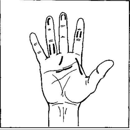
Fig. 73. Incisions for felon
Some authors recommend applying primary sutures in the postoperative period after abscess incision and necrotomy, infiltrating the tissues several times with a benzylpenicillin-novocaine solution.
If general clinical symptoms are present, detoxification therapy is performed.
Paronychia is an inflammation of the nail fold. It is most commonly caused by microtraumas on the dorsal surface of the terminal phalanx near the nail, or by hangnails. Paronychia is characterized by painful Swelling of the perionychium and hyperemia of the surrounding tissues. The affected fold overhangs the nail plate. Palpation of the fingertip, especially at the Base of the nail, is acutely painful. Sometimes pus can penetrate under the nail plate, resulting in subungual felon. Pressure causes pus to ooze from beneath the skin fold. The patient's general condition remains virtually unchanged.
Based on the localization of pus, superficial (subepidermal) and deep paronychia are distinguished. In the latter case, pus accumulates within the thickness of the nail fold.
Treatment of paronychia begins with conservative measures. Warm sodium bicarbonate FOOT/hand baths are given several times a day. Physiotherapy Procedures are carried out. To ensure the outflow of pus, the nail fold can be slightly pulled upward with tweezers or small scissors. When this fails to help, surgical treatment is indicated. Two incisions of the nail fold (0.5 cm each) are made as an extension of the two edges of the nail plate. The flap of the nail fold is separated. The proximal end of the nail plate is incised and excised at a distance of 0.5 cm where it has detached from the bed. It is important to well deflect the nail plate in this process and thoroughly remove the pus without damaging the nail bed and nail fold. Otherwise, the regrowing nail and nail fold will be deformed and split. After the manipulations described above, the flap of the nail fold is put back in place, and an aseptic dressing is applied (Kanavel operation).
Subungual felon is the result of trauma and infection of the distal edge of the nail (the nail bed). It is caused by various hangnails, pricks, and minor injuries. Subungual felon can be primary (the process usually begins directly as a subungual felon) and secondary (infection gets under the nail in the presence of paronychia).
The clinical picture is characterized by throbbing pain in the finger. Palpation of the nail is extremely painful. Edema of the dorsal surface of the nail phalanx appears. The general systemic reaction manifests as fever and chills. Accumulation of pus is visible through the nail.
Treatment of subungual felon is surgical. Near the free edge of the nail and in its distal part, if the nail ROOT is not involved in the process, the distal affected part is resected. If the process is localized at the base of the nail without detachment of the rest of it, the Kanavel operation is performed. If the subungual felon involves the entire nail bed and separates the nail, its complete removal is indicated. After surgery, ointment dressings, biological film dressings, or placental membrane dressings are applied.
Tendon felon (tenosynovitis) occurs when the inflammatory process spreads from the subcutaneous base or due to direct damage to the tendon sheath. Of all inflammatory processes, this type is the most dangerous because, when digits I and V are affected, it can spread to the palm and forearm, subsequently leading to impaired function of not only the fingers but the entire hand. Tendon inflammation occurs easily because tendons have a relatively poor blood supply, while the closed cavity of the sheath creates favorable conditions for microbial proliferation.
The tendons of the back of the hand are more resistant to infection and, as a rule, are not involved in the inflammatory process.
An unfavorable disease course is often caused by late recognition and inadequate treatment.
The clinical picture and course of tendon felon of digits II, III, IV differ from those of digits I and V, which is explained by their Anatomical Structure.
In tendon felon of digits II, III, IV, the fingers are flexed. This position reduces tension on the tendons. The soft tissues are severely edematous, and the slightest movement, especially extension, is accompanied by severe pain. Sharp pain also occurs when a probe touches the skin over the PROJECTION OF THE tendons. Attempts to straighten the finger cause very severe pain, which is the main symptom of tendon felon. The patient's general condition is also impaired: body Temperature rises to 39–40 °C, weakness, chills, and headache appear, and later edema of the back of the hand develops.
In tenosynovitis of digits I and V, all local manifestations rapidly spread to the palm and even to the forearm. The disease is accompanied by lymphangitis and lymphadenitis.
Treatment of tendon felon is surgical. The sooner the operation is performed, the better the outcomes. The fact is that the exudate accumulating in the tendon sheath compresses the blood vessels supplying the tendon, leading to its rapid necrosis.
In the phase of serous exudation, some surgeons recommend puncturing the tendon sheath with aspiration of 1–2 drops of pus and injection of a benzylpenicillin sodium salt solution into it. Puncture of the tendon sheath is performed within the middle phalanx. The antibiotic is injected in a high concentration (100,000–200,000 IU of benzylpenicillin sodium salt and 1 ml of novocaine).
The operation is performed under various types of anesthesia. Many incisions have been proposed for accessing the tendons. Most often, two parallel incisions are used at the level of the middle phalanges, made on the palmar-lateral surfaces of the finger. Additionally, one incision is made on the palm to open the blind pouch of the tendon sheath. Pus is evacuated, the sheath cavity is washed with Antibiotics, and the area above the tendon is drained with a rubber strip. The finger and hand are immobilized with a plaster cast.
When the tendon has a dull yellow color, it indicates its necrosis. In this case, preserving it is impractical; it must be removed and a tendoectomy performed.
Some surgeons recommend suturing the wound after opening suppurative tenosynovitis and washing the tendon sheath. The tissues around it are infiltrated with an antibiotic. This manipulation is repeated several times in the postoperative period.
It is more difficult to treat tendon felons of digits I and V. Palmar-lateral incisions in the region of the middle phalanx of the finger are also performed for them. In addition, additional incisions are made on the palm (along the inner margins of the thenar and hypothenar eminences with the opening of the ulnar and radial synovial bursae). If the process spreads along the synovial sheaths to the forearm, Pirogov-Parona cellular space must be opened.
The duration of treatment for patients with tendon felon is 20–40 days.
Tendon felon often leads to persistent impairment of finger function due to tendon necrosis. Isolated cases of death from Sepsis complications are observed.
Articular felon is a purulent inflammation of the interphalangeal and metacarpophalangeal joints. It can be primary or secondary. Primary felon is the result of joint trauma (puncture, wound), most often from the dorsal side. In the case of secondary articular felon, infection penetrates into the joint from adjacent tissues (in subcutaneous, tendon, or bone felons).
Clinically, articular felon manifests as severe pain in the area of the affected joint, in which movements become impossible. The joint acquires a spindle shape, and the dorsal interphalangeal creases are smoothed out. Local temperature elevation is observed. Swelling and skin hyperemia are most pronounced on the DORSAL SIDE OF the finger. When pressing on the tip of the finger, the patient feels pain in the area of the affected joint. The finger is slightly flexed to maximize relaxation of the articular capsule. The inflammatory process can destroy the collateral ligaments, and then non-physiological movements appear in the joint. In case of destruction of the interarticular cartilages, crepitus is felt in the joint. The Diagnosis is confirmed by X-ray Examination and joint puncture, from which a small amount of cloudy fluid can be obtained from its cavity.
Conservative treatment of articular panaritium in the stage of serous exudation may include joint puncture, aspiration of pus, irrigation of the cavity with an antiseptic, administration of an antibiotic into the cavity, immobilization of the finger and hand with a plaster splint, and systemic administration of antibiotics.
Surgical Treatment of articular panaritium involves making two parallel incisions on the dorsolateral surface of the finger. The joint is opened, and granulations and affected areas of the capsule are excised, followed by irrigation and drainage of the joint cavity. During joint exposure, it is advisable to preserve the collateral ligaments. When the articular Cartilage is affected by the pathological process, conservative joint resection is performed. Immobilization of the finger and hand is required for 2 weeks.
In cases of articular panaritium, preserving finger function (especially the thumb) is of utmost importance. Therefore, after acute symptoms subside, early Therapeutic Exercises and physical therapy procedures are necessary.
Bone panaritium (Osteomyelitis of the phalanges) is a purulent inflammation of the finger involving the Bone tissue.
Similar to previous forms of panaritium, bone panaritium can be primary (most commonly resulting from direct trauma), secondary (when the inflammatory process spreads from the soft tissues of the finger to the periosteum and bone), and hematogenous. The latter type is rare.
Primary bone panaritium most frequently affects the distal phalanx, whereas secondary osteomyelitis develops in the proximal and middle phalanges.
Bone panaritium accounts for 5 to 6.3% of all inflammatory processes affecting the fingers.
The distal phalanx is most commonly affected due to its Anatomical and physiological features. Additionally, subcutaneous panaritium on the distal phalanx frequently progresses to involve the bone.
Depending on the bone changes, the course of bone panaritium is divided into three stages: the first is characterized by phalanx Osteoporosis, the second by periosteal reaction, and the third by bone destruction, tissue necrosis, and sequestration.
Radiologically, Three types of sequestration are distinguished: marginal, subtotal, and total, the latter representing complete Necrosis of the phalanx involving the joint.
In the early stages, the clinical course of bone panaritium resembles that of subcutaneous panaritium. The patient complains of severe pain in the phalanx region, swelling, and skin hyperemia, accompanied by localized hyperthermia. Depending on the affected phalanx, it acquires a club-like or spindle-shaped appearance. Palpation causes sharp pain, and finger mobility is limited.
In secondary panaritium, when the inflammatory process spreads to the bone from the soft tissues, the patient's general condition deteriorates sharply, and both local and systemic clinical signs progress.
During the first few days, radiological examination of the bone reveals no pathological changes. They typically appear no earlier than the 10th–12th day, at which point bone destruction becomes visible on the radiograph. Invoecrum formation (sequestral capsules) is not observed in bone panaritium.
The treatment of bone panaritium is surgical, and the promptness of the Procedure directly correlates with a better outcome. The surgery is performed under anesthesia using the same incisions as for subcutaneous panaritium. Soft tissues are dissected down to the bone, exercising caution to avoid joint injury. If the bone phalanx is compromised and contains sequestra, its removal along with necrotic tissues is indicated. However, if a sequestrum maintains even minor attachment to the periosteum, it should be preserved.
In the postoperative period, antibiotics are administered via intramuscular, intravenous, perifocal (surrounding soft tissues), intraosseous routes, or by iontophoresis. Tetracycline antibiotics yield good results due to their ability to deposit in bone tissue. Calcium Electrophoresis is performed, followed later by quartz ultraviolet irradiation of the affected phalanx.
Pandactylitis is a severe form of panaritium involving all tissues of the finger—skin, subcutaneous tissue, joint, tendon, and bone. As a primary condition, pandactylitis is rare; it is more commonly a result of the spread of Other types of panaritium to adjacent finger tissues. Pandactylitis typically presents as wet gangrene with tissue liquefaction, except in cases of bone panaritium of the distal phalanx, which manifests as dry necrosis.
In pandactylitis, the affected phalanx is significantly enlarged, and the skin is hyperemic with a cyanotic-purple hue. Swelling extends to the dorsum of the hand. Severe pain develops in the finger. Surgical wounds from prior interventions or fistulous tracts from ruptured subcutaneous, articular, or tendon panaritiums may be visible on the skin, discharging thick pus. The clinical course is characterized by marked intoxication (headache, high fever, tachycardia). The condition may be complicated by epitrochlear and axillary lymphadenitis.
The treatment of pandactylitis must be comprehensive. Surgery is performed under anesthesia (intravenous or inhalation mask anesthesia). Parallel incisions of all soft tissues down to the bone are made on the lateral surfaces of the finger, sometimes encompassing all three phalanges. The affected joint and tendon sheaths are opened. Particular attention must be paid to the management of bone fragments during surgery: only clearly necrotic bone areas that have lost their connection with the periosteum are removed. Necrotic tissues and granulations are excised. Afterward, the wound is irrigated with an antiseptic (or antibiotic) and drained. The finger and hand are immobilized with a plaster cast.
In the postoperative period, systemic and local (intravenous, intraosseous) antibiotic therapy is administered, along with physical therapy. Proteolytic Enzymes are used during dressings.
In certain cases (longstanding or neglected processes), amputation or disarticulation of the finger is performed. Indications for either procedure on the thumb (I finger) must be absolute. Even in the absence of postoperative joint mobility, the thumb retains its vital function of opposition to the other fingers. Therefore, every effort must be made to preserve the thumb.
Timely and thorough management of any finger injury plays a major role in the Prevention of panaritium. To this end, a meticulous toilet of the skin around the injury site is recommended. A warm antiseptic bath is beneficial, after which the wound area and surrounding skin should be treated with a 2% alcoholic iodine solution and covered with a moist-drying alcohol dressing, or a dressing soaked in furacilin or diakrin. The affected finger is immobilized for 2–3 days.
If the microtrauma is superficial and confined to the epidermal layer, it can be covered with a film dressing (collodion, BF-6 glue, or bactericidal film dressings such as Syntocol, Syntoplast, or Furaplast).
Eliminating the inflammatory process in the finger is only half the task facing the surgeon in this pathology. Preserving the functional capacity of the fingers and restoring the patient's working ability are of critical importance, with rehabilitation therapy playing the primary role.
Hand function must be considered even during the course of treatment. During the acute inflammatory stage, immobilization of the affected finger(s) in a functionally advantageous position is indicated. Immobilizing all fingers, except the thumb, in full extension is an error, as it invariably leads to joint stiffness. A single finger should be immobilized in a functional position while requiring active movement of all healthy fingers. The duration of immobilization should not exceed 10–14 days (covering the acute inflammatory phase). Functional treatment should begin the day after surgery. During dressing changes (by soaking the hand or finger in warm Water), light active Movements of the affected finger are recommended.
Early mobilization of all fingers is the best exercise to stimulate Blood Circulation and combat pain and swelling. Movements must be dosed and adequate in both nature and intensity, and they should not provoke pain.
Thermal treatments must also be carefully regulated, as prolonged hot baths stimulate blood flow to the hand and increase swelling, which in turn sustains joint stiffness.
It is a mistake to avoid all movement of the injured finger until the plaster cast is removed, postponing functional treatment until the wound has completely healed.
Moderate massage of the hand fingers, postoperative physiotherapy, and therapeutic exercises stimulate finger and hand mobility. They not only prevent the development of contractures but also counteract immobilization and toxic tissue atrophy by improving circulation and neurotrophism.
Last update: 08/08/2026
Editorial and Educational Adaptation: This material has been compiled based on the primary/original source text. The project team performed an editorial review, corrected technical inaccuracies, structured sections, and adapted the content for an educational format.
What was processed:
- elimination of formatting defects (OCR errors, structural breaks, corrupted characters);
- editorial organization of content;
- standardization of terminology in accordance with academic sources;
- verification of factual statements against the original source text.
All mentions of the author, publication year, and origin of the primary text have been preserved in accordance with the source.