Antibiotics (Properties, Applications, Interactions) - M.P. Cherenko 1999
Acute Suppurative Infection
Purulent Bone and Joint Diseases
OSTEOMYELITIS
The term «osteomyelitis», first introduced into medical practice in 1831 by the French surgeon Reynaud, refers to an infectious inflammatory process involving all bone elements and adjacent Tissues, which is accompanied by osteolysis and osteonecrosis.
The clinical picture of this disease was first described in 1852 by the French scientist Chassaignac, who referred to it as «essential osteomyelitis».
The infectious Etiology of osteomyelitis was established in the late 19th century, during which time its causative agents were also isolated.
To properly understand the Pathogenesis of osteomyelitis, it is helpful to briefly review the Water/140.html">Anatomical Structure of a bone.
Every bone consists of compact bone on the outside and cancellous (spongy) bone on the inside. The dense outer layer is formed by bone lamellae interspersed with Haversian canals, which contain loose Connective Tissue housing bone-forming Cells (osteoblasts), Blood and Lymphatic vessels, and nerves. Running along the center of tubular bones is the medullary cavity filled with Bone Marrow. The bone is externally covered by a connective tissue membrane known as the periosteum, which is also rich in osteoblasts responsible for bone growth in thickness as well as regeneration following injury and inflammation. In children (until the end of the growth period), the ends of the epiphyses are covered with epiphyseal Cartilage, which drives longitudinal bone growth.
Osteomyelitis is most commonly caused by Staphylococcus aureus. Other non-specific pathogens (such as Streptococcus, Salmonella typhi, Proteus, and Pseudomonas aeruginosa) and specific pathogens (such as Mycobacterium tuberculosis, Salmonella typhi, and Treponema pallidum) may also be encountered.
Osteomyelitis most frequently affects individuals aged 12 to 17 years.
Classification of osteomyelitis. A number of classifications have been proposed for osteomyelitis.
Based on the etiological factor, osteomyelitis is divided into non-specific and specific forms. Non-specific osteomyelitis is caused by common pyogenic flora, whereas specific-type osteomyelitis is caused by specific pathogens (such as tuberculosis or brucellosis bacilli, Treponema pallidum, etc.).
Depending on the route of pathogen entry into the bone, a distinction is made between hematogenic osteomyelitis, in which microorganisms reach the bone via the bloodstream, and exogenous osteomyelitis, where infection enters the bone from the outside through open wounds or spreads from adjacent infected tissues.
Based on its clinical course, osteomyelitis is classified as acute or chronic. The acute form is most frequently hematogenic. Chronic osteomyelitis can be either primary chronic (the disease takes a chronic course from the outset) or secondary chronic (developing as a sequel to acute osteomyelitis or open wounds).
ACUTE Hematogenous osteomyelitis
As noted previously, infection plays a crucial role in the etiology of osteomyelitis. In acute hematogenous osteomyelitis, microorganisms are transported to the bone via the bloodstream from any purulent focus within the body. These may include acute inflammatory processes (such as furuncles, carbuncles, erysipelas, or Phlegmon), injuries (abrasions or wounds), or occult latent infections (carious Teeth, chronic tonsillitis, chronic Urinary Tract infections, etc.). However, the mere presence of microbes in the bone does not necessarily lead to osteomyelitis; additional predisposing factors are required.
While the etiology of acute hematogenous osteomyelitis is well understood, its pathogenesis remains not fully elucidated to this day. A number of theories have been proposed, each explaining the course of acute hematogenous osteomyelitis from a different perspective while taking into account the anatomical and PHYSIOLOGICAL CHARACTERISTICS OF the body, its immunological status, the MORPHOLOGICAL STRUCTURE OF tubular bones, their blood supply, and factors promoting disease development.
The Study of the pathogenesis of acute hematogenous osteomyelitis began to evolve in the late 19th century, when E. Lexer and O. Bobrov hypothesized that the onset of an acute inflammatory process in bone is triggered by the occlusion of its Blood Vessels by bacterial emboli (the embolic theory). According to this theory, proliferating microbes induce inflammation of the vascular wall, which subsequently spreads to the Bone tissue. The authors considered Skin lesions, Pyoderma, furunculosis, Sepsis, and occult infections to be the primary sources of infection.
Lexer and Bobrov attributed the high susceptibility of children and adolescents to hematogenous osteomyelitis to the Anatomical Features of bone blood supply during childhood. They believed that the diaphyses, metaphyses, and epiphyses of growing bones possess functionally isolated circulations, causing the terminal vessels in these bone regions to branch into blind endings without anastomoses. This anatomical arrangement facilitates the trapping, deposition, and proliferation of microorganisms within these vascular terminations.
The authors emphasized the fact that children predominantly have Cytology/practical/86.html">Red bone marrow, which is less resistant to infection than the yellow bone marrow found in adults.
Over time, the Lexer-Bobrov theory proved inadequate, as critics pointed out that it took a mechanical view of the complex process of acute hematogenous osteomyelitis.
In 1925, V. Moskalenko suggested that in children, the Arteries supplying blood to the bone branch off from the main trunk at an acute angle, which facilitates the rapid penetration of microbes into the bone. In adults, this branching angle is right (perpendicular), causing microbes to be intercepted.
A novel explanation for the pathogenesis of acute hematogenous osteomyelitis was introduced by S. Derizhanov in 1937. Through animal experiments, he investigated the disease through the lens of allergic sensitization, concluding that hematogenous osteomyelitis is a process driven by organismal sensitization and manifests as a hyperergic inflammation. The author sensitized rabbits by administering horse serum, which was subsequently injected into their medullary cavity, resulting in aseptic allergic osteomyelitis. When the same experiment was conducted using a mixture of serum and a small number of microbial bodies, the animals developed a clinical picture of acute purulent osteomyelitis.
Based on his experimental findings, the author concluded that bacterial emboli play no role in the onset of hematogenous osteomyelitis, and that the disease develops exclusively against the Background of a sensitized Organism.
This theory was subsequently subjected to criticism as well. First, other researchers were unable to replicate the author's animal experiments. Furthermore, based on his experimental results, Derizhanov dismissed The Significance of the Anatomical and physiological peculiarities of Bone Structure in children—an erroneous Conclusion, given that acute hematogenous osteomyelitis is predominantly a pediatric disease. Derizhanov's theory also fails to explain why the inflammatory process predominantly localizes in the metaphysis. Additionally, clinical observations of patients frequently fail to confirm the presence of a prior source of sensitization.
According to the neuro-reflex theory advocated by M. Yelansky (1954), the primary mechanism underlying acute hematogenous osteomyelitis is prolonged reflex vasospasm resulting in impaired Blood Circulation within the bone.
In 1977, M. Grinyov proposed his own theory regarding the pathogenesis of acute hematogenous osteomyelitis. He postulated that osteonecrosis is caused not by intravascular circulatory disturbances (such as emboli or thrombi), but rather by the external compression of blood vessels by inflammatory infiltrates. According to the author's hypothesis, microflora from a primary infection focus penetrates the bone, localizing in the metaphyses due to the rich network of branching vessels and sluggish blood flow there. From the vessels, the infectious agent invades the paravascular space, provoking inflammation. Because the edema and infiltration of the paravascular connective tissue are constrained by the rigid walls of the bone cavity, this results in the compression of Veins and arteries, impaired intraosseous blood circulation, and the subsequent development of necrosis.
According to the author, the immune factor plays a certain role in the aforementioned interpretation of the pathogenesis of acute hematogenous osteomyelitis. It is emphasized that this condition suppresses cellular Immunity while activating humoral immunity. As the patient's condition improves, an increase in The activity of the humoral link is observed.
L. Prokopova and A. Tatur (1979) synthesize several ideas presented in the theories mentioned above. They believe that the antigen–antibody reaction occurring within a sensitized organism leads to microcirculatory intravascular and extravascular disorders, as well as damage to capillaries and Connective Tissues. This causes impairments in blood rheology and hemostasis, increased intravascular coagulation and permeability of capillary and venule walls, and the adhesion of erythrocytes, leukocytes, and platelets. The pathological reactions taking place within the rigid tubular bone cause compression and closure of the bone vessels, thereby contributing to cellular destruction—the ultimate consequence of microcirculatory failure—and The Development of the inflammatory process. All of this occurs against the background of reduced nonspecific resistance of the organism and its prior sensitization.
The aforementioned concept forms The basis of the recommended therapeutic measures, which include the administration of Antibiotics, anticoagulants, Proteolytic Enzymes, and bone decompression.
Clinical observations indicate that factors provoking acute hematogenous osteomyelitis or contributing to its development play an important role. Foremost among these are chronic endogenous foci of infection. Localized in caries-affected teeth, Tonsils, urinary tracts, and FEMALE REPRODUCTIVE Organs, they sensitize the body and render it susceptible to the development of osteomyelitis. Closed injuries (which suppress the local defense mechanisms of the bone marrow), hypothermia, overwork, vitamin deficiency, and physical exhaustion resulting from prior illnesses can directly trigger the inflammatory process in the bone.
Pathomorphological Changes in the bone during acute hematogenous osteomyelitis begin with The formation of an inflammatory focus, most frequently localized in the metaphysis. It is characterized by local vasodilation, slowed blood flow, increased vascular wall permeability, migration of cellular elements, and leukocyte infiltration of the bone marrow.
The serous, and subsequently purulent, inflammatory exudate is held under high pressure within the bone. It seeks an exit both longitudinally and transversely. Edema encompasses the Haversian canals and compresses the venous vessels, which become thrombosed after a certain time. All of this disrupts the bone's Blood supply and leads to necrosis. Spreading through the Haversian canals, the pus loosens the bone tissue. Areas of bone of varying sizes that have completely lost their connection to the parent base and died become sequestered—these are sequestra. Depending on the site of formation, G. Akzhigitov (1986) classifies them as cortical (localized in the compact bone layer), central (formed from the endosteum and circularly surrounding the medullary canal), penetrating (Necrosis of the entire thickness of the compact layer, but limited to a specific sector along the bone circumference, with one end remaining in the cavity and the other penetrating into soft tissues), and total (necrosis involving the entire circumference of the bone).
Following final demarcation, the sequestrum is surrounded by pus on all sides. A so-called involucrum (sequestral capsule) forms around it. Sequestra can vary in size (ranging from 0.5 cm to 20 x 3 cm in diameter). Occasionally, the entire bone undergoes necrosis.
Pus spreading along the bone, upon reaching a joint, can destroy the Joint Capsule, penetrate the joint, and cause Purulent Arthritis. Reaching the periosteum, it strips it away, accumulating underneath as a subperiosteal abscess. The pus then breaks through the periosteum and enters the intermuscular space, forming an intermuscular phlegmon. Eventually, the pus penetrates through the thinned skin, resulting in the formation of fistulas. The affected bone becomes deformed and thickened (Fig. 74).
After the acute process in the bone subsides and transitions into a chronic stage, the vessels in the newly formed bone tissue become obliterated, and calcium salts are deposited in the intercellular substance, imparting exceptional density to the bone. The longer the destruction focus persists, the more pronounced the sclerotic process. Over time, the bone acquires extraordinary strength and thickness, and the distinction between compact and spongy substances becomes blurred.
Clinical presentation. Acute hematogenous osteomyelitis is predominantly a disease of young individuals, mostly males.
Acute hematogenous osteomyelitis occurs more frequently in spring and autumn, which can be associated with common colds, decreased reactivity of the organism, and increased sensitization.
Class="center">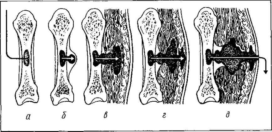
Fig. 74. Selection/3.html">Stages of development of acute hematogenous osteomyelitis: a — bone marrow phlegmon; b — formation of a subperiosteal abscess; c — intermuscular phlegmon; d — subcutaneous phlegmon; e — rupture of pus to the skin surface and fistula formation
For several days prior to the onset of the disease, the patient experiences weakness, lethargy, and chills.
The acute inflammatory process primarily affects long tubular bones—the Femur, humerus, and Tibia. The area of the bone most involved in growth, namely the metaphysis, is most frequently damaged. Multiple bone lesions are occasionally observed.
The clinical picture of acute hematogenous osteomyelitis is diverse. T. Krasnobaev distinguishes the following clinical forms: 1) toxic (adynamic); 2) septicopyemic; 3) with predominant local manifestations.
The toxic form of acute hematogenous osteomyelitis resembles an acute generalized infection in its course, accompanied by high body fever, unconsciousness, rapid pulse, and low blood pressure. Death may occur within the first 24 hours. The Temperature elevation is accompanied by chills and profuse sweating. The face is hyperemic, with a toxic rash or petechiae on the skin. Respiration is rapid and shallow. Death results from cardiovascular failure.
The disease develops very rapidly, and local manifestations do not have time to develop. Even at autopsy, it is impossible to detect any changes in the bones characteristic of this form of osteomyelitis. According to some authors, this type of osteomyelitis resembles septic Shock in its clinical presentation.
The septicopyemic form of hematogenous osteomyelitis has an acute course: elevated body temperature, chills, vomiting, tachycardia, hypotension, blood composition changes, and pronounced local signs (edema, tenderness of the affected area). Purulent metastatic foci form in various tissues and organs.
It develops against the background of reduced organism reactivity and high microbial virulence. Metastases are most frequently detected in the Lungs. Body temperature is persistent, sometimes accompanied by vomiting and diarrhea.
Hematogenous osteomyelitis with isolated or multiple bone lesions, but with a predominance of local manifestations, is also characterized by an acute onset and pronounced intoxication. Patients complain of severe, bursting pain in the bone that deprives them of Sleep, general weakness, loss of appetite, headache, and elevated body temperature. The face is pale, the Lips are cyanotic, the sclera and skin are icteric, and the Tongue is coated and dry. The Liver and Spleen are enlarged. The patient is lethargic, with tachycardia and tachypnea present. Meningeal symptoms are occasionally observed.
Under METABOLISM/18.html">The Influence of Treatment, intoxication symptoms rapidly subside, and local manifestations of the disease come to the fore: limb pain, Swelling of the affected area, a semi-flexed position of the limb, and restricted mobility. The patient guards the limb, lies motionless, and fears touching it. Concurrently with the pain, soft tissue swelling appears, spreading to adjacent areas without clear boundaries. After some time, the skin at the site of the swelling becomes hyperemic. Palpation reveals a local temperature rise and diffuse tissue infiltration. When tapping along the longitudinal axis of the limb, the patient feels localized pain in the bone. In limb segments covered by a thick layer of Muscles, local changes may initially be difficult to notice. The pain is associated with the development of bone Hypertension and persists until the intraosseous pressure decreases (due to the rupture of pus outward or surgical intervention). The pain is severe and excruciating. The child cannot sleep.
Sometimes the inflammatory process spreads from the bone to an adjacent joint. In such cases, its contours become smoothed, movements within it are restricted, and severe pain and fluctuation signs appear. The Diagnosis is confirmed by joint puncture, which yields pus.
Blood changes are characterized by leukocytosis, a left shift of the leukocyte formula, signs of anemia, and an increased ESR. In this type of osteomyelitis, other tissues and organs are not affected by the purulent process. This form of osteomyelitis is the most common.
Additional Diagnostic Methods assist in diagnosing acute hematogenous osteomyelitis. The most accessible is X-ray imaging. It allows confirmation or exclusion of the diagnosis and Determination of the localization of the lesion focus. However, During the first 10–14 days of the disease, X-ray changes may be absent on radiographs. This is explained by the fact that initially the inflammatory process is localized in the bone marrow, while structural changes in the compact bone layer are not yet detectable.
The rate at which radiographic changes appear is influenced by the clinical course of acute osteomyelitis (the more severe the disease, the faster they develop) and The Use of antibiotic therapy (in which case they appear later).
The earliest radiographic changes affect the soft tissues. Muscles near the lesion become enlarged and denser, and the border between them and the subcutaneous tissue disappears. Within 2 weeks, The structure of the cancellous and cortical bone layers is erased. Subsequently, foci of localized Osteoporosis and destruction emerge. Periosteal thickening is observed in the area of the affected bone. These are important radiological signs of osteomyelitis. Over time, bone osteoporosis progresses; as the process advances, the entire bone becomes porous and translucent.
One month after the onset of the disease, distinct cavities containing sequestra become visible within the bone. When the acute process transitions into a chronic one, areas of bone osteoporosis on the radiograph begin to alternate with foci of osteosclerosis. If the joint is involved, widening of the joint space becomes apparent.
Hematological findings indicate blood changes typical of an inflammatory process.
Special diagnostic methods include rheovasography, arterial oscillography, ultrasound echolocation, skin thermometry, osteotonometry (measurement of intraosseous pressure), radionuclide imaging, thermography, and Cytological examination methods, among others. Some of these have not yet found widespread application despite their high diagnostic value.
Bone puncture plays a crucial role in diagnosing acute hematogenous osteomyelitis. However, in the first hours of the disease, it may be uninformative due to the absence of pus in the bone. Therefore, it is advisable to combine it simultaneously with diagnostic methods such as intraosseous thermometry, osteotonometry, and cytological and MICROBIOLOGICAL ANALYSIS OF the punctate. Bone puncture has not only diagnostic but also therapeutic value, as it reduces intraosseous pressure.
Complications of acute hematogenous osteomyelitis include generalized blood infection (sepsis), metastatic purulent foci in the lungs, liver, Brain, Pleura, and Kidneys, secondary arthritis, and spontaneous bone fractures and dislocations.
Treatment for acute hematogenous osteomyelitis must be timely and comprehensive. It should be initiated within the first hours of the disease onset. The earlier treatment is started, the better the therapeutic outcome. S. Popkirov points out that when treatment begins within the first 8 hours, complete recovery occurs in 100% of cases.
Management of acute hematogenous osteomyelitis should be pursued in three main directions: 1) direct action against the pathogen; 2) enhancement of the body's resistance; 3) Treatment of the local lesion.
1. Currently, antibiotics are primarily used to combat the infection. They are administered in the first hours of the disease. This helps eliminate systemic intoxication, significantly reduces the incidence of arthritis, and noticeably shortens the duration of treatment.
Antibiotic therapy should be targeted, based on the microbial sensitivity to antibiotics.
To date, it is recommended to administer large doses of benzylpenicillin sodium salt combined with one of the broad-spectrum agents (kanamycin, gentamicin, mefoxin, osteotropic lincomycin, etc.).
There are various routes of antibiotic administration: intramuscular, intra-arterial, intravenous, intraosseous, and oral. The intraosseous administration method, developed by K. Alexyuk, has found widespread use. Its essence lies in introducing one or more needles with lateral openings into the bone infection site under local anesthesia (or general anesthesia in young children). To secure them properly, the needles are passed through the entire thickness of the bone to the opposite cortical layer. Through these needles, antibiotics, other antiseptics, or proteolytic enzymes are delivered directly into the affected zone. Treatment lasts for 2–4 weeks, subject to periodic rotation of antibiotics.
To prevent candidomycosis, which may develop during prolonged antibiotic therapy, large doses of Vitamins, nystatin, levorin, and desensitizing agents are administered.
To achieve a high concentration of antibiotics within the bone, intra-arterial administration is also recommended. M. Kovalyshyn, studying penicillin concentration in the bone marrow of rabbits, established that with intra-arterial administration, the concentration of the antibiotic in the bone is 1.2–4 times higher than with intramuscular or intravenous injection. Treating patients using this method yielded favorable results. Following 4–6 intra-arterial infusions of benzylpenicillin sodium salt, patients' conditions visibly improved: septic manifestations sharply decreased, temperature approached normal levels, and leukocyte counts in the blood decreased.
2. To enhance the body's resistance and nonspecific immunity in the treatment of acute hematogenous osteomyelitis, immunotherapy is employed. Active immunization
(using specific Vaccines and staphylococcal toxoid) is indicated for the subacute and chronic courses of the disease.
In the acute phase, passive immunization is recommended (hyperimmune antistaphylococcal gamma-globulin, antistaphylococcal plasma).
For general strengthening, blood transfusions from Donors previously immunized with staphylococcal toxoid are also prescribed.
An important role in the comprehensive treatment of patients with acute hematogenous osteomyelitis is assigned to vitamin therapy (ascorbic acid, B-group vitamins, Vitamin P) and the administration of proteolytic enzymes (trasylol, contrical).
For detoxification and improvement of microcirculation, low-molecular-weight dextrans (hemodes, rheopolyglucukin, neocompensan, rheoglucuman), 10% glucose solution, and protein preparations (native plasma, protein, aminochrome, aminopeptide, hydrolysin) are infused.
Infusion therapy for patients with acute hematogenous osteomyelitis must include preparations that normalize water-electrolyte balance and acid-base status. Electrolyte disturbances are corrected by administering 1%, 3%, or 7.5% potassium chloride solutions or balanced combination solutions such as Ringer-Locke, lactasol, trisol, or acesol, which contain sodium, potassium, and chloride electrolytes.
Acute hematogenous osteomyelitis is typically characterized by metabolic or mixed acidosis, which is corrected by the administration of sodium bicarbonate.
In severe conditions with pronounced intoxication, many authors recommend including exchange blood transfusion and hemisorption in the complex therapy.
Proper attention must be paid to patient care and symptomatic treatment. Nutrition should be high in calories, with a preference for a dairy and plant-based diet. Careful skin care (Prevention of pressure ulcers) and Oral Cavity care (prevention of stomatitis and parotitis) should be maintained.
3. Local treatment begins with the immobilization of the limb using a plaster cast. This ensures rest for the affected area and prevents the generalization of the pathological process. After acute manifestations subside, patients are prescribed physiotherapeutic Procedures (UHF therapy, Electrophoresis of potassium iodide and calcium).
An important role in the management of patients with acute hematogenous osteomyelitis belongs to Surgical treatment, although surgeons hold diverse views regarding its nature. T. Krasnobaev and T. Vengerovsky recommend limiting the intervention solely to periosteal incision. They strongly oppose opening the medullary canal, believing that this leads to a worsening of the disease course.
M. Grinyov, based on his theory of the pathogenesis of acute hematogenous osteomyelitis, is conversely convinced that the earlier the medullary canal is opened, the better the treatment outcomes. The medullary canal should be opened using an electric drill or a triangular awl. Several holes are made in the cortical layer of the bone, which prevents vascular damage and bone marrow necrosis. Osteoperforation is also advisable in later Stages of the disease when destructive foci with sequestra are present. It facilitates pus drainage and helps prepare the patient for radical surgery.
Indications for osteoperforation include: lack of effect from 48–72 hours of conservative treatment, an increase in soft tissue edema during this time, the onset and progression of joint inflammation, the presence of fluctuation in the affected area, and increasing local tenderness.
The surgery is performed under general anesthesia. The incision is made in the area of maximum tenderness.
In recent years, lasers have been used to open the medullary cavity. A laser beam Burns through the organic part of the bone at the designated site, exposing the medullary canal. The advantages of this surgical method are its speed and low invasiveness. Furthermore, the beam "scalpel" exhibits strong bactericidal and hemostatic effects.
Depending on the timeliness and comprehensiveness of treatment, the outcomes for patients with acute hematogenous osteomyelitis can vary. If the patient is hospitalized within 24 hours of the onset of the disease, recovery is observed in 91.86% of cases, whereas transition to the chronic stage occurs in 8.15%.
CHRONIC OSTEOMYELITIS
Chronic osteomyelitis (osteomyelitis chronica) has two forms: secondary chronic and primary chronic.
Secondary chronic osteomyelitis (osteomyelitis chronica secundaria) is most commonly a consequence of acute hematogenous osteomyelitis (chronic hematogenous osteomyelitis), open fractures (traumatic osteomyelitis), and gunshot wounds (gunshot osteomyelitis). Primary chronic osteomyelitis (osteomyelitis chronica primaria) includes Brodie's abscess, Ollier's albuminous osteomyelitis, Garré's sclerosing osteomyelitis, and antibiotic osteomyelitis.
Chronic hematogenous osteomyelitis. The use of antibiotics and early osteoperforation enable favorable treatment outcomes in a significant percentage of patients with acute osteomyelitis. In the remaining patients, the condition progresses to secondary chronic osteomyelitis. After the acute symptoms subside, the individual may feel satisfactory and resume their routine work. However, after some time, under the influence of triggering factors (hypothermia, trauma, malnutrition), a relapse occurs. Pain returns in the affected area, accompanied by edema, skin hyperemia, fever, elevated WHITE BLOOD Cell counts, and an increased ESR. All of this indicates that a latent infection has reactivated within the bone, leading to the formation of an abscess. Following its drainage (either spontaneous or surgical), the patient's condition improves, body temperature drops, edema subsides, and pain decreases. A sinus tract remains, discharging pus over an extended period. Its persistence is maintained by bone sequestra, which occasionally exit spontaneously through the fistulous tract. At this stage, radiographs reveal signs of bone destruction, a involucrum, and areas of necrotic bone of varying sizes. Following their spontaneous discharge or surgical removal, the process subsides, the sinuses close, and the patient feels well until the next exacerbation.
This form of osteomyelitis typically lasts for years or even a lifetime.
All patients with the fistulous form of chronic osteomyelitis require fistulography (Fig. 75). This Procedure makes it possible to trace the course of the sinus tract, identify cavities within the bone, and determine the size of the sequestra. Fistulography is performed by introducing a contrast oily (iodolipol) or water-soluble agent into the fistulous tract followed by radiography.
Traumatic and gunshot chronic osteomyelitis complicates open fractures and gunshot wounds. This category also includes forms of osteomyelitis that develop following metal osteosynthesis.
While all open fractures and gunshot wounds are infected, not all of them become complicated by osteomyelitis. As past wartime experience shows, osteomyelitis occurred in every fourth wounded soldier, and in every second case involving bone fractures.
Traumatic and gunshot osteomyelitis differs to some extent from hematogenous osteomyelitis. In hematogenous osteomyelitis, infection spreads from the inside of the bone outwards, whereas in traumatic and gunshot forms, it spreads in the reverse direction. Hematogenous osteomyelitis is more frequently caused by a monoinfection, whereas traumatic and gunshot forms involve polyinfection. These types of osteomyelitis often feature Foreign bodies in the tissues, which do not occur in hematogenous osteomyelitis. Furthermore, larger areas of sequestration are observed in hematogenous osteomyelitis compared to traumatic and gunshot forms. Traumatic and gunshot osteomyelitis are not accompanied by the severe deterioration of the patient's general condition that is typical of hematogenous osteomyelitis.
The Clinical presentation of traumatic and gunshot osteomyelitis is characterized by a sluggish, torpid course, which is associated with the limited zone of the traumatized bone area.
The prolonged course of chronic osteomyelitis can cause numerous complications, namely: internal organ amyloidosis, bone deformities, impaired liver and Kidney function, weight loss, anemia, hypoproteinemia, pathologic fractures, pseudarthrosis, and malignant transformation of the sinus tract walls.
The treatment of secondary chronic osteomyelitis is complex, prolonged, and accompanied by frequent surgical interventions.
Surgical intervention is preceded by conservative therapy, which essentially serves as preoperative preparation. It includes the following measures: high-calorie nutrition, blood and blood substitute transfusions, administration of anabolic Hormones (nerobol, retabolil), and immunological agents (y-globulin).
Infusion therapy plays an important role in the management of patients with chronic osteomyelitis: 5% glucose solution with Insulin, B vitamins, ascorbic acid, panangin, trental or curantyl, potassium chloride solution; rheopolyglucukin, albumin, and protein.
Intensive infusion therapy helps improve the functional activity of organs and tissues, correct Homeostasis disorders, and facilitate a smoother postoperative period. It provides prophylaxis against thromboembolic complications, stimulates reparative processes, and reduces the likelihood of osteomyelitic relapses.
A certain role in the treatment of chronic osteomyelitis is assigned to antibacterial therapy, although it is less effective than in acute hematogenous osteomyelitis due to the restricted penetration of antibiotics into the bone.
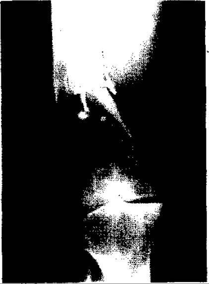
Fig. 75. Chronic osteomyelitis of the femur (fistulous form)
Local treatment is carried out by irrigating the focus of inflammation with antiseptic solutions (chlorhexidine, hydrogen peroxide) and applying physiotherapeutic procedures (UHF therapy, mud therapy, paraffin and ozokerite therapy). To achieve rapid lysis of necrotic areas, proteolytic enzymes (Trypsin, Chymotrypsin) are used.
Surgical treatment is indicated when an involucrum (sequestral capsule) has formed and clear demarcation of the sequestrum is evident.
A number of surgical interventions have been proposed for the treatment of chronic osteomyelitis.
1. Opening of the osteomyelitic phlegmon. When a fistula closes, pus becomes trapped and, lacking outflow, infiltrates adjacent tissues, causing intermuscular and subcutaneous phlegmons. Retained pus triggers a systemic bodily response (elevated body temperature, increased pain, worsening edema, skin redness, and blood count alterations). In the majority of patients, surgery is palliative in nature because the infection focus remains and may eventually cause a new flare-up.
2. Sequestrectomy (removal of the primary source of the purulent-necrotic process, i.e., the sequestrum). This surgery is also palliative in nature.
3. Excise of the fistula is applied in the presence of its branching, blind tracts, and wall sclerosis. Contrast fistulography is performed prior to surgery.
4. Sequestronecrectomy — complete removal of the inflammatory focus (sequestra, granulations, pus, and the sequestral capsule within healthy bone boundaries, followed by management of the residual cavity).
It is crucial to fill the bone cavity formed after surgery, as it does not tend to collapse. Various Materials have been proposed for this purpose, including plaster of Paris, a pedicle Muscle flap, a blood clot, a bone graft, egg yolk, bovine Peritoneum, antibiotic-impregnated Collagen sponge, etc. However, these methods proved ineffective. Many of the proposed materials rapidly suppurate and sustain inflammation. Among them, the best results are achieved by packing the bone cavity with a pedicle muscle flap—provided There is a well-developed muscle mass near the bone from which a graft can be taken—as well as with cancellous (spongy) bone tissue.
In cases of local complications of chronic osteomyelitis (bone fracture, nonunion, large bone defects), the Ilizarov compression-distraction method is widely used.
In the postoperative period, the comprehensive conservative therapy initiated before surgery is continued. This includes limb immobilization, repeated blood and protein plasma substitute transfusions, detoxification therapy, antibiotic therapy, immunotherapy, physical therapy, Therapeutic Exercises, etc.
Primary chronic osteomyelitis. This disease develops insidiously and gradually. However, some clinicians believe that even in this case, osteomyelitis begins acutely or subacutely, but the clinical picture is subtle and frequently goes unnoticed.
Primary chronic osteomyelitis develops due to low microbial virulence and reduced bodily reactivity.
There are several forms of primary chronic osteomyelitis, named after the authors who first described them.
Brodie's intraosseous abscess (abscessus Brodie) is a localized bone abscess filled with purulent, hemorrhagic, or serous fluid, most commonly located in the tibial metaphysis or epiphysis. The disease typically begins at a young age and may manifest itself many years later. Patients complain of a dull ache in the limb that worsens at night or during physical exertion, along with localized tenderness upon Percussion over the affected area. Diagnosis is established based on radiographic findings. A round cavity of varying size with clear margins is revealed within the bone. A sclerotic rim surrounds the edges of the cavity. There is no temperature reaction. Treatment is surgical: opening the abscess cavity, curettage, irrigation with antibiotics, and packing with a pedicle muscle flap.
Garre's sclerosing osteomyelitis (osteomyelitis scleroticans Garre). In this form of bone inflammation, osteosclerotic processes predominate, accompanied by the gradual obliteration of the medullary canal. The disease also runs a torpid, prolonged course with an unpronounced clinical picture: minor nocturnal pain in the limb, pain during physical exertion, limb thickening, and local tenderness upon palpation. Body temperature is moderately elevated. The inflammatory process primarily affects the diaphysis. Diagnosis is established on the basis of radiographic findings: a fusiform thickening of the bone diaphysis, against which small cavities with tiny sequestra may be visible. Rarely, the disease begins acutely, accompanied by fever and intoxication. These symptoms quickly subside, and the process assumes a chronic course.
Treatment of sclerosing osteomyelitis is conservative (antibiotics, physical therapy, trypsin electrophoresis).
Ollier's albuminous osteomyelitis (osteomyelitis albuminosa Ollier) is less common than Other types of osteomyelitis. It is characterized by the formation of subcortical and subperiosteal cavities filled with mucous, proteinaceous, or oily exudate. Small sequestra are sometimes present. The femur and humerus are most frequently affected. The onset of the disease is insidious, and its course is sluggish. Patients complain of pain in the affected limb. The process is typically localized in the distal femur. Swelling gradually develops and adds to the pain. Radiographs reveal regular and irregular hollow formations in the bone with periosteal layering.
Treatment of albuminous osteomyelitis is surgical—sanation of the focus of chronic purulent inflammation.
Antibiotic-induced osteomyelitis occurs in debilitated individuals who have undergone prolonged, ineffective antibiotic treatment for some underlying condition. Antibiotics alter the morphological STRUCTURE OF THE bone and the clinical course of the disease. Bone destruction in such patients is unpronounced, periosteal reaction and sequestral involucrum are absent, and areas of osteoporosis alternate with areas of osteosclerosis.
Clinically, this form of osteomyelitis also runs a sluggish course, without fever and with mild local manifestations. Suppuration and fistulae do not occur. There is no sequestral capsule or granulation tissue. Treatment is surgical—radical necrectomy is performed.
It must be emphasized that this disease is characterized by a prolonged course, unfavorable treatment outcomes, and frequent relapses. From this perspective, prevention is of paramount importance. In acute hematogenous osteomyelitis, early and effective treatment of any inflammatory processes in children (boils, pyoderma, tonsillitis, dental caries, respiratory infections, infected wounds, etc.) is essential.
Prevention of chronic hematogenous osteomyelitis consists of early diagnosis of the acute process, urgent hospitalization of patients, and timely treatment. Thoroughness of treatment is also of great significance.
Prevention of traumatic osteomyelitis consists of preventing wound infection, timely primary surgical debridement of wounds and open fractures, prophylactic antibiotic administration, and immobilization.
PURULENT INFLAMMATION OF JOINTS (ARTHRITIS)
Each joint consists of the articular ends of bones covered with cartilage, and the articular capsule (joint capsule) with joint ligaments.
The articular capsule consists of two layers: the outer, or fibrous, layer and the inner, or synovial, layer. Ligaments run within the thickness of the fibrous capsule. They may also be present within the joint cavity (e.g., in the knee or hip joints).
The synovial membrane is composed of loose epithelium-like tissue containing a significant number of BLOOD AND Lymph vessels. It secretes a viscous, yellowish fluid that lubricates the articular surfaces of the bones and serves as a favorable culture medium for microorganisms. It also possesses absorptive properties, which plays a role in the penetration of toxins into the bloodstream in cases of purulent joint inflammation.
In some articular ends, additional structures are present—such as menisci, mucous bursae, and synovial bursae, which frequently communicate with the joint cavity.
The etiology of acute arthritis (arthritis purulenta) involves: 1) open joint injuries caused by contaminated objects; 2) the spread of an inflammatory process from adjacent tissues or bone epiphyses into the joint cavity; 3) hematogenous dissemination of infection. In some cases, joint inflammation is aseptic in nature (resulting from closed trauma).
Infectious arthritis is most commonly caused by streptococci and staphylococci.
The pathological changes occurring in a joint upon infection depend on the virulence of the MICROORGANISMS AND THE reactivity of the organism.
In mild cases, the process is limited to serous inflammation of the joint capsule with the accumulation of synovial fluid (synovitis). It contains a varying amount of fibrin. As the condition progresses, the effusion becomes seropurulent or even purulent. The process may remain confined to the synovial membrane or spread to the tissues surrounding the joint (periarthritis) or to the articular ends of the bones (osteoarthritis).
Following the subsidence of the acute phase, thanks to timely and comprehensive treatment, granulations form on the walls of the joint capsule and fibrin is deposited, leading to joint stiffness (contracture) or complete immobility (ankylosis).
When all elements of the joint and its adjacent tissues are involved in the inflammation, the condition is termed panarthritis.
The clinical presentation of acute arthritis is characterized by the sudden onset of pain in a specific joint and the inability to perform active and passive movements. The joint is swollen, extremely tender upon palpation, the skin overlying it may be hyperemic, and passive movements are severely painful. In the presence of a substantial joint effusion, signs of fluctuation and patellar tapotement ("balottement") may be elicited. The limb often assumes a Bonnet position (slight flexion), which maximizes joint volume and minimizes pain. The contours of the joint are smoothed, and local temperature is elevated. Mild percussion along the axis of the bone causes pain in the affected joint (for example, in purulent gonitis, striking the heel with the leg extended elicits pain).
For diagnostic purposes—to determine The Nature of the effusion and to perform bacteriological Analysis of the exudate—joint puncture (arthrocentesis) is performed. To ascertain whether the inflammatory process has spread to the articular ends of the bones, an X-ray Examination is indicated. Radiographs reveal widening of the joint space, signs of osteoporosis, and destruction of the bone ends.
In severe cases of purulent arthritis, pus may breach the joint capsule and extend into the surrounding tissues, causing phlegmonous abscesses.
Treatment for patients with purulent arthritis is provided in an inpatient Setting and depends on the stage of the inflammatory process. It begins with joint immobilization using a fenestrated plaster cast in a physiologically advantageous position, encompassing two adjacent joints. In cases of serous inflammation, joint aspiration is indicated to evacuate the exudate and administer antibiotics directly into the cavity. This procedure is repeated until sterile fluid is obtained. A pressure bandage is applied to the joint. Dry heat, physical therapy modalities (UHF therapy, Sollux lamp, quartz lamp), and antibiotic therapy are indicated.
After acute symptoms subside, gradual active and passive movements in the joint should be encouraged to prevent ankylosis.
If the condition progresses and spreads to adjacent tissues, surgical intervention is indicated. This may be of three types: opening of the joint (arthrotomy), joint resection, or limb amputation.
The purpose of an arthrotomy is to establish a continuous outflow of exudate (pus) from the joint cavity, followed by lavage and the Introduction of antiseptic solutions. Optimal surgical incisions have been established for each joint to ensure the best possible drainage of pus. Following arthrotomy, the joint is drained, and the limb is immobilized.
When articular cartilages—and especially the articular surfaces of the bones—are involved, as confirmed radiologically, joint resection is indicated. During the procedure, the bone epiphyses are removed along with the synovial membrane. This surgery results in joint ankylosis.
In critical patient conditions accompanied by severe intoxication in The Setting of sepsis, an exhausted organism, and panarthritis, limb amputation is performed as a life-saving measure. This is a procedure of last resort.
In recent years, compression-distraction apparatuses developed by G. Ilizarov, M. Volkov, O. Oganesyan, and V. Kalnberz have been utilized in the treatment of acute arthritis. They prevent the development of contractures and fibrous ankylosis.
Surgical intervention is combined with intensive corrective therapy.
The outcomes of purulent arthritis vary. With timely and comprehensive treatment and minimal pathological changes in the joint, recovery with full restoration of joint function may be achieved. In more severe cases, varying degrees of functional impairment occur, ranging from contractures to ankylosis and limb amputation.
Last update: 08/08/2026
Editorial and Educational Adaptation: This material has been compiled based on the primary/original source text. The project team performed an editorial review, corrected technical inaccuracies, structured sections, and adapted the content for an educational format.
What was processed:
- elimination of formatting defects (OCR errors, structural breaks, corrupted characters);
- editorial organization of content;
- standardization of terminology in accordance with academic sources;
- verification of factual statements against the original source text.
All mentions of the author, publication year, and origin of the primary text have been preserved in accordance with the source.