ZOOLOGY OF CHORDATES: STUDY GUIDE - Zakharenko M.O. - 2015
CHAPTER 5. HIGHER VERTEBRATES
5.4. Class Mammals - Mammalia
Mammals are the most highly organized warm-blooded (homeothermic) amniotes, characterized by a Hair-covered body, live-bearing reproduction, and feeding their young with milk.
The main Organizational features of mammals are:
1. a high level of Brain development and complex behavior;
2. a body covered with hair;
3. viviparity (except for monotremes) and Lactation (feeding young with milk);
4. the appearance of the Placenta in higher mammals;
5. A large number of sebaceous and Sweat Glands in the Skin, some of which are modified into mammary and scent glands;
6. the presence of three auditory ossicles in the Middle ear (incus, malleus, stapes);
7. The formation of the External ear with an external auditory meatus;
8. the Formation of the spinal Column consisting of platycoelous vertebrae;
9. a constant number of cervical vertebrae;
<10. the presence of a synapsid tropibasal Skull, two occipital condyles, and autostyly;
11. a highly differentiated dentition (thecodont-type Teeth);
12. the division of the body cavity into thoracic and abdominal sections by a Diaphragm;
13. refinement of the digestive tract and the formation of a multichambered Stomach;
14. a four-chambered Heart and two completely separated circulatory loops;
15. the retention of only the left aortic arch;
16. anucleated erythrocytes;
17. metanephric Kidneys.
Mammals inhabit all ecological environments. The Class comprises about 4,500 species. Structural Features of mammals
Body shape. Highly diverse in size and appearance (Fig. 281).
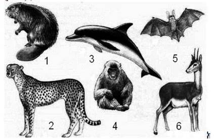
Fig. 281. Body shape in mammals
1 - beaver, 2 - leopard, 3 - dolphin, 4 - monkey, 5 - bat, 6 — antelope
The smallest modern mammal is the Etruscan shrew or pygmy white-toothed shrew (genus Suncus) (an insectivore), weighing 1.2–1.7 g with a body length of 3.8–4.5 cm. The largest terrestrial mammal is the African bush elephant (Loxodonta africana), standing 3.5 m tall and weighing up to 4–5 t, whereas the largest aquatic mammal is the blue whale (Balaenoptera musculus), with individual specimens reaching a length of 33 m and a mass of 150 t (equivalent to 30–35 elephants). The mammalian body is divided into the HEAD, neck, trunk, paired limbs, and tail (Fig. 282).

Fig. 282. Main body parts of mammals
1 - head; 2 - neck; 3 - trunk; 4 - forelimb; 5 - hindlimb; 6 - chest; 7 - abdomen; 8 - groin; 9 - flank; 10 - back; 11 - withers; 12 - sacrum; 13 - scapula; 14 - shoulder; 15 - forearm; 16 - wrist; 17 - hand; 18 - thigh; 19 - lower leg; 20 - FOOT.
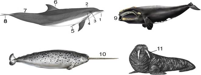
Fig. 283. External anatomy of marine mammals 1 - rostrum; 2 - blowhole; 3 - eye; 4 - ear; 5 - pectoral flippers; 6 - dorsal fin; 7 - caudal peduncle; 8 - fluke; 9 - baleen of a right whale; 10 - narwhal tusk; 11 - walrus tusks
Pinnipeds, sirenians, and especially cetaceans develop a streamlined body shape, with limbs modified into flippers; a leather-like caudal fin gives sirenians and whales a fish-like appearance. Semi-aquatic mammals (platypus, Russian desman, beaver, nutria, otter) develop a dense coat that prevents waterlogging, a shortened neck, webbing between the digits, and a flattened tail to varying degrees. Many perissodactyls and artiodactyls share a similar appearance: long legs, a compact trunk, and a long, flexible neck that facilitates long-distance travel and helps evade common predators, namely large carnivores. Lagomorphs and rodents exhibit similar body forms. Adaptation to rapid locomotion on hind limbs in open biotopes has led to a convergent body plan—weak forelimbs, powerful hind limbs, and a long counterbalancing tail—seen in kangaroos (marsupials), African elephant shrews (insectivores), and various rodents such as jerboas and gerbils. In fossorial (burrowing) species, the body becomes barrel-shaped, and the limbs and tail are shortened (gophers, marmots, voles, etc.). With the transition to a subterranean lifestyle, alongside a barrel-shaped trunk, the forelimbs involved in soil excavation undergo significant development (marsupial and common moles, etc.).
In arboreal species, adaptations for locomotion include short, strong limbs with sharp claws, and a long, bushy tail that AIDS in leaping (squirrels, martens, etc.). The long limbs of primates enable movement through the forest canopy. Opossums, certain monkey species, arboreal anteaters, pangolins, and porcupines use a prehensile tail for grasping. In flying phalangers, flying squirrels, and colugos, a lateral skin fold (patagium) enables prolonged gliding leaps. Notably, various life forms among marsupials closely parallel those of many placental mammals.
Integument. All vertebrates possess a relatively thick skin consisting of two layers: the epidermis and the corium (dermis) (Fig. 284).

Fig. 284. Structure of mammalian skin
1 - outer layer of the epidermis; 2 - Malpighian layer; 3 - dermis (true skin); 4 - hair; 5 - sweat gland; 6 - opening of the sweat duct; 7 - sebaceous gland; 8 - arrector pili Muscle; 9 - Connective Tissue fibers of the dermis; 10 - Blood vessel; 11 - hair papilla.
The epidermis is stratified. At its base lies the Malpighian layer, formed by cylindrical or prismatic, living Cells capable of Cell Division. Closer to the surface, these cells lose their nuclei and proliferative capacity. They flatten, keratinize, and form a dead outer layer. These cells slough off the surface and are gradually replaced by new cells originating from the Malpighian layer. Skin coloration is determined by pigments in the form of melanin granules located within the Cells of the germinative layer, intercellular spaces, and specialized pigment cells (melanoblasts, melanophores).
The dermis (corium) is the Deep Layer of mammalian skin. It is rich in Collagen and Elastin fibers that intertwine to form a reticular structure. Adipose tissue—the subcutaneous fat layer—is deposited in the deep layers of the dermis and is exceptionally well developed in some mammals (whales, dolphins).
The epidermal layer reaches its greatest thickness in areas subject to constant friction during the animal's locomotion; calluses may form in these regions (soles of the feet, ischial callosities in certain monkeys, knee calluses in camels, etc.) (Figs. 285, 286).
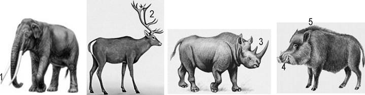
Fig. 285. Cornified and bony structures of mammals
1 - tusks; 2 - horns; 3 - tusks; 4 - canines; 5 - wild boar bristles
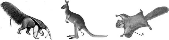
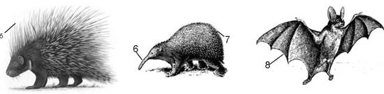
Fig. 286. Cutaneous modifications in mammals
1, 2 - tail and snout of an anteater, 3 - kangaroo pouch, 4 - gliding skin fold of a flying squirrel,
5, 7 - quills of a porcupine and spines of an echidna, 6 - "bill" of an echidna, 8 - patagium of bats
The terminal Phalanges of the digits in most mammals are protected by epidermal cornified claws (Fig. 287).

Fig. 287. Mammalian claws, Nails, and hooves
1 - claw plate; 2 - bone process of the terminal phalanx; 3 - dermis; 4 - digital pad;
5 - sole plate; 6 - nail plate; 7 - hoof frog.
In arboreal forms, they are sharp and strongly curved, whereas in fossorial (burrowing) forms, they are elongated and flatter. In all felids (except the cheetah), claws are retractable; the claw, together with the terminal phalanx, is pulled toward the dorsal surface of the penultimate phalanx by specialized tendons, preventing it from blunting during locomotion. In many primates, claws have evolved into nails, covering only the upper surface of the digital tips, while the underside features a soft pad that enhances tactile sensitivity. The elaboration of claws led to the formation of hooves—thick, keratinized structures almost entirely encasing the terminal phalanx. Hooves are particularly well-developed in cursorial (fast-running) species (horses, antelopes, goats, etc.) (Fig. 287).
Through the pronounced proliferation of cornified epithelium, horns develop in rhinoceroses and bovid artiodactyls, forming hollow horn sheaths that fit over bony cores and fuse with the frontal bones (Fig. 288).

Fig. 288. Mammalian horns: 1 - unbranched/simple, 2 - spiraled, 3 - branched, 4 — pointed
Deer antlers are bony structures of dermal origin; they are shed annually (Fig. 289).

Fig. 289. Longitudinal section diagrams of mammalian horns: 1 - bovid horn; 2 - deer antler with velvet skin;
3 - deer antler without skin; 4 - pronghorn horn; 5 - giraffe ossicone/horn; 6 - rhinoceros horn
In many mammals, horny scales similar to those of reptiles and birds develop on the tail and limbs (marsupials, insectivores, rodents) (Fig. 290).
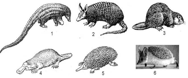
Fig. 290. Modifications of the mammalian integument
1 - Sunda pangolin; 2 - armadillo; 3 - beaver; 4 - platypus; 5 - echidna; 6 - hedgehog
In pangolins, large, overlapping, tile-like rhombic horny scales cover the entire body. In armadillos (xenarthrans), the armor consists of bony scutes (derived from the corium) covered dorsally by epidermal keratin plates.
The pelage (hair coat) is a defining characteristic of mammals. Mammalian hair is heterogeneous. The largest hairs, typically standing out distinctly above the general fur coat as solitary bristles, are known as vibrissae (Fig. 291).

Fig. 291. Mammalian vibrissae
1 - dog; 2 - hare; 3 - cat; 4 - walrus; 5 — seal
Vibrissae function as tactile Organs; they are located on PARTS OF THE body that the animal most frequently uses to Touch surrounding objects (tip of the snout, abdomen, limbs) and are richly supplied with nerve endings within the hair follicles. The upper tier of the fur coat is formed by guard hairs. The lower tier consists of thread-like, often spirally curled, fine underfur hairs that play a particularly crucial role in thermal insulation. In a few mammals, the fur consists of a single category of hair—the summer coat of deer and wild boars comprises only guard hairs, whereas the pelage of subterranean mammals (mole, mole-rat) consists exclusively of underfur. Bristles (pigs) and spines/quills (echidnas, hedgehogs, porcupines) are modified guard hairs.
In most therians, the hair coat is developed across all body regions (absent on the Lips and, in some species, on the soles of the feet) and performs thermoregulatory Functions, serves as tactile receptors, protects the skin from injury and certain parasites, improves the aero- and hydrodynamic Properties of the body, and ensures species-specific coloration. Only a few mammals (cetaceans, elephants, rhinoceroses, hippopotamuses) are practically devoid of body hair; they are transiently covered with hair primordia during embryonic development within the female's body.
Hair on the body is typically arranged in an orderly fashion. As a rule, it is inclined in a specific direction, which ensures the streamlining of the body against air and Water currents. The primary direction of the hair tract is from the head to the tail. The orientation of the hair changes in areas where skin folds and stretches frequently form. The hair coat is closely linked to the animal's lifestyle and mode of locomotion. For instance, sloths hang upside down from trees continuously, and their hair grows in a direction from the abdomen to the back, which is related both to gravity and The impact of rain. In subterranean dwellers (moles, mole-rats), which frequently move both forward and backward, the short hair stands erect. A similar pelage is found in inhabitants of dense grassy thickets and leaf litter (shrews).
Fur wears out and needs to be replaced, which occurs through shedding. In the tropics, this process is continuous as the hair wears down (in monkeys, it can last year-round). A similar phenomenon in temperate latitudes is observed in burrowing animals, whose fur quickly wears thin in certain areas, leading to localized partial shedding. A complete replacement of the coat in most temperate-zone species typically occurs twice a year—in autumn and spring—accompanied by Changes in the fur's structure and frequently its coloration. When transitioning from summer to winter coats, the hair density increases, sharply raising the winter coat's thermal insulation compared to that of summer.
The mammalian skin contains a significant number of glands. Based on The structure of their excretory ducts, glands are classified as tubular or alveolar. In most cases, sweat glands have a tubular structure (with walls formed by a single-layered epithelium) and open primarily onto the skin's surface, though occasionally into hair follicles, terminating in a coiled knot. The walls of these glands consist of a single-layered epithelium, the cells of which secrete actual sweat. Sweat consists of 97–99% water, with dissolved urea, creatinine, volatile Fatty acids, and salts (which are also present in urine). Thus, sweat helps excrete Metabolic waste products, but its primary function is thermoregulation: evaporating sweat cools the body. Sweat glands are well developed in primates and ungulates, relatively poorly developed in canids, felids, lagomorphs, and rodents, and entirely absent in cetaceans, sloths, and pangolins. Species with poorly developed sweat glands use alternative thermoregulation mechanisms. For example, when overheating, dogs enhance heat dissipation by accelerating shallow breathing and evaporating water from the saliva on their protruding Tongue and oral mucosa.
The second type of glands comprises alveolar glands, which include Sebaceous Glands. They have a grapelike (acinar) structure, and their ducts open into hair follicles. The walls of these glands are formed by stratified squamous epithelium. Their cells undergo fatty degeneration to produce an oily secretion that lubricates the skin and hair surface, helping maintain elasticity and preventing the penetration of microbes and Fungi. Such glands are absent in cetaceans, sloths, and certain insectivores.
Musk glands are modified cutaneous glands that secrete a scented substance—musk—which serves primarily to attract mates, mark territory, and facilitate species recognition. These glands are modifications of sweat or sebaceous glands, occasionally exhibiting a mixed structure that combines alveolar and tubular elements. Scent glands include the anal glands of many carnivores (especially mustelids), the musk glands of musk deer, beavers, desmans, and muskrats, the preorbital glands of many artiodactyls (deer, antelopes, sheep), and the foot glands of goats. The strong, pungent secretion of the anal glands in American skunks and certain ferrets is used for self-defense. The combined scents of secretions from musk, sebaceous, and sweat glands help animals distinguish conspecifics from other species and facilitate encounters between males and females.
Mammary Glands—which are modified sweat glands—are developed in the females of all mammals. In monotremes, mammary glands retain a tubular structure and are arranged in groups known as glandular fields: on the abdomen in platypuses, and within the brooding pouch in echidnas. There are no nipples, and the gland ducts open directly into hair follicles; the young lick up the protruding droplets of milk from the fur. In all other mammals, mammary glands have a more complex, acinar structure, with their ducts opening onto nipples. In some species, nipples are arranged in two rows from the pectoral region to the groin (insectivores, carnivores, rodents), while others retain only a single pair of nipples (primates, sirenians, elephants, bats) or possess nipples restricted to the inguinal region. In most ungulates, the mammary Glands of the right and left sides fuse into an udder located in the groin region, bearing 2 or 4 nipples.
Skeleton. The mammalian skeleton exhibits considerable structural diversity. THE Vertebral Column is divided into cervical, thoracic, lumbar, sacral, and caudal regions (Figs. 292, 293).
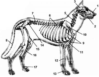
Fig. 292. Mammalian skeleton
1 - upper jaw; 2 - lower jaw; 3 - neurocranium (braincase); 4 - teeth; 5 - cervical vertebrae; 6 - thoracic vertebrae; 7 - lumbar vertebrae;
8 - caudal vertebrae; 9 - thoracic Ribs; 10 - scapula; 11 - humerus; 12 - BONES OF THE forearm; 13 - bones of the manus (hand); 14 - pelvic bone; 15 - Femur; 16 - bones of the crus (lower leg); 17 - bones of the pes (foot); 18 - masseter muscle.

Fig. 293. Regions of the mammalian vertebral column
The vertebrae are platymysial/amphicoelous/platyspondylous (with flat surfaces), separated by cartilaginous intervertebral discs. The neural arches are well developed (Fig. 294).
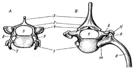
Fig. 294. Vertebrae of a fox (anterior view).
A - cervical vertebra; B - thoracic vertebra; 1 - vertebral body, 2 - neural arch, 3 - spinous process, 4 - articular surface of the neural arch (for articulation with adjacent vertebrae), 5 - spinal canal, 6 - transverse process, 7 - cervical rib rudiment, 8 - rib,
9 - foramen for Blood Vessels, 10 - rib head, 11 - rib tubercle
The cervical region contains seven vertebrae, with exceptions such as the manatee (6) and the sloth (8–10). Ribs attach to the thoracic vertebrae, forming the rib cage. The thoracic region contains 9–24 (most commonly 12–15) vertebrae, with the last 2–5 thoracic vertebrae bearing "false ribs" that do not reach the Sternum. The lumbar region consists of 2 to 9 vertebrae with well-developed transverse processes. The sacral region is formed by 4–10 fused vertebrae, of which only the first two are true sacral vertebrae, while the remaining ones are caudal in origin. The number of free caudal vertebrae ranges from 3 (in gibbons) to 49 in the long-tailed pangolin.
The mammalian skull is of the synapsid type. In the braincase, four occipital bones fuse into a single Occipital bone, and an unpaired Ethmoid bone develops (Fig. 295).

Fig. 295. SKULL OF A fox
A - lateral view; B - ventral view: 1 - occipital bone, 2 - foramen magnum, 3 - occipital condyle, 4 - interparietal bone, 5 - Parietal bone, 6 - Frontal bone, 7 - supraorbital process of the frontal bone, 8 - Nasal bone, 9 - Temporal bone, 10 - zygomatic process of the temporal bone, 11 - Zygomatic bone, 12 - basisphenoid bone, 13 - presphenoid bone, 14 - orbitosphenoid bone, 15 - pterygoid bone, 16 - Vomer, 17 - incisive bone (premaxilla), 18 - palatine process of the incisive bone,
19 - Maxilla, 20 - palatine process of the maxilla, 21 - Palatine bone, 22 - ethmoid bone with nasal conchae, 23 - Lacrimal bone, 24 - tympanic bulla, 25 - external acoustic meatus, 26 - dentary bone, 27 - coronoid process of the dentary bone, 28 - mandibular articulation site, 29 - incisors, 30 - canines, 31 - premolars, 32 - molars
The floor of the skull is formed by the unpaired basisphenoid and presphenoid bones. The interorbital septum and the anteroventral part of the braincase are formed by paired foundational bones: the orbitosphenoid and alisphenoid. The roof of the skull is formed by paired dermal bones—nasals, lacrimals, frontals, parietals—and the unpaired interparietal. The floor of the skull is reinforced by paired dermal bones (palatines and pterygoids) and a small unpaired vomer. The lower jaw is formed by a single paired bone—the dentary. All mammals are characterized by The Development of a secondary bony palate that separates the nasal passages from the Oral Cavity. The auditory bones fuse into a paired petrosal bone.
The ancestral limb structure across various mammalian orders underwent significant modifications in adaptation to different modes of locomotion: the relative length of limb segments, bone proportions and thickness change, the number of digits decreases, and so forth. The Skeleton of the paired limbs retains the typical pentadactyl features of terrestrial vertebrates. Unlike birds and similarly to amphibians, the mammalian forelimb features a mobile joint located between the forearm bones and the proximal row of Carpal Bones, whereas the hindlimb features a joint between the lower leg and the proximal row of Tarsal Bones (the tibiotarsal joint). The ancestral limb structure across various mammalian orders underwent significant modifications in adaptation to different modes of locomotion: the relative length of limb segments, bone proportions and thickness change, the number of digits decreases, and so forth.
The Pectoral Girdle is simplified and connected to the Axial Skeleton solely by Muscles and ligaments. The scapula features a spine. The coracoid is reduced and fused to the scapula (Fig. 296).
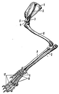
Fig. 296. Mammalian forelimb
1 - scapula; 2 - scapular spine; 3 - acromion process; 4 - glenoid cavity; 5 - coracoid process;
6 - humerus; 7 - ulna; 8 - radius; 9 - carpus; 10 - metacarpus; 11 - phalanges of the digits
The Pelvic Girdle consists of two hip bones formed by the fusion of the ilium, pubis, and ischium, while the limb exhibits the typical structure characteristic of terrestrial vertebrates (Fig. 297).

Fig. 297. Pelvic girdle of a fox
A - pelvic girdle: 1 - ilium; 2 - ischium; 3 - pubis; 4 - acetabulum.
B - hindlimb: 1 - femur; 2 - Patella; 3 - Tibia; 4 - Fibula;
5 - tarsus; 6 - metatarsus; 7 - phalanges of the digits; 8 - talus; 9 - calcaneus
In bats, the unusually elongated phalanges of the second to fifth digits support a stretched membrane, forming a wing. The front paws of a mole serve as specialized digging tools; the metacarpals and metatarsals of monkeys are adapted for grasping, whereas the hindlimbs of kangaroos and jerboas are built for leaping. A horse's single-toed legs are adapted for rapid running on firm ground, while the flippers of cetaceans and sirenians, characterized by shortened limb segments and an increased number of phalanges, resemble the fins of lobe-finned Fishes (Fig. 298).

Fig. 299. Structural features of limbs in mammals from different ecological groups
1 - bat wing; 2 - mole paw; 3 - kangaroo hind leg; 4 - horse leg; 5 - orangutan hand; 6 - dolphin flipper
The musculature of mammals is highly differentiated and varies significantly across orders and families depending on their mode of life (Fig. 300).
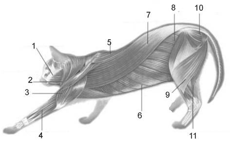
Fig. 300. Muscular System of mammals
1 - jaw muscles; 2 - deltoid muscles; 3 - triceps; 4 - digital extensors; 5 - trapezius muscle; 6 - external oblique muscles;
7 - back muscles; 8 - sartorius muscles; 9 - rectus abdominis muscle; 10 - gluteal muscles; 11 - gastrocnemius muscle
Masticatory muscles, associated with capturing and mechanically Processing food, achieve a high degree of development and differentiation. The complex subcutaneous musculature is involved not only in thermoregulation (adjusting hair position, curling the body into a ball) but also in social communication: it controls the movement of tactile hairs (vibrissae) and is responsible for facial expressions, which play a crucial role in information transfer, particularly in carnivores and primates. The dome-shaped diaphragm—a muscular wall—separates the thoracic and abdominal cavities; its evolution allowed for a dramatic intensification of pulmonary ventilation.
Internal Structure of mammals. The Topography of the Internal Organs in mammals is very similar to that of birds (Fig. 301).
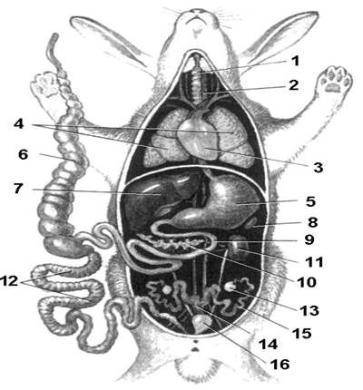
Fig. 301. Topography of internal organs in mammals 1 - Trachea; 2 - Esophagus; 3 - heart; 4 - Lungs; 5 - stomach; 6 - cecum; 7 - Liver;
8 - Spleen; 9 - Small Intestine; 10 - Pancreas; 11 - Kidney; 12 - Large Intestine; 13 - Ovary; 14 - Uterus; 15 - oviduct;
16 - Urinary Bladder.
The Digestive System of mammals is highly diverse and depends on their feeding habits. Mammalian feeding exhibits remarkable variety—no other class of vertebrates utilizes plant food as comprehensively as mammals. Vegetative parts of trees, shrubs, and herbaceous plants form the dietary foundation for ungulates, proboscideans, lagomorphs, and rodents (voles, marmots, susliks, beavers); rodents (especially jerboas, zokors, mole-rats, etc.) also extensively consume underground plant parts. Sirenians feed on aquatic plants. Seeds and fruits sustain mice, squirrels, chipmunks, and dormice, while large quantities of seeds and fruits are eaten by bears, deer, wild boars, and martens. The population size of sables is determined not only by the availability of animal prey (small rodents), but also by the yield of cedar nuts and other fruits. Fruit-bats feed on fruits. Certain specialized groups of bats and some marsupials utilize floral nectar, while South American vampire bats feed on the blood of large mammals. Invertebrates are consumed by most insectivores, bats (except fruit-bats), mice, jerboas, susliks, squirrels, and small or even large predators (bears). Marine plankton serves as the primary food source for the largest modern mammals—baleen whales. Among carnivorous mammals, only a few survive exclusively on large live prey (primarily felines), whereas the majority diversify their diet with plant matter. Predators frequently attack prey larger than themselves and successfully capture it by employing individual advantages—strength, surprise attack, agility (mustelids, leopards, etc.)—or by hunting in groups (wolves, lions). In general, the mammalian digestive system is characterized by the following sections (Fig. 302).

Fig. 302. Internal structure of mammals
1 - oral cavity; 2 - trachea; 3 - esophagus; 4 - lung; 5 - liver; 6 - stomach; 7 - small intestine; 8 - pancreas; 9 - kidney; 10 - rectum; 11 - urinary bladder; 12 - large intestine; 13 - caecum
The digestive system begins with the oral cavity, into which the Salivary Glands empty (Fig. 303).

Fig. 303. Salivary glands of a dog
1 - parotid; 2 - submandibular; 3 - long-duct sublingual; 4 - sublingual; 5 - short-duct sublingual; 6 - orbital; I-IV - excretory ducts of the salivary glands
The oral cavity begins with the vestibule—the space between the fleshy lips (which are unique to mammals), Cheeks, and jaws. In hamsters, chipmunks, and monkeys, cheek pouches form here, where food is gathered and subsequently transported to storage sites. Lips typically possess tactile receptors. Fleshy lips are absent in monotremes and cetaceans.
The jaws of modern mammals bear teeth of various shapes and functions. They are subdivided into incisors, canines, premolars, and molars (Fig. 304).
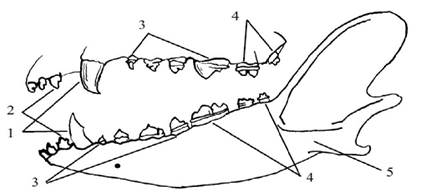
Fig. 304. Types of teeth in mammals 1 - canines; 2 - incisors; 3 - premolars; 4 — molars
Incisors, canines, and premolars have two generations (milk teeth replaced by permanent ones), whereas molars have only one. In pinnipeds and odontocetes (toothed whales), the teeth are undifferentiated. Mammalian heterodonty is an important adaptation that improves feeding and Digestion (Fig. 305).
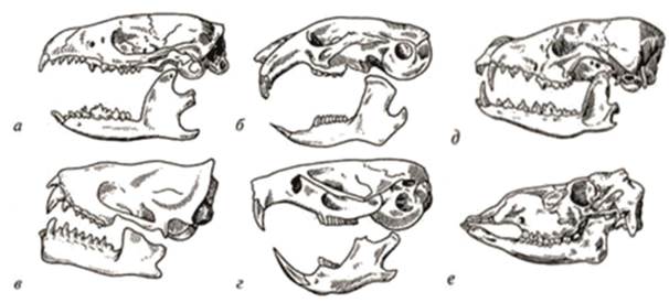
Fig. 305. Various types of mammalian dentition
a - insectivores (hedgehog); b - lagomorphs (pika); c - chiropterans (noctule bat); d - rodents (vole); e - carnivores (wolf); f - artiodactyls (deer)
Different mammalian orders vary in their dental Composition and Structure. The ancestral condition was likely a continuous row of relatively weakly differentiated teeth. To some extent, this has been preserved in insectivores, bats, and partially in primates and carnivores. However, in each group, it underwent adaptive modifications. In small insectivores (shrews), which feature an elongated, narrow snout, the dental rows with their forward-projecting incisors form a peculiar "forceps" well-suited for grasping and holding small prey (insects, earthworms, etc.), while their cuspate molars with sharp peaks crush the chitinous exoskeleton. The dentition of carnivores underwent greater differentiation: sharp incisors, large canines, and molars with cutting edges. Canines, serving as tools for capturing and subduing prey, reached their greatest development in the extinct saber-toothed tiger. To determine population numbers and species identification, researchers use "dental formulas," which indicate the number of teeth in one half of the upper and lower jaws. Tooth types are denoted by the initial letters of their Latin names. A dental formula looks as follows: wild boar I 3/3 C 1/1 PM 4/4 M 3/3 = 44 teeth; hare I 2/1 C 0/0 PM 3/2 M 3/1 = 28 teeth. In baleen whales, teeth form during the Embryonic Stage but later disappear. Simultaneously, on the sides of the oral cavity along the outer margins of the upper jaws, epidermal ridges develop, which subsequently proliferate, keratinize, and divide into a series of triangular plates whose bases are embedded in the gum tissue; their number ranges from 160 to 500 in different species. These plates, known as "whalebone" or baleen, bear fringes of intertwined keratin fibers and serve to filter out planktonic organisms.
Masticated and thoroughly moistened with saliva, food passes from the oral cavity through the Pharynx and Esophagus into The Stomach. The esophagus in most mammals is a thin-walled tube equipped with smooth musculature. Only in ruminant ungulates does it possess striated muscles, which allow food (the "cud") to be voluntarily regurgitated from the stomach back into the oral cavity for further chewing. The stomach is separated from both the esophagus and the intestines and is well-developed in all mammals. Its size, shape, and structure vary across different orders (Fig. 306).

Fig. 306. Types of mammalian stomachs
A - echidna; B - kangaroo; C - human; D - hare; E - hyrax; F - dolphin; G - bull:
1 - Stratified Epithelium; 2 - Simple Epithelium; 3 - epithelium with fundic glands; 4 - epithelium with pyloric glands
In monotremes, the stomach has the shape of a simple sac and lacks digestive glands.
The simple stomach of carnivores, primates, and insectivores resembles a retort in shape, and the epithelium of its walls contains numerous digestive glands. The diet of ruminants is dominated by coarse plant forage, which is why their stomach consists of the rumen, reticulum, omasum, and abomasum (Fig. 307).

Fig. 307. STRUCTURE OF THE sheep's stomach and diagram of food passage
1 - esophagus; 2 - abomasum; 3 - omasum; 4 - reticulum; 5 - rumen; 6 - pyloric region
The first three chambers (the rumen, reticulum, and omasum) form the so-called forestomach and are lined with stratified epithelium; the forestomach lacks digestive glands and only undergoes bacterial Fermentation involving symbionts, which can exist exclusively in a neutral or slightly alkaline environment. The breakdown of plant matter by symbionts takes place in the rumen, where only partially chewed food accumulates; fermentation is enhanced after regurgitation and re-chewing of the cud, accompanied by moistening with weakly alkaline saliva. Fermentation and mechanical grinding of food continue in the reticulum and omasum. Treatment with gastric juice occurs solely in the abomasum, which has an acidic environment.
The gastric glands are varied. In the region closest to the esophagus, branched "cardiac" glands are found; the middle section is dominated by digestion-essential "fundic" glands, which are sparsely branched, while the pyloric region adjacent to the intestine contains "pyloric" glands. The gastric juice secreted by these glands contains Hydrochloric acid (up to 0.4–0.5%) and Enzymes such as Pepsin (which breaks down Proteins), lipase (which breaks down fats), and several others.
The mammalian intestine is divided into the small, large, and rectum; the initial section of the small intestine is called the duodenum. At the border between the small and large intestines, a large cecum branches off, which is reduced in size or entirely absent in a few carnivorous species. Like the stomach, the intestinal walls consist of three layers: an inner mucous layer with digestive glands, a muscular layer containing longitudinally and transversely arranged smooth muscle fibers, and an outer serous membrane. The glands of the small intestine wall secrete various digestive enzymes; it is also here that the DIGESTION AND ABSORPTION of the primary nutrients take place. Partially digested food with hard-to-digest particles enters the large intestine. There, particularly in the cecum, fermentation processes occur involving Bacteria, fungi, and Protozoa, analogous to those in the complex stomach of ruminants. In the rectum, water is reabsorbed and fecal masses are formed.
The mammalian circulatory system is represented by two circulatory loops (Fig. 308).

Fig. 308. The mammalian circulatory system
1 and 2 - external and internal carotid Arteries, 3 - Subclavian Artery, 4 - left aortic arch, 5 - pulmonary artery, 6 - left atrium,
7 - right atrium, 8 - left ventricle, 9 - right ventricle, 10 - dorsal aorta, 11 - celiac artery, 12 - renal artery,
13 - iliac artery, 14 - jugular vein, 15 - subclavian vein, 16 - left hemiazygos vein, 17 - right azygos vein, 18 - posterior vena cava, 19 - hepatic vein, 20 - HEPATIC PORTAL VEIN, 21 - liver, 22 - kidney, 23 - iliac vein
The mammalian heart is four-chambered (Fig. 309).

Fig. 309. Structure of the mammalian heart
1 - carotid arteries, 2 - pulmonary arteries, 3 - right atrium, 4 - right ventricle, 5 - left atrium, 6 - left ventricle,
7 - venae cavae, 8 - Pulmonary Veins, 9 - subclavian artery, 10 - dorsal aorta, 11 - left aortic arch
A single left aortic arch extends from the left ventricle of the four-chambered heart. In most species, a short brachiocephalic artery branches from it, dividing into the right subclavian and carotid (right and left) arteries; the left subclavian artery branches off independently. The dorsal aorta, which is a continuation of the left arch, branches out to supply the musculature and internal organs.
Only in a few mammals are both venae cavae equally developed; in most species, the right anterior vena cava receives the brachiocephalic vein, formed by the merger of the jugular and left subclavian veins. The remnants of the posterior cardinal veins of lower vertebrates—the so-called azygos (vertebral) veins—are asymmetrical and characteristic exclusively of mammals. In most species, the left hemiazygos vein connects to the right azygos vein, which empties into the right anterior vena cava. A notable feature is the absence of a renal portal system, which is associated with the specifics of excretory processes. Hematopoietic organs are specialized. The Bone Marrow produces erythrocytes, granulocytes, and platelets; the spleen and Lymph Nodes produce lymphocytes; the reticuloendothelial system produces monocytes. Substances such as agglutinins, lysins, precipitins, and antitoxins neutralize or destroy harmful substances entering the bloodstream. The small erythrocytes of mammals are anucleate, which enhances their oxygen-carrying efficiency.
Respiratory organs and gas exchange. Lungs play the primary role in mammalian gas exchange (Fig. 310).
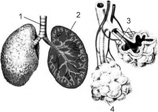
Fig. 310. The mammalian Respiratory system
1 - trachea; 2 - lung; 3 - Bronchi; 4 — bronchioles
Mammalian respiration also involves the mucous surface of the respiratory tract and, to a small extent, the skin; the latter accounts for only about 1% of the animal's required oxygen. The Nasal cavity is divided into the vestibule, respiratory region, and olfactory region. The function of the vestibule is to trap relatively large particles (dust) entering with the air; it is more developed in inhabitants of steppe and desert regions where the air is particularly dusty. Finer dust particles are trapped in the respiratory region of the nasal cavity, which is lined with a ciliated epithelium and rich in blood capillaries. Here, the air undergoes not only further purification from mechanical impurities but is also disinfected by the bactericidally active substances of the mucus. Simultaneously, as the air passes between the folds of the nasal conchae, it is moistened and warmed by the blood flowing through the capillaries, while also giving off a portion of its oxygen to the blood. The olfactory region forms a maze of cavities lined with olfactory epithelium, created by expansions and outgrowths of the lateral walls. Passing through the choanae into the pharynx, air enters the Larynx. The Base of the larynx is supported by a ring-shaped cricoid Cartilage, which is characteristic of all terrestrial vertebrates. The anterior and lateral walls of the larynx are formed by the thyroid cartilage, found only in mammals. Arytenoid cartilages are located on the sides of the dorsal aspect of the larynx. A thin epiglottis (Fig. 310) adjoins the anterior edge of the thyroid cartilage, closing the entrance to the larynx when food passes through the pharynx. The vocal cords are paired folds of the mucous membrane situated between the thyroid and arytenoid cartilages. The vocalizations of most mammals are largely confined to a low-frequency range; however, this does not preclude the transmission of complex and substantial information. The larynx is followed by the trachea, supported by cartilaginous rings. The trachea divides into two bronchi, which differ from it only in having a smaller diameter; within the lungs, they branch into even finer tubes, forming the bronchial tree. The smallest tubes—the bronchioles—open into expansions lined with respiratory epithelium known as alveoli, measuring 25–400 µm, the walls of which feature a dense network of capillaries. This lung structure significantly increases their respiratory surface area, making it 50 to 100 times greater than the body surface area. The relative magnitude of the gas-exchange surface in the lungs is greater in animals with high activity and mobility. A relative enlargement of the lungs is observed in high-altitude and aquatic mammals. Whales possess circular musculature that closes the entrance to the alveoli, allowing them to retain air even at considerable depths. During costal (rib) breathing, the volume of the thoracic cavity changes with the help of the intercostal muscles; during diaphragmatic breathing, this same volume changes through the lowering and raising of the musculotendinous partition known as the diaphragm. Both mechanisms operate in various combinations in all mammals; the first mechanism predominates in carnivores, while the second prevails in ungulates. The respiratory rate is higher in smaller animals with a higher metabolic rate compared to larger ones: it is 8–16 breaths per minute in horses, 100–150 in rats, and ~200 in mice. Breathing frequency increases during locomotion. Respiration also plays a role in thermothermoregulation. Rapid, shallow breathing in carnivores increases evaporation from The surface of the Upper Respiratory Tract, facilitating heat loss. Conversely, accelerating deep breathing enhances pulmonary gas exchange, thereby increasing the body's heat production at low temperatures.
Excretory Organs and Water-Salt Balance
The excretory organs of mammals are represented by pelvic kidneys (Fig. 311).
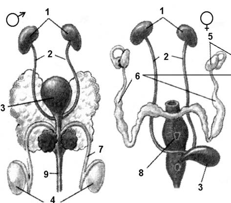
Fig. 311. Urinary System of mammals
1 - kidneys; 2 - Ureters; 4 - Testes; 5 - ovary; 6 - oviduct; 7 - vas deferens; 8 - uterus; 9 - ejaculatory duct
Urea is excreted as the primary nitrogenous waste product in mammals, placing them closer to amphibians in this characteristic. Water-salt balance is maintained primarily through the kidneys and regulated by Hormones from the posterior Pituitary Gland, which affect diuresis (urine output) and the reabsorption of water from the primary urine (filtrate) in the renal tubules. In addition, the skin with its sweat glands and the intestinal tract also participate in water-salt regulation in mammals.
Mammalian kidneys, like those of other amniotes, are metanephric and possess their own excretory ducts—ureters—that empty into the urinary bladder. The duct of the latter opens into the copulatory organ in males and the Vestibule of the Vagina in females. In monotremes (egg-laying mammals), the excretory ducts empty into the cloaca. The kidneys are bean-shaped and lie on either side of the vertebral column. A Cytology/practical/54.html">Longitudinal section of the kidney reveals that it consists of two distinct layers. The outer (cortical) layer contains the filtering apparatus—glomeruli, which consist of a capillary tuft surrounded by Bowman's capsule. This capsule gives rise to the excretory tubule, divided into four sections: the proximal convoluted tubule, the Loop of Henle, the Distal convoluted tubule, and the collecting duct. Together, these structures form a nephron (Fig. 312).
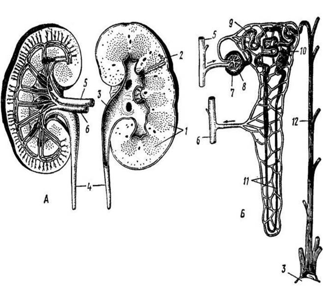
Fig. 312. Mammalian kidney
A - Cross section of a human kidney; B - diagram of Blood Circulation in the kidney:
1 - renal pyramids; 2 - position of the nephron; 3 - renal pelvis; 4 - Ureter; 5 - renal artery; 6 - renal vein; 7 - Bowman's capsule; 8 - glomerulus; 9 - proximal and 10 - distal convoluted sections of the renal tubule; 11 - loop of Henle; 12 - collecting duct.
Nervous system. The brain is characterized by its large size and consists of five distinct regions (Fig. 313).
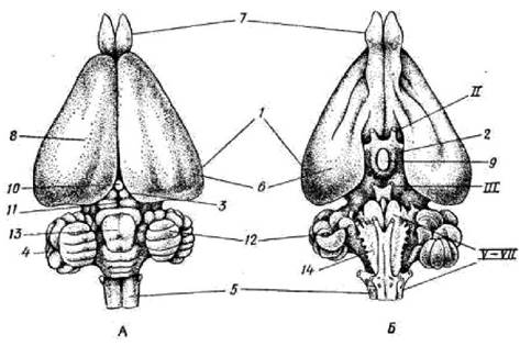
Fig. 313. Brain of a rabbit
A - dorsal view; B - ventral view: 1 - Forebrain, 2 - Diencephalon, 3 - Midbrain, 4 - Cerebellum, 5 - Medulla Oblongata,
6 - cerebral hemispheres, 7 - olfactory bulbs, 8 - neocortex, 9 - pituitary gland, 10 - Pineal Gland, 11 - corpora quadrigemina, 12 - cerebellar hemispheres, 13 - cerebellar vermis,
14 - pyramids, II, III, V-VII - Cranial Nerves
The forebrain features relatively large hemispheres. The upper surface of these hemispheres is covered by grey matter—the Cerebral Cortex—which consists of Nerve Cells and Unmyelinated nerve fibers. It is also referred to as the neopallium. The second component of the forebrain is White matter, which is a collection of nerve fibers. In higher mammals, these fibers form a commissure between the hemispheres known as the corpus callosum. Through these nerve fibers, the cerebral cortex connects with other parts of the brain and, consequently, with all organs and Tissues of the body. The surface of the cerebral hemispheres may be smooth in some animals (such as the rabbit) or folded with sulci and gyri in others. The hemispheres contain two cavities—the right and left Lateral ventricles—which communicate with each other and with the Third ventricle located further back.
Behind the forebrain lies the diencephalon, the main bulk of which is formed by the thalamus (optic thalami). It contains the pineal gland (a vestige of the reptilian parietal organ), the infundibulum (which forms the floor of the third ventricle), and the pituitary gland, an important endocrine gland. Posterior to the diencephalon is the midbrain, divided by two perpendicular furrows into four colliculi (optic lobes). The anterior colliculi receive visual stimuli, while the posterior ones receive auditory stimuli. The fourth region of the brain is the cerebellum. It consists of a median unpaired lobe, the vermis, and two lateral lobes, the cerebellar hemispheres. The primary function of the cerebellum is the coordination of movement and muscle tone. The fifth region—the medulla oblongata—regulates The activity of several vital neural centers, particularly the respiratory center. The ventricle of the medulla oblongata (the Fourth ventricle) forms the Rhomboid fossa and continues into the spinal canal. The Spinal Cord is a direct continuation of the medulla oblongata. As in the latter, the grey matter is located internally within the spinal cord, surrounded by white matter. Mammals possess 12 pairs of cranial nerves.
Sense Organs. Olfactory organs are well-developed in mammals and play a crucial role in their lives. The progressive Evolution of the olfactory system is evident in the enlarged volume of the nasal capsule and its structural complexity through the formation of a system of turbinate bones (nasal conchae) (Fig. 314).

Fig. 314. Mammalian olfactory organs
1 - olfactory center; 2 - nasal mucosa; 3 - Hypothalamus; 4 - nasal passage; 5 - vomeronasal organ (Jacobson's organ); 6 - oral cavity; 7 — tongue
The actual olfactory function is performed by a region in the upper part of the nasal cavity associated with the labyrinth of the ethmoid bone. Typically, the nasal cavity communicates with the surrounding bone sinuses.
The mammalian Organ of Hearing consists of the internal and middle ear, the external auditory meatus, and the auricle (pinna) (Fig. 315).

Fig. 315. Diagram of The Mammalian Ear structure
1 - semicircular canals; 2 - cochlea; 3 - stapes; 4 - incus; 5 - malleus;
6 - oval window; 7 - tympanic membrane; 8 - external ear
The external auditory meatus is an auditory tube surrounded by the tympanic bone, with one end opening outwardly and the other sealed by the tympanic membrane, behind which lies the middle ear cavity. Unlike amphibians, reptiles, and birds, mammals possess not a single auditory ossicle, but three: the malleus (homologous to the articular bone), the incus (homologous to the quadrate bone), and the stapes (homologous to the hyomandibular bone). They are movably connected and arranged in a chain such that the malleus rests against the tympanic membrane, the incus is movably attached to it, and the incus, in turn, connects with the stapes. The latter abuts against the oval window of the membranous labyrinth of the Inner ear. The inner ear contains a highly developed, spirally coiled outgrowth—the cochlea—inside of which lie the finest fibers that, much like strings of varying length, resonate in unison with sounds of different pitches. This is The Organ of Corti.
Visual organs are of less importance to mammals than to birds and feature a comparatively simple structure (Fig. 316).
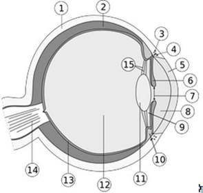
Fig. 316. Structure of the mammalian eye
1 - sclera; 2 - choroid; 3 - canal of Schlemm; 4 - ROOT of the iris; 5 - cornea; 6 - iris; 7 - pupil; 8 - anterior chamber of the eye;
9 - posterior chamber of the eye; 10 - ciliary body; 11 - lens; 12 - vitreous body; 13 - retina; 14 - Optic nerve; 15 - zonular fibers
They have acquired several progressive features. Mammals exhibit binocular stereoscopic Vision—the focusing of both eyes on a single object, which has improved their spatial orientation. Accommodation is achieved solely by altering the shape of the lens through the action of the ciliary muscle. Visual acuity is enhanced due to the expansion of the lens. Organs of touch in mammals are represented by specialized corpuscles and sensitive hairs, or vibrissae, located across the entire body surface, particularly on the snout, around the eyes, and elsewhere.
Mammalian reproduction. The reproductive organs of mammals are more complexly organized than those of other amniotes (Fig. 317).
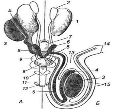
Fig. 317. Mammalian Reproductive System
1 - kidney, 2 - Adrenal gland; 3 - Testis; 4 - Epididymis; 5 - vas deferens; 6 - Seminal Vesicle; 7 - rectum; 8 - urinary bladder; 9 - Prostate Gland; 10 - Cowper's glands; 11 - Penis; 12 - preputial glands; 13 - Inguinal Canal; 14 - abdominal wall;
15 - walls of the Scrotum
The testes are paired, located either in the posterior region of the Abdominal cavity (monotremes, certain insectivores, xenarthrans, proboscideans, cetaceans, sirenians, rhinoceroses) or having descended into the scrotum—a cutaneous pouch communicating with the body cavity via the inguinal canal (marsupials, carnivores, ungulates, primates). An axially elongated body, the epididymis—an aggregation of efferent testicular ducts (a remnant of the anterior mesonephric kidney)—adjoins the testis. Extending from the epididymis is the Wolffian duct, or vas deferens, which empties near the base of the penis into its urogenital canal. Before entering this canal, the vasa deferentia form lobular expansions known as Seminal Vesicles, the secretion of which contributes to the formation of semen. In rodents, this secretion is released after the ejaculation of sperm into the female reproductive tract, solidifying to occlude the tract following copulation.
The size of the Ovaries is noticeably smaller than in other vertebrates. The paired oviducts, homologous to the Müllerian ducts, open into the body cavity near the ovaries via funnels lined with ciliated epithelium. The upper, slender and convoluted section of the oviduct is called the Fallopian tube, while the lower section, which is expanded and has muscular walls, is the uterus. In monotremes, the uteri open into the urogenital sinus of the cloaca; in all other mammals, the cloaca disappears. In marsupials, the lower part of the oviducts transforms into paired vaginae, which sometimes fuse at their ends and open into the vaginal vestibule (urogenital sinus). In placental mammals, the lower regions of the oviducts merge into a single, unpaired vagina. Egg cells, or oogonia, develop in the ovaries from the germinal epithelium; they grow and transform into oocytes, with each oocyte becoming surrounded by follicular cells to form a primary follicle. By the time of reproduction, the oocyte begins to grow, accumulating yolk, while its envelope expands, creating a fluid-filled space between it and the egg cell (oocyte). The follicle transforms into a Graafian follicle, which protrudes On the surface of the ovary. Subsequently, the follicle wall ruptures, and the egg cell is released into the body cavity (ovulation occurs), from where it enters the Fallopian tube and is fertilized. The scar formed on the ovarian surface fills with a blood clot and is invaded by lutein cells containing a yellow pigment and possessing hormonal functions. Thus, the corpus luteum develops in the ovary, secreting the hormone progesterone. Upon egg Fertilization and the onset of Pregnancy, the corpus luteum persists, while progesterone stimulates the implantation of the embryo into the uterine wall and simultaneously inhibits the formation of new Graafian follicles and ovulation until the end of pregnancy.
During development, the embryonic membranes of marsupial mammals merely appose the uterine wall, inducing partial histolysis of its superficial layers. In some species, a rudimentary embryonic part of the placenta forms through the fusion of the outer wall of the allantois with the serosa, yet without the formation of villi penetrating deep into the uterine wall. Embryonic Nutrition is sustained by the egg yolk and secretory products of the uterine wall cells. In placental mammals, a true placenta arises: the fused regions of the serosa and the outer wall of the allantois form the chorion (the embryonic component of the placenta), whose outgrowths (villi) penetrate the uterine wall, where the latter loosens to form the maternal component of the placenta. This process enhances the histolysis of uterine tissues, bringing embryonic blood vessels into closer contact with those of the maternal Organism. This facilitates embryonic nourishment via maternal blood components (hemotrophic nutrition). The formation of the placenta is termed embryonic implantation. The placenta may exhibit various structures: it is termed diffuse when villi are distributed evenly across the entire chorion, cotyledonary when gathered into separate groups, or discoid when villi are concentrated in a single area forming a disk, and so forth. The degree of attachment between the embryonic and maternal placentas varies, on The basis of which placentas are distinguished as deciduate or non-deciduate. In the former case, the connection is so profound that at parturition, a portion of the uterine wall is torn away and expelled as the afterbirth.
Mammals are distinguished by a high degree of parental care at all stages of offspring development. Only monotreme mammals lay large, yolk-rich eggs. In all mammals, the young are nourished with milk after birth.
Evolutionary origin of mammals. The earliest mammals—descendants of cynodonts—are known from the Late Triassic (approximately 200 million years ago); it was likely then that forms belonging to the subclass Allotheria (literally "other beasts") emerged, notably the Multituberculata, named for the numerous cusps on their molars arranged in two parallel rows. These animals lacked canine teeth, but featured prominent long incisors and likely occupied the same ecological niche as modern rodents. At the same time, numerous structural features distinguish them from modern mammals, precluding them from being ancestral to the latter. They are believed to have existed from the middle Jurassic period (160 million years ago) to the Early Oligocene (35 million years ago). Another mammalian Lineage is linked to the Symmetrodonta—animals that acquired the characteristic mammalian molar structure with cusps arranged in a triangular pattern; these animals apparently became the ancestors of the extant subclasses Prototheria and Theria, as well as an extinct subclass represented by the family Kuehneotheriidae.
As for the Prototheria, by the end of the Jurassic period, the Monotremata had already appeared; their most ancient fossil ancestor, *Teinolophos*, according to paleontological data, inhabited Australia as early as 120 million years ago. Meanwhile, the Theria continued to diverge, splitting during the Jurassic and Cretaceous periods into several infraclasses. The first to branch off were the extinct Triconodonta, followed by the likewise extinct Dryolestoidea, and finally the extant Metatheria and Eutheria—the latter three infraclasses are united into the group Cladotheria. The split between marsupials and placentals occurred in the Early Cretaceous period. Fossil specimens of *Sinodelphis* and *Eomaia*—primitive marsupial and placental mammals that lived 120–110 million years ago—have been discovered in China. Initially, both infraclasses were fairly widespread, but subsequently, marsupial evolution shifted largely to Australia and South America, which remained isolated from other continents for prolonged periods. Placental mammals never reached Australia, and only a few representatives, which were predatory, appeared in South America, allowing marsupials to exist and evolve relatively undisturbed.
By the end of the Cretaceous period, a multitude of their orders existed, both extinct and modern. Within Eurasia and North America, early marsupial representatives vanished, unable to withstand competition with placentals. Among extant marsupial orders, the American opossums (Didelphimorphia) are considered the most ancient; they originated in North America and later migrated to South America, while another lineage continued its migration via Antarctica to Australia, where it successfully evolved in the absence of competitors and gave rise to five orders united into the superorder Australidelphia. Placental mammals initially presented as small insectivorous creatures resembling modern moles, hedgehogs, and shrews. These were the earliest Insectivora.
According to current views, the most ancient placentals initially divided into two branches: Atlantogenata and Boreoeutheria. The former trace their origin to the shores of the Atlantic Ocean; from them, Representatives of the superorder Xenarthra likely separated first—currently represented by the orders Cingulata and Pilosa—and migrated to South America. The remaining forms in Africa constituted the superorder Afrotheria. The second placental branch originates from ancient Eurasia and North America, where two additional branches emerged: Laurasiatheria and Euarchontoglires, which later also split into several orders. Genetic studies have demonstrated that various groups of placentals that appear outwardly similar acquired their traits not through descent from a common ancestor, but via convergence. Conversely, genetically closely related orders are entirely disparate in both structure and lifestyle. Artiodactyls and perissodactyls are indeed relatives, yet relatively distant ones. Paradoxically, the closest living relatives of cetaceans are artiodactyls, whereas the closest relatives of perissodactyls are carnivores and bats. According to paleontological and genetic data, the majority of extant mammalian orders already existed by the Late Cretaceous to Paleocene epochs. Subsequent epochs witnessed their divergence into families and finer taxonomic groups.
Significance of mammals to humans
Useful mammals are numerous and diverse. Among long-domesticated farm animals, they account for over 60% (15 species). To these should be added fur-bearing animals bred in captivity, which are at an early stage of domestication (approximately 20 species), as well as numerous laboratory animals (rats, mice, guinea pigs, and many others). Breeding in captivity is accompanied by the DEVELOPMENT OF NEW breeds of these animals. The number of dog breeds reaches 200 and continues to grow; over a hundred breeds of rabbits are known; among laboratory animals, the number of genetically pure lines of mice and rats is increasing. The domestication of new species continues: the moose as a transport animal, the maral deer for antler velvet; new fur-bearing and laboratory animals. To improve existing breeds and obtain new ones, Hybridization of domestic animals with wild species is employed. Game and fur animals provide considerable profit in many regions. Successful conservation of their stocks and the restoration of populations nearly decimated by predatory exploitation—such as the sable, beaver, sea otter, fur seals, and other animals—enhance the hunting wealth of Ukraine and neighboring countries; the successful acclimatization of American rodents, the muskrat and nutria, has also contributed to this. The main commercial species of the forest zone remain the squirrel, sable, marten, ermine, foxes, and hares. Due to the population growth of valuable mustelids, the squirrel has yielded its leading position (in terms of pelt cost). In the tundra zone, the arctic fox and mountain hare retain primary importance; in steppes and deserts—foxes, hares, small mustelids, ground squirrels; in river valleys—the muskrat, water vole, otter, and in the south, the nutria; in mountainous regions, marmots are of significant importance. Other species (bears, felines, etc.) make up only a negligible fraction of the harvested pelts. Wild ungulates are an important source for meeting meat and hide needs: reindeer in the tundra, moose and deer in the forest zone, saiga antelope in steppes and semi-deserts. A special place is held by marine mammal harvesting, either at rookeries (fur seals), where the animals come to breed, or from sea vessels (whaling and sealing). In the first case, harvesting focuses predominantly on young males.
The list of harmful animals includes predators that attack livestock and humans, pests of forests and agricultural crops, as well as vectors and reservoirs of human and livestock diseases. Attacks by large predators (tiger, lion, leopard, wolves, hyenas, bears, etc.) on humans are quite rare and are usually committed by individuals that have lost The ability to obtain natural prey. Such "man-eaters" must undoubtedly be eliminated. Wolf attacks on humans are almost always associated with rabies. It is necessary to control the population of wolves and other predators in areas where they attack farm animals; however, their harmfulness should not be exaggerated. Wolves are often driven to this by a decrease in the population of ungulates, their natural food. A sharp decline in the numbers of large predators necessitates conservation measures: hunting the polar bear, tiger, and snow leopard is strictly prohibited.
Crops, plantations, and pastures suffer damage from rodents and lagomorphs. Their harmful impact is further compounded by the fact that many mice and voles are prone to population explosions. Gardens are damaged by dormice and mice.
Many mammals are reservoirs and vectors of dangerous Human and Animal diseases, and they also host vectors of these diseases, such as ticks, fleas, lice, and mosquitoes. Marmots, ground squirrels, gerbils, certain voles, mice, and other rodents (totaling about 200 species) harbor and transmit the dangerous bacterium that causes plague. Water voles (over 60 species in total) spread tularemia. Viral infections, especially encephalitis,
transmitted by ticks, are associated with small mammals (larval and nymphal stages of ticks) and large mammals—predators and ungulates (adult ticks). A closely related circle of reservoirs exists for hemorrhagic fever, tick-borne typhus, and relapsing fever. Desert rodents are hosts of cutaneous leishmaniasis, while canids host visceral leishmaniasis. Without the involvement of vectors, mammals preserve and transmit infections such as rabies (carnivores, certain bats), erysipeloid, leptospirosis, and Listeriosis (voles, rats, mice, insectivores, carnivores, and ungulates). Many of these infections have natural foci, meaning they exist permanently in nature. A person can become infected by contacting a sick animal or an infected vector (flea, tick, etc.) upon entering the territory of a natural focus.
Systematics of the Class
Class MAMMALIA - MAMMALIA
Subclass PROTOTHERIA - PROTOTHERIA
Order MONOTREMATA - MONOTREMATA
Subclass Theria - THERIA
Infraclass METATHERIA - METATHERIA
Order MARSUPIALIA - MARSUPIALIA
Infraclass PLACENTALIA OR HIGHER MAMMALS - PLACENTALIA seu EUTHERIA)
Order EDENTATA - EDENTATA
Order HYRACOIDEA - HYRACOIDEA
Order Proboscidea - PROBOSCIDEA
Order TUBULIDENTATA - TUBULIDENTATA
Order PHOLIDOTA - PHOLIDOTA
Order DERMOPTERA - DERMOPTERA
Order Insectivora - INSECTIVORA
Order Rodentia - RODENTIA
Order Carnivora - CARNIVORA
Order Primates - PRIMATES
Order Chiroptera - CHIROPTERA
Order Cetacea - CETACEA
Order SIRENIA
Last update: 19/08/2026
Editorial and Educational Adaptation: This material has been compiled based on the primary/original source text. The project team performed an editorial review, corrected technical inaccuracies, structured sections, and adapted the content for an educational format.
What was processed:
- elimination of formatting defects (OCR errors, structural breaks, corrupted characters);
- editorial organization of content;
- standardization of terminology in accordance with academic sources;
- verification of factual statements against the original source text.
All mentions of the author, publication year, and origin of the primary text have been preserved in accordance with the source.