Fundamentals of Biochemistry - Filippovich, Y. B. 1999
Coenzymes, Vitamins, and Certain Other Bioactive Compounds
Vitamins
General concept of Vitamins AND THEIR Classification. Vitamins constitute a chemically diverse group of Organic compounds; therefore, from the perspective of their chemical Structure, a single general definition cannot be formulated. The Physical Properties of substances classified as vitamins are as varied as their chemical nature. The Physiological effects of vitamins on animals, plant Tissues, and microorganisms also differ significantly, and individual vitamins bear virtually no resemblance to one another in this regard.
Vitamins are grouped together as a distinct category of naturally occurring organic compounds due to their absolute necessity for heterotrophic organisms as an essential dietary component supplementary to Proteins, fats, CARBOHYDRATES, and minerals.
In most cases, vitamins are not synthesized by heterotrophic organisms, and their deficiency leads to pathological conditions.
The quantitative requirement for vitamins is extremely small: for instance, an average human must consume about 600 g of major nutrients daily (calculated on a dry matter basis) compared to only 0.1–0.2 g of supplementary nutritional factors—vitamins.
Hence, it is evident that vitamins perform catalytic Functions within the body. In many instances, as will be demonstrated below, vitamins serve as integral components of Enzymes and are essential for their functioning.
Recently, The Role of vitamins in autotrophic organisms, particularly plants where vitamins are synthesized, has been under investigation. It turns out that they are also absolutely vital for plant life activity and similarly perform predominantly catalytic functions there. Consequently, the primary characteristic of vitamins as a unique group of compounds is their ability in minute concentrations to ensure the execution of enzymatic processes.
Thus, Vitamins can be defined as a group of organic substances with diverse structures and physicochemical properties that are strictly essential for the normal life activity of any Organism, performing catalytic and regulatory functions either directly or as part of more complex compounds.
Vitamins were discovered in 1880 by our compatriot N. I. Lunin. They attracted attention precisely as supplementary nutritional factors for animals. In his dissertation entitled "On The Significance of Inorganic Salts for Animal Nutrition," defended on September 18, 1880, at the University of Dorpat (now Tartu University), N. I. Lunin concluded that In addition to proteins, fats, sugars, salts, and Water, animals (mice) also require other, yet unknown substances. In his opinion, discovering these substances and studying their significance in nutrition would be a research topic of great interest. Subsequently, the work of N. I. Lunin was confirmed and expanded by other scientists. In 1912, the Polish researcher C. Funk proposed calling these unknown substances vitamins, meaning amines of life (from the Latin vita—life), since one of them, isolated and studied by him, contained an amino group. This term subsequently came to be applied to all mandatory supplementary dietary factors. Although many of them contain no amino groups or nitrogen whatsoever, the name "vitamins" remains firmly established in biology and medicine to this day.
Thanks to the efforts of numerous biochemists and physiologists over the more than century-long history of vitaminology, about three dozen vitamins have been isolated, their Composition and Structure studied, their physiological effects determined, and in the vast majority of cases, the Chemical synthesis of the corresponding preparations accomplished. Among the research of the Soviet period, notable contributions were made by V. N. Bukin, A. V. Palladin, L. A. Cherkes, M. N. Shaternikov, A. V. Trufanov, V. V. Efremov, K. M. Leutsky, B. A. Kudryashov, M. I. Smirnov, Yu. M. Ostrovsky, R. V. Chagovets, and others.
During the initial study of vitamins, each was named after the disease that developed when that vitamin was absent from the diet. The prefix "anti-" was added to the corresponding disease name, as introducing the vitamin into the diet led to a rapid cure (Table 12). Diseases developing due to a complete lack of vitamins in the diet came to be designated as avitaminoses, those caused by insufficient intake as hypovitaminoses, and those resulting from excess as hypervitaminoses. Later, at McCollum's suggestion (1913), individual vitamins were designated by letters of the Latin alphabet as they were isolated: A, B, C, etc. Finally, once the Chemical Nature of A number of vitamins had been investigated, their chemical names began to be introduced. Currently, all three nomenclature systems for vitamins are in use. There is a discernible trend toward adopting chemical names, which have been recognized internationally by the IUPAC (International Union of Pure and Applied Chemistry) Biochemistry Section since 1956. The most important vitamins and their names are listed in Table 12; some of them (Q, F, B15, U) are sometimes classified as Vitamin-like substances.
Class="center">Table 12 Major vitamins and their nomenclature
|
Nomenclature |
Human daily requirement, mg |
||
By letter |
Chemical (official international) |
Physiological (in relation to humans) |
|
|
Fat-soluble |
|||
А |
Retinol |
Antixerophthalmic |
2,5 |
D |
Calciferol |
Antirachitic |
0,0025 |
Е |
Tocotrienol |
Antisterility (tocopherol) |
15,0 |
К |
Phylloquinone |
Antihemorrhagic |
0,25 |
Q |
Ubiquinone |
— |
— |
F |
Complex of Unsaturated Fatty acids (linoleic, linolenic, and arachidonic acids) |
1000 |
|
Water-soluble |
|||
B1 |
Thiamine |
Antineuritic |
2,0 |
В2 |
Riboflavin |
Growth vitamin |
2,0 |
В3 |
Pantothenic acid |
Antidermatitic factor |
12 |
PP(B5) |
Nicotinic acid and nicotinamide |
Antipellagra |
25 |
В6 |
Pyridoxine |
Antidermatitic |
2,0 |
B12 |
Cyanocobalamin |
Antihemopoietic / Antianemic |
0,003 |
B15 |
Gluconodimethylaminoacetate |
Antianoxic |
2,0 |
Bс |
Pteroylglutamic acid |
Antianemic |
0,2 |
ВТ |
Carnitine |
— |
— |
С |
Ascorbic acid |
Antiscorbutic |
75 |
Н |
Biotin |
Antiseborrheic |
0,15 |
Р |
Rutin, bioflavonoid |
Capillary-strengthening vitamin |
50 |
U |
S-methylmethionine |
Antiulcer |
— |
The significance of vitamins is particularly profound for the developing child's body, as illustrated in Table 13.
Table 13 Daily requirement (mg) for certain vitamins in children and adolescents (according to M. I. Smirnov, 1974)
Age, years |
В1 |
В2 |
В6 |
С |
РР |
7—10 |
1,4 |
1,9 |
1,7 |
60 |
15 |
11—13 14—17 |
1,7 |
2,3 |
2,0 |
72 |
19 |
boys |
1,9 |
2,5 |
2,2 |
79 |
21 |
girls |
1,7 |
2,2 |
1,9 |
69 |
18 |
Based on their solubility in water and fat Solvents, vitamins are divided into two groups: water-soluble and fat-soluble.
Fat-soluble and certain Water-Soluble Vitamins exhibit vitamerism. This phenomenon consists in the fact that the physiological effect characteristic of a given vitamin is produced not by a single compound, but by several chemically similar compounds known as vitamers.
According to their physiological effect on The Human Body, vitamins are conventionally divided into the following groups (Table 14).
Vitamins exert a similar influence on life processes in animals. The absence or deficiency of vitamins in feed leads to impaired normal development, retarded growth, reduced productivity, and other undesirable consequences. Vitamins D, A, and B12 are particularly deficient in the vast majority of feeds. Introducing them into animal diets makes it possible to dramatically increase livestock productivity.
Table 14 Group characteristics of certain vitamins (according to P. I. Shilov and T. N. Yakovlev, 1974)
Vitamin group (by therapeutic and prophylactic effect) |
Brief clinical and physiological characteristics |
Names of major vitamins |
Enhancing general body reactivity |
Regulate the functional state of the Central Nervous system, METABOLISM, and tissue trophism |
В1, В2, РР, А, С |
Antihemorrhagic1 |
Ensure normal vascular permeability and stability, enhance Blood clotting |
С, Р, К |
Antianemic |
Normalize and stimulate hemopoiesis |
В12, Вс, С |
Anti-infective |
Increase the body's resistance to infection: stimulate antibody production, enhance the protective Properties of the epithelium |
С, А |
Vision-regulating |
Enhance visual acuity, widen the field of Color Vision |
А, В2, С |
1 Hemorrhage (from Greek haima—blood, and rhae—bursting forth) refers to bleeding, blood effusion, or the escape of blood from Blood Vessels.
Vitamin A (retinol). The Study of this vitamin began in 1909, and its synthesis was accomplished in 1933. Vitamin A has several vitamers, of which vitamin A1 is considered the most common (it is abundant in marine fish Liver):

Vitamin A2 differs from A1 by an additional double bond between the 3rd and 4th carbon atoms of the six-membered ring (found in freshwater fish liver). Both forms (A1 and A2) exist as a series of geometric isomers, but only some of them are physiologically active. Thus, vitamin A consists of a mixture of cyclic unsaturated alcohols with a characteristic chemical structure featuring A large number of conjugated double bonds.
These are lemon-yellow crystalline bodies with a melting point ranging from 59 to 64° C (depending on the geometric isomer type), highly soluble in fats and fat solvents such as gasoline, diethyl ether, chloroform, acetone, etc.
Group A vitamins are easily oxidized both under laboratory conditions (using MnO2) and within the organism. Upon Biological Oxidation mediated by a biocatalyst, retinol (an alcohol) is converted into retinal (an aldehyde), which also exhibits vitamin A activity:

However, in the absence of O2, retinol remains stable even at 100° C. In animal tissues, such as the liver, vitamin A frequently occurs as esters of palmitic acid. In this form, it is more stable and can thus be stored for future use, being released as needed. Retinol is transported to other tissues and Organs by binding to the blood retinol-binding protein, first isolated in 1968 (M = 21000, composed of 181 amino acid residues; its Primary Structure was elucidated in 1974).
A lack of dietary vitamin A leads to a range of specific pathological changes in animals and humans (Vitamin A deficiency): impaired vision (dim-light or night blindness), damage to Epithelial Tissues (dryness and desquamation of the epithelium), including the Cytology/practical/76.html">Cornea of the eye (dryness and inflammation known as xerophthalmia, hence the alternative name for vitamin A — antixerophthalmic vitamin). Furthermore, vitamin A deficiency results in retarded growth, weight loss, and general emaciation of the organism.
Dryness of the Skin and mucous membranes facilitates The entry of pathogenic microorganisms, leading to dermatitis, Bronchitis, and respiratory catarrh. Because vitamin A protects against these infectious diseases, it is classified among the anti-infective vitamins.
In plants, normal pollen germination and Fertilization occur only when precursors of vitamin A (carotenoids) are present in sufficient quantities.
The exact mechanism by which vitamin A helps maintain the normal state of epithelial tissues remains unknown. However, its role in maintaining visual acuity has been elucidated: the oxidized form of vitamin A (retinal) as a cis-isomer serves as the prosthetic group of the protein opsin, forming rhodopsin—the primary light-sensitive pigment of the retinal rod Cells (hence the name retinol). Rhodopsin was discovered over a century ago (1876) by F. Boll.
Opsin has an M of 38850 and contains two oligosaccharide fragments linked to a polypeptide chain of 348 amino acid residues, The sequence of which has been determined. It is embedded in the disc membrane of the outer segment of rod photoreceptor cells, spanning the membrane via seven compactly arranged α-helices. 11-cis-retinal is attached to one of these helices through an imine bond formed by the interaction of its aldehyde group with the ε-NH2 group of Lys296 (Fig. 60). Upon the action of a light quantum, cis-retinal is converted into the trans-form (the sensitivity of this reaction is a single photon):
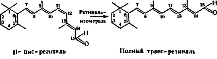
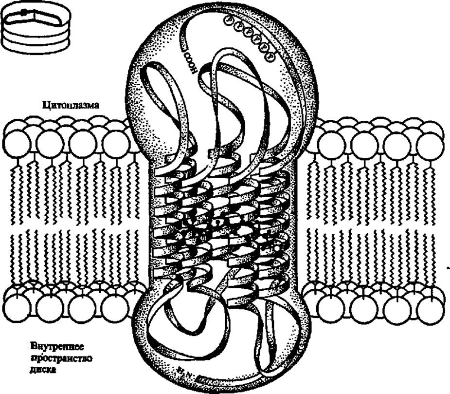
Fig. 60. STRUCTURE OF THE photoreceptor protein rhodopsin and its arrangement in the disc membrane of a photoreceptor Cell (details in the text):
in the upper left corner of the figure is a fragment of the outer segment of a photoreceptor cell, consisting of approximately two thousand discs; the square indicates the Location of rhodopsin within the membrane
This, in turn, stimulates rhodopsin activity, causing several hundred molecules of Transducin (~ 500)—a protein (M = 85,000) belonging to the G-protein family (see p. 457) and acting as the second component of the biochemical light-signal Amplification cascade (occurring within 1 ms)—to dissociate into an α-subunit (M = 39,000), in which guanosine diphosphate is simultaneously replaced by guanosine triphosphate (see p. 458), and a $\beta\gamma$-subunit dimer (M = 35,000 and 8,000, respectively). The complex of the transducin α-subunit with guanosine triphosphate interacts with the third component of the amplification cascade: cyclic guanosine monophosphate phosphodiesterase (composed of four subunits: α — 88 kDa, β — 84 kDa, and two γ subunits of 11 kDa each). In the presence of its activating protein (which is an integral protein of the retinal membrane), this enzyme converts several hundred molecules of cGMP into linear form within a second. The Hydrolysis of cyclic guanosine monophosphate is accompanied by the closure of sodium channels in The Plasma Membrane of the photoreceptor cell, its hyperpolarization, and the generation of an electrical impulse that travels to the synapse of the inner segment and is transmitted to the central nervous system. The simultaneous phosphorylation of the C-terminal region of the rhodopsin molecule (see Fig. 60) terminates its further action on transducin dissociation, thus completing the photoreceptor cycle.
Quantum-chemical calculations of retinal's structure provide deeper insight into its potential role in the visual process. The conjugated double bond system in the retinal molecule creates conditions for The formation of geometric isomers, primarily (as indicated by bond order values — see p. 191) at the double bonds between the 9–10 and 11–12 carbon atoms (where they are minimal compared to those at the 7–8 and 13–14 bonds):
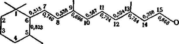
If the interaction of cis-retinal (within rhodopsin) with light quanta induces electron excitation—which can be viewed as the initial phase of electrical impulse generation—then the cis-trans transition likely serves as a unique gating mechanism ensuring the unidirectional flow of light energy utilization.
Dietary sources of vitamin A for humans include fish oil, fish and livestock liver, egg yolk, butter, green plant tissues, and red-fleshed vegetables (carrots, bell peppers, tomatoes, etc.). In the latter two, vitamin A is present as a provitamin, namely β-carotene. A β-carotene molecule is cleaved in the intestinal wall of humans and animals to yield two molecules of vitamin A1 (see p. 418).
The Use of vitamin A in animal husbandry yields tangible benefits. When pastures wither during the hot summer and the content of vitamin A—or more precisely, its provitamin, carotene—in grasses drops sharply, Karakul sheep develop vitamin A deficiency, which impairs their fertility. Vitamin A supplementation increases offspring yield by 5–7 lambs per 100 ewes, amounting to approximately 3 million lambs in the southern regions of our country. Moreover, adding vitamin A or carotene to the feed of young animals (chicks, calves, piglets) ensures better survival rates and faster growth, while including it in fattening rations for cattle increases live weight gain by 12–15%.
Vitamin D (calciferol). Research on this vitamin began in 1916; it was first synthesized in 1931.
Like vitamin A, vitamin D exists in several vitamers. The most widespread are vitamins D2 and D3, which can be regarded as sterol derivatives (see Ch. IX):
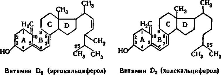
The provitamins for D2 and D3 are ergosterol and Cholesterol, respectively, which are converted into their active forms through the Cleavage of the bond between carbon atoms 9 and 10 of ring B under The Influence of solar radiation (cholesterol is first dehydrogenated to 7-dehydrocholesterol, which serves as the direct provitamin). Consequently, in the presence of the appropriate provitamins (e.g., 7-dehydrocholesterol in humans), vitamin D3 can be synthesized within the body, making dietary intake optional.

Fig. 61. Transformations of vitamin D3, their regulation, and The Effect of dioxy derivatives on phosphorus-calcium metabolism
Vitamins D2 and D3 are colorless crystals that melt at 115–116° C, are insoluble in water, but readily soluble in fats and fat solvents (chloroform, benzene, sulfuric and ethyl acetate ethers, acetone, alcohol). Both are unstable and rapidly destroyed by oxidizing agents (decomposition occurs at the double bond between the 7th and 8th carbon atoms of ring B) and mineral acids.
A lack of vitamin D in the diet leads to the well-known disease Rickets. Its cause lies in disordered phosphorus-calcium metabolism and impaired normal deposition of calcium phosphate in Bone tissue. It is hypothesized that D-avitaminosis disrupts the absorption of Ca and P in the gastrointestinal tract and the formation of phosphoric esters of various organic compounds; these two processes are likely interrelated. Recently, it has been demonstrated that the absorption, transport of Ca, and bone calcification are regulated not directly by vitamin D3, but by its hormonally active metabolite containing hydroxyl groups at the 1st and 25th positions. It is this metabolite that, by binding to nuclear receptors, ensures The Biosynthesis of Messenger RNA for The production of Ca-binding proteins and Hormones (Calcitonin and parathyroid hormone) that regulate calcium metabolism (Fig. 61).
Sources of vitamin D for humans include fish oil, butter, egg yolk, animal liver, and milk.
Vitamin D is especially important for laying hens and dairy cows. It has been calculated that 1/10 of all Ca contained in a hen's body goes into the eggshell, and with every liter of milk, more than 1 g of Ca is removed from a cow's body. Even the slightest impairment of Ca Absorption in the intestines of animals during D-avitaminosis has a detrimental effect on their condition and productivity. Therefore, vitamin D is widely used in livestock farming to increase the productivity of poultry and cattle, providing a 12–15% increase in weight gain.
Vitamin E (tocopherol). The first information about the existence of a vitamin regulating the reproductive process appeared in 1922. However, it was not until 1936 that three benzopyran derivatives, which turned out to be vitamers of vitamin E—a-, ß-, and y-tocopherols (from the Greek tokos — offspring, phero — to bear)—were isolated from wheat germ oil and cottonseed oil. In 1938, a-tocopherol was synthesized:

ß-Tocopherol differs from a-tocopherol in lacking a methyl group at position 7, and y-tocopherol lacks it at position 5. Later, four more tocopherols were isolated, differing in the number and arrangement of methyl groups in the benzene ring.
Tocopherols are colorless, oily liquids that are highly soluble in vegetable oils, alcohol, sulfuric ether, and petroleum ether. Chemically, they are quite stable; they withstand heating up to 100° C with concentrated HC1 and 170° C in air; they are destroyed by ultraviolet radiation and are optically active.
Vitamin E can be oxidized to a-tocopherylquinone, whose structure is very close to that of vitamins K and Q (see below):

The similarity in the Chemical Structure of vitamins E, K, and Q determines the resemblance of their Mechanisms of action in the body.
For a long time, it was believed that the significance of vitamin E was limited solely to its effect on reproduction, since in the absence or deficiency of vitamin E in humans and animals, Embryogenesis (fetal development in the maternal organism) is disrupted and degenerative Changes in the reproductive organs are observed. In plants, vitamin E promotes pollen germination. However, a deeper study of E-avitaminosis proved this view to be mistaken. E-avitaminosis manifests as a disruption in the normal functioning and structure of many tissues: muscular dystrophy, Spinal Cord degeneration and limb paralysis, fatty degeneration, etc., develop—i.e., a generalized disease of the organism.
The MECHANISM OF ACTION of vitamin E in the body is twofold. On the one hand, vitamin E is a crucial intracellular agent that protects fats and other easily oxidizable compounds from oxidation; it is one of the strongest natural antioxidants, primarily for Lipids. By reacting with lipid peroxy radicals and being oxidized themselves in the process, tocopherols terminate oxidation chains. On the other hand, vitamin E functions as a structural component of Introduction/36.html">Biological Membranes, forming molecular complexes with unsaturated Higher Fatty Acids of Phospholipids via its hydrocarbon radical and stabilizing (protecting against oxidation) the membranes. Since this ensures the normal course of biochemical processes, the multiple functional disorders observed in E-avitaminosis become understandable. Recently, another perspective on The Mechanism of action of vitamin E has been put forward—its possible involvement in the Regulation of the Biosynthesis of certain enzymes at the Transcription level in the GENETIC APPARATUS OF The Cell for their messenger RNAs. In addition, there is evidence that vitamin E controls the metabolism and functions of ubiquinone and is thus related to the Coupling of oxidation with ADP phosphorylation, i.e., to the Bioenergetics of the organism.
Sources of vitamin E for humans include vegetable oils (sunflower, corn, cottonseed, soybean, hemp, etc.), lettuce, cabbage, and grain products. The requirement for this vitamin is negligible, so E-avitaminoses and hypovitaminoses are very rare occurrences, especially since vitamin E is stored in many tissues (mainly adipose tissue). Its reserves ensure the replenishment of losses even in the complete absence of the vitamin in the diet for several months.
Vitamin K (phylloquinone). The first observations indicating the existence of a specific vitamin regulating blood clotting were made in 1929. Subsequent work led to the discovery of two natural vitamins, K1 and K2, which proved to be naphthoquinone derivatives. Vitamin K1 was synthesized in 1939:
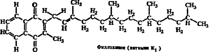
Vitamin K2 differs in The structure of its side chain, which contains from 30 to 45 carbon atoms and carries from 6 to 9 double bonds, respectively. It is specific to Bacteria and has also been synthesized (K2(35)). Its general formula is as follows (where n takes values from 5 to 8):

In addition to vitamins K1 and K2, many naphthoquinone derivatives possess a similar physiological effect. Among them, the preparation "vicasol" (a water-soluble bisulfite compound of methylnaphthoquinone), synthesized in 1942 by A. V. Palladin, has found wide Practical Application. It is a derivative of vitamin K3 (methylbenzoquinone):
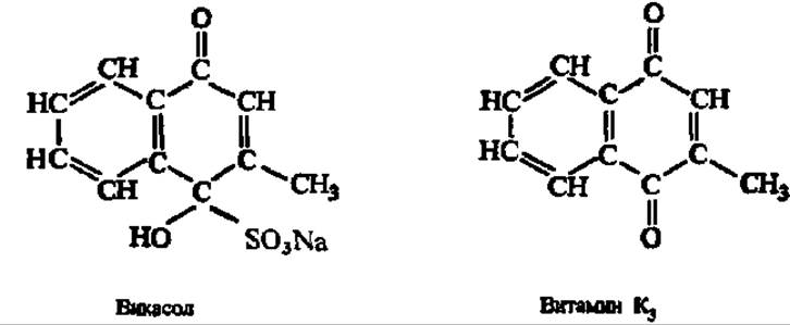
Vitamin K1 is a yellowish oily liquid with a boiling point of 115–145° C, insoluble in water. It is very unstable when heated in an alkaline medium and upon irradiation. Vitamin K2(35) consists of yellow crystals with a melting point of 54° C, and is even more unstable than vitamin K1. Vitamin K3 is a yellow crystalline powder with a melting point of 106° C, insoluble in water but soluble in alcohol and ether. Vicasol is a colorless, finely crystalline powder.
Vitamin K promotes the synthesis of components involved in blood clotting and positively affects the condition of the endothelial lining of blood vessels. Its deficiency in the diet can lead to spontaneous hemorrhages (nosebleeds, bloody vomit, internal bleeding, etc.). It is believed that vitamin K takes part in the synthesis of prothrombin and several other protein factors necessary for clotting. Prothrombin is converted into Thrombin, and the latter causes The conversion of fibrinogen into fibrin—i.e., it directly ensures Blood Coagulation. Vitamin K1, therefore, stands at the very roots of this complex system.
The main purpose of vitamin K in plants and microbes is The transfer of electrons during The process of Photosynthesis. It has recently been shown that the Post-translational Modification of Proteins via the conversion of glutamyl radicals into y-carboxyglutamyl radicals (see p. 301) is carried out by a vitamin K-dependent carboxylase localized in the membrane of The Endoplasmic reticulum. The role of vitamin K in this process boils down to the removal of a hydrogen atom from the y-carbon atom of the glutamic acid radical.
Sources of vitamin K for humans include tomatoes, cabbage, pumpkin, green parts of plants, and animal liver. In addition, vitamin K is synthesized by microbes that normally inhabit the intestine. The intestinal microflora is a constant supplier of vitamin K for humans and animals.
Vitamin Q (ubiquinone). This group of Fat-soluble vitamins was discovered quite recently. It is very close in structure and, probably, in functions to vitamins E and K, which served as the formal basis for classifying ubiquinones as vitamins. In 1955, ubiquinone was first isolated from animal fat.
Vitamin Q is widespread in nature. It has been found in microorganisms, plants, the Human and Animal body, as well as in foodstuffs. Consequently, it is exceptionally difficult to establish its dietary essentiality and prove that it cannot be synthesized by the animal organism itself. Nevertheless, experiments involving nutritional deficiency in monkeys, rats, rabbits, chickens, turkeys, and hamsters have demonstrated the vitamin activity of ubiquinones. It is believed that while the polyisoprenoid side chain of vitamin Q can be readily synthesized in the animal body, the cyclic quinoid moiety apparently is not. The chemical structure and mechanism of action of ubiquinone are discussed on p. 121, and its role in the functioning of the Respiratory Chain in Chap. X.
PLANT AND ANIMAL tissues characterized by intensive oxidation-reduction processes serve as sources of vitamin Q. For instance, high concentrations of ubiquinone (n = 10) are found in The Heart Muscle, liver, and brown adipose tissue of hibernating animals. Ubiquinone (n = 10) is used in the Treatment of cardiovascular diseases.
Vitamin F (a complex of unsaturated fatty acids). This complex includes linoleic, linolenic, arachidonic, and possibly certain other higher unsaturated acids. Arachidonic and linoleic acids are biologically the most active; linolenic acid enhances the effect of linoleic acid. In 1928, Evans and Burr (historically referred to as Hoogen and Gunter in Russian literature) proposed considering these three acids as a vitamin. Linoleic and linolenic acids have been obtained synthetically.
The classification of higher unsaturated fatty acids as vitamins is not universally accepted, since their catalytic function in the organism remains unknown and overt signs of avitaminosis in humans are absent. However, when linoleic, linolenic, and arachidonic acids were excluded from the diet of rats and dogs, striking symptoms of vitamin F deficiency were observed: dry and scaly skin, Hair loss, Necrosis of the tail tip, growth retardation, and weight loss.
Vitamin F is involved in the Regulation of Lipid Metabolism. Of particular importance is the fact that higher unsaturated fatty acids promote The excretion of cholesterol from the animal and human body, thereby preventing The Development of atherosclerosis. A beneficial effect of the vitamin on the condition of the skin and hair coat has also been noted.
The mechanism of action of vitamin F remains unknown. A special study using a series of synthetic unsaturated acids revealed that the biological activity of unsaturated fatty acids is associated with the presence of double bonds between the 6th–7th and 9th–10th carbon atoms.
In recent years, the biochemical effect of arachidonic acid has been elucidated: it turned out to be the precursor of a new type of hormones—Prostaglandins:

About 20 different prostaglandins are synthesized from arachidonic acid and other polyenoic acids, exerting a powerful influence on metabolism and physiological functions in humans and animals. In particular, a number of prostaglandins affect The activity of smooth Muscles of the uterine blood vessels and other organs and tissues, which is why they are used for treating Hypertension, facilitating labor, terminating Pregnancy, etc. (see p. 463).
Vitamin B1 (thiamine). Vitamin B1 holds a special place in the History of Vitamin research—it is the first crystalline vitamin obtained in the laboratory. It was first investigated by the Polish scientist C. Funk in 1912, already isolated in crystalline form in 1913, and synthesized in 1936. The structure of vitamin B1 is as follows:
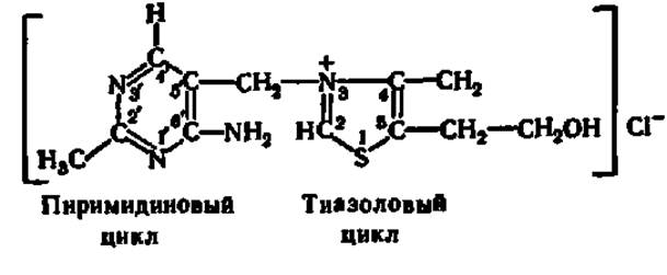
Since vitamin B1 contains a sulfur atom (S) in its molecule along with an amino group, it was given the chemical name thiamine (from the Greek theion meaning sulfur).
In the form shown above, i.e., as a salt of a quaternary ammonium base (thiamine chloride), vitamin B1 exists in an acidic environment. Neutral and alkaline environments favor a different structure—with an opened thiazole ring; in this case, free aldehyde and sulfhydryl groups appear in the thiamine molecule. The structure of the thiamine molecule has been confirmed by synthesis.
Thiamine occurs as small, colorless, bitter-tasting crystals that are readily soluble in water. Its solutions in acidic environments are stable and withstand heating to high temperatures. In neutral and especially in alkaline environments, thiamine is rapidly destroyed. Upon oxidation, it converts into thiochrome—a compound exhibiting a bright blue fluorescence in ultraviolet light, which makes its quantitative determination easy.
Vitamin B1 deficiency leads to a disease known as polyneuritis (beriberi). It involves the progressive degeneration of nerve endings and conduction pathways, resulting in loss of cutaneous sensitivity, impaired gastrointestinal motility, cardiac pain, and the like; eventually, paralysis and death ensue. In addition to humans, birds, rabbits, dogs, rats, guinea pigs, and many other animals are susceptible to this disease.
The mechanism of action of vitamin B1 has been studied in detail. Mediated by thiamine pyrophosphokinase (an oligomer of subunits with molecular weights of 27,000 and 30,000 Da and maximum activity in the tetramer), which transfers a pyrophosphate residue from ATP to thiamine, it is converted into thiamine pyrophosphate:

It has recently been discovered that thiamine pyrophosphate can be further phosphorylated with the participation of thiamine diphosphokinase; the resulting thiamine triphosphate is considered a storage form of thiamine pyrophosphate, although there is already evidence of its absolute indispensability for the functioning of Nerve Tissue. Thiamine pyrophosphate acts as a coenzyme for keto acid Decarboxylases. During the decarboxylation of pyruvic acid, in particular, an intermediate compound is first formed, which subsequently breaks down with the release of CO2:
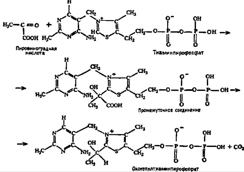
The formation of an intermediate compound between pyruvic acid and other α-keto acids and thiamine pyrophosphate is attributed to the peculiar features of its electronic structure: the second carbon atom of the thiazole ring possesses an increased electron density due to the dissociation of a proton from it; therefore, the α-carbon atom of the keto acid, which is characterized by an electron density deficit, readily adds to it.
Hydroxyethylthiamine pyrophosphate, in turn, breaks down with the release of thiamine pyrophosphate and a breakdown product of pyruvic acid, either as acetaldehyde (which is further converted into ethyl alcohol) or as acetyl-CoA (see p. 352). Most importantly, this eliminates pyruvic acid itself, which accumulates in large quantities during The breakdown of carbohydrates (and partly Amino Acids). It acts as a potent toxin to The Nervous System, leading to the severe consequences noted above in connection with vitamin B1 deficiency.
Thiamine pyrophosphate also catalyzes two-carbon fragment transfer reactions by serving as a coenzyme for the corresponding enzymes. The disruption of these processes due to vitamin deficiency affects the state of the organism and likewise manifests as vitamin B1 deficiency. Recently, non-coenzyme Functions of the phosphoric derivatives of vitamin B1 have been attracting attention: their participation in phosphorylation reactions, The regulation of Phospholipid Metabolism, and others.
For humans, the primary sources of vitamin B1 are bread and cereals, provided that the milling process does not strip away the germ and bran, which contain the bulk of the thiamine (rye flour, unpolished rice, etc.). Baker's and brewer's Yeasts are exceptionally rich in vitamin B1.
Vitamin B2 (riboflavin). Solutions of this vitamin, bright yellow in color and characterized by a yellow-green fluorescence, were obtained as early as the last century, but it was not until 1932 that the preparation was isolated in a concentrated form and subsequently obtained as orange needle-like crystals aggregated into clusters (melting point tm = 282°C) or plates (tm = 290°C). Vitamin B2 has now been synthesized.
The core of the riboflavin molecule is isoalloxazine, which combines benzene, pyrazine, and pyrimidine rings. The isoalloxazine derivative methylated at positions 6 and 7, and bearing a pentahydric alcohol residue—ribitol—at position 9, constitutes vitamin B2. Its chemical name, "riboflavin," reflects the presence of the ribitol residue in the molecule and the yellow color of the oxidized form of the preparation. Structurally, it is 6,7-dimethyl-9-ribitylisoalloxazine:

Riboflavin is chemically unstable and is easily destroyed by boiling and exposure to light. Under the influence of light, it decomposes into ribitol and 6,7-dimethylalloxazine, or lumichrome. The ability of riboflavin to undergo easy Oxidation and reduction is particularly important, as this underlies the biological action of this vitamin. The highest ability to attach H atoms is exhibited by the N atoms located at positions 1 and 10 in the isoalloxazine molecule; they have the maximum free valence indices (1.470 and 1.035, respectively), which characterize the reaction capabilities at that specific point of the molecule.
Vitamin B2 deficiency in humans manifests as growth retardation, hair loss, lesions of the mucous membranes (especially at the corners of the Mouth), rapid visual fatigue, decreased work capacity, impaired normal Hemoglobin synthesis, and pathological changes in the nervous system as well.
The mechanism of action of vitamin B2 has been well studied. In the form of a phosphoric ester (at the terminal hydroxyl group of ribitol) or even more complex compounds (particularly with NUCLEOTIDES), riboflavin acts as a coenzyme for oxidoreductases (see p. 119). About 30 of them are known. They carry out a number of important reactions in the organism—The oxidation of L- and D-amino acids, aldehydes, monoamines, purine bases (xanthine oxidase), carbohydrates (glucose oxidase), etc. Fe, Mo, and Co ions are frequently present in their active centers; some Flavoproteins oxidize substrates with molecular oxygen, meaning they function as oxidases, whereas others include Primary and secondary dehydrogenases.
Sources of vitamin B2 for humans include milk and green vegetables; vitamin B2 is abundant in animal liver and Kidneys, as well as in brewer's and baker's Yeast.
Vitamin B3 (pantothenic acid). This vitamin, first discovered in 1933, was obtained in crystalline form several years later (1939). In 1940, its chemical structure was elucidated and synthesis was achieved. It turned out to be D(+)-(α,γ-dihydroxy-β,β-dimethylbutyryl)-β-Alanine:

Pantothenic acid is found in all animal, plant, and microbial sources (from the Greek pantothen, meaning "everywhere"). It is a viscous, pale-yellow, oily liquid that is miscible with water and acetic acid. Only the dextrorotatory (+) optical isomer possesses biological activity. Pantothenic acid is relatively unstable and is easily oxidized and hydrolyzed in the presence of acids and alkalis at the peptide (—CO—NH—) bond.
A deficiency of pantothenic acid in humans and animals leads to various pathological phenomena: lesions of the skin and mucous membranes of Internal Organs, degenerative changes in a number of organs and tissues (Endocrine glands suffer particularly), hair loss, hair depigmentation, etc. The most prominent symptom of B3 deficiency in humans is numbness of the toes accompanied by a tingling sensation, followed by a burning pain in the toes and soles that spreads up to the lower leg ("burning feet syndrome"). All this is explained by the fact that pantothenic acid is a component of an exceptionally important organic compound—coenzyme A—which occupies a key position in the Synthesis and Breakdown of Fatty Acids and ensures the reactions necessary for the Interconversion of Carbohydrates and fats:
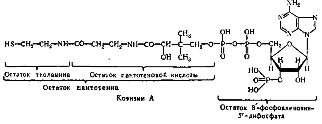
Rich sources of pantothenic acid include yeast, liver, egg yolk, and green parts of plants; it is also present in small amounts in all foods. In addition, pantothenic acid is synthesized by the intestinal microflora.
Vitamin PP (nicotinic acid and nicotinamide). Nicotinic acid and its amide (nicotinamide, niacin) have been known for a very long time:

However, it was only in 1937 that these substances were shown to be vitamins, as they prevent pellagra and cure the already established disease: pellagra (Italian) means "hard or rough skin." The Initial Stages of pellagra manifest as inflammation of the mucous membranes of the gastrointestinal tract, and subsequent stages involve skin inflammation (dermatitis) on areas of the body exposed to sunlight. Therefore, it was named vitamin PP, from the initial letters of preventive pellagra (Italian), meaning "preventing pellagra."
Nicotinic acid is a white crystalline substance (tm — 235.5°C) with a slightly acidic taste, highly soluble in water (especially upon heating); it is very stable and is not destroyed by the action of usual chemical and physical agents. Nicotinamide behaves similarly, forming colorless needles with tm = 131–132°C.
Strictly speaking, only nicotinamide possesses antipellagric action, whereas nicotinic acid itself acts as a provitamin. The mechanism of action of nicotinamide has been elucidated. It is a component of the essential dehydrogenase coenzyme, nicotinamide adenine dinucleotide (see p. 118), and its derivative, nicotinamide adenine dinucleotide phosphate.
A certain amount of nicotinic acid is synthesized in the bodies of animals and humans from The amino acid Tryptophan. This synthesis proceeds with the participation of vitamin B6. Thus, PP deficiency develops under conditions of inadequate protein nutrition (low tryptophan) and a lack of vitamin B6. Therefore, pellagra is currently regarded not purely as a vitamin PP deficiency, but as a polyavitaminosis—a disease caused by the absence of several vitamins and dependent on The amount of tryptophan in the diet.
Nicotinic acid and its amide are widely distributed in plant and animal tissues. Sources of vitamin PP for humans include wheat bread, animal liver and kidneys, potatoes, and many other foods.
Vitamin B6 (pyridoxine). Vitamin B6 is currently considered as a combination of three individual substances: pyridoxol, pyridoxal, and pyridoxamine. Each of them possesses vitamin properties because in the body it can be converted into Pyridoxal phosphate, which is precisely what participates in the Chemical Reactions associated with the activity of this vitamin:

Of the three aforementioned substances forming the pyridoxine complex, pyridoxol is the most studied; it was discovered in 1934 and synthesized in 1939. It forms colorless crystals (tm = 160°C), bitter in taste, and readily soluble in water and alcohol. Solutions of pyridoxol are stable to heating with acids and alkalis, but rapidly lose activity upon exposure to light.
Pyridoxal phosphate serves as a coenzyme in the decarboxylation reactions of a number of amino acids, as well as in Transamination reactions between Amino Acids and keto acids. The mechanism of the transamination reaction and the involvement of pyridoxal phosphate in it are discussed in detail in Chapter III (see pp. 124–127).
The absence of pyridoxine in the diet is accompanied by a severe disruption of Protein metabolism, since transamination reactions between amino acids and keto acids maintain the pool of free amino acids required for the Biosynthesis of Proteins. The main symptom of B6 deficiency is impaired hematopoiesis and the development of Various Forms of dermatitis that do not respond to treatment with nicotinic acid. Growth arrest occurs in young animals. Recently, it has been discovered that B6 deficiency is accompanied by Lipid Metabolism disorders, leading to the development of atherosclerosis.
Sources of vitamin B6 for humans include beef, fish, peas, egg yolk, and green plant parts. Since vitamin B6 is very widely distributed in food products, B6 deficiency is generally not observed in humans under normal conditions.
Vitamin B12 (cyanocobalamin). Vitamin B12 was first obtained in crystalline form in 1948. Its chemical structure is exceptionally complex; it was elucidated in 1953, and subsequently, As a result of a decade of efforts (1961–1971) by the distinguished organic chemist R. B. Woodward, the total synthesis of vitamin B12 was achieved.
The vitamin B12 molecule consists (Fig. 62) of a so-called planar group, which contains reduced pyrrole rings with a Co atom in the center, and two nucleoside groups positioned perpendicularly to it, containing dimethylbenzimidazole and adenine as nitrogenous bases and a-D-ribofuranose as a carbohydrate.
The planar group of vitamin B12 acts as a chromophore, which is why needle-shaped cyanocobalamin crystals have a ruby-red color, while its aqueous solutions are light lilac. Vitamin B12 crystals darken at 210–220°C and melt at t ~ 300°C. Vitamin B12 is readily soluble in water, alcohols, lower fatty organic acids, and phenols, but is insoluble in benzene, diethyl ether, chloroform, and acetone. It loses its activity in the light, but can be stored for a long time in the dark, remaining a very stable substance. Vitamin B12 is optically active. It has been established that a number of compounds with vitamin B12 activity exist in nature. The structure of several of them has been elucidated: pseudovitamin B12 contains adenine instead of benzimidazole, factor A contains 2-methyladenine, factor B lacks the nucleotide moiety, and so on.
A deficiency of vitamin B12 in the diet of humans and animals leads to impaired normal hematopoiesis in the Bone Marrow, resulting in a disease known as anemia; therefore, vitamin B12 is referred to as the anti-anemic vitamin.
The mechanism of action of vitamin B12 is that some of its forms function as Coenzymes and, by combining with various apoenzymes, give rise to a family of cobamide enzymes that accelerate vital reactions of nitrogen, carbohydrate, nucleic, and lipid metabolism. For example, mediated by methylcobalamin as a coenzyme, Methyl group transfer reactions are accelerated, enabling processes such as Methionine biosynthesis. In this reaction, vitamin B12 acts in tandem with vitamin Bc. Adenosylcobalamin is a component of enzymes that facilitate the intramolecular transfer of H atoms and various chemical groups (hydroxyl, amine, carbonylthioether, etc.) in accordance with the following scheme:

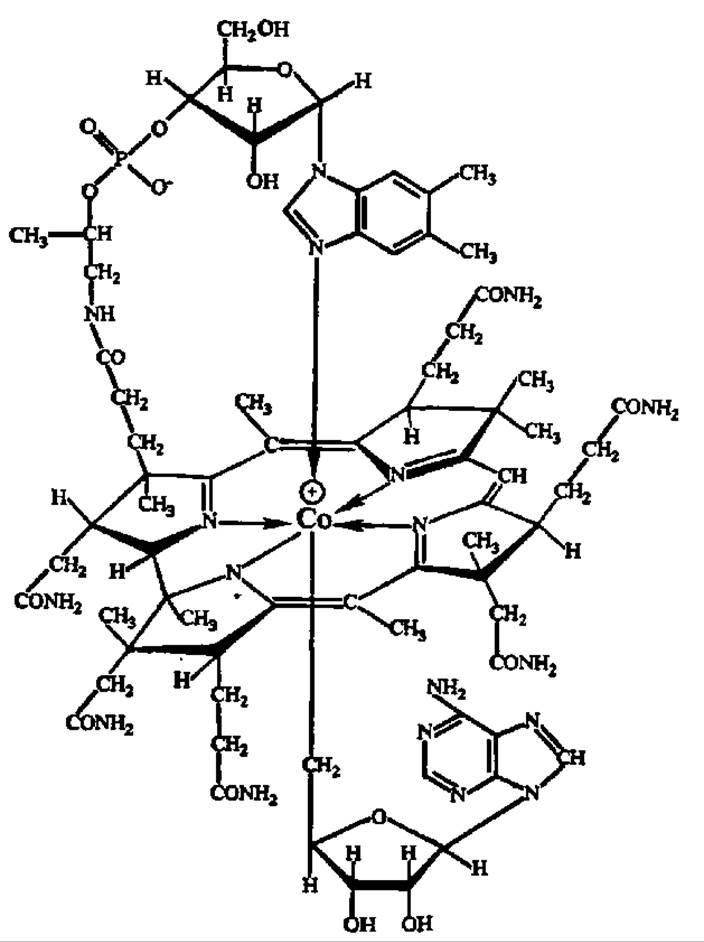
Fig. 62. Spatial configuration of the vitamin B12 molecule
Examples of such enzymatic processes will be discussed below.
The vitamin B12 depot in humans is located in the liver, where it accumulates in amounts of several milligrams. A protein compound that specifically binds the vitamin—the so-called intrinsic factor—takes part in transporting vitamin B12 across the intestinal wall. Consequently, impaired synthesis of this factor leads to B12 avitaminosis even when adequate amounts of the vitamin are present in the diet. Part of the vitamin B12 enters the human and animal organism as a result of the activity of microbes that are symbionts of the intestinal tract. Plants do not contain vitamin B12, so the sources for humans are meat, milk, and eggs, but it is synthesized exclusively by microorganisms.
Vitamin B12 finds very wide practical application in animal husbandry. For instance, adding it to the feed of pigs and poultry increases weight gain by 15%. In poultry, egg production also increases. In our country, a cheap method for producing vitamin B12 via microbiological synthesis (methane Fermentation using wastes from the acetone-butanol production) has been developed. The production of crystalline vitamin B12 for medical purposes has also been established, and a portion of the crystalline vitamin B12 is exported.
Vitamin B15 (pangamic acid). In 1950, T. Tomiyama discovered a compound in bovine liver extract that he named vitamin B15. In 1951, a similar substance was found (by E. Krebs et al.) in an aqueous extract of apricot kernel pits and was termed pangamic acid. Later, this compound was isolated in crystalline form from rice sprouts, brewer's yeast, the liver, and other sources. It turned out that this vitamin is very widely distributed in nature and is invariably present in plant seeds, which is THE ORIGIN OF its name (from the Greek *pan* meaning all and *gamos* meaning seed). The Chemical Composition and structure of pangamic acid have been elucidated and confirmed by synthesis:

Pangamic acid is a hygroscopic, white crystalline powder that is readily soluble in water, but insoluble in ether, chloroform, and benzene.
Pangamic acid has a positive effect on tolerance to oxygen deprivation (anoxia) and can be characterized as an anti-anoxic vitamin. Furthermore, it protects against fatty degeneration of the liver. However, it remains unknown whether pangamic acid is synthesized in the body or must necessarily be supplied from outside.
The mechanism of action of pangamic acid consists in the catalytic acceleration of methyl group transfer reactions. In particular, it ensures the normal course of biosynthesis of choline, methionine, creatine, and creatine phosphate. Since the latter is rapidly broken down during functional overload of the heart, calcium pangamate is used to treat pre-infarction and post-infarction states.
Vitamin Bc (pteroylglutamic acid). The first information regarding the existence of vitamin Bc was obtained in 1940 in experiments on chicks (hence the index c, derived from the English word *chicken*). By 1945, the identity of vitamin Bc with folic acid, isolated from spinach leaves and obtained synthetically, was established. This vitamin is better known as folic acid because it is found in significant quantities in leaves (from the Latin *folium* meaning leaf). However, several folic acids have been isolated, and today each member of this vitamin group is given a precise name corresponding to its chemical structure. The structure of one of the folic acids, pteroylmonoglutamic acid, is as follows:
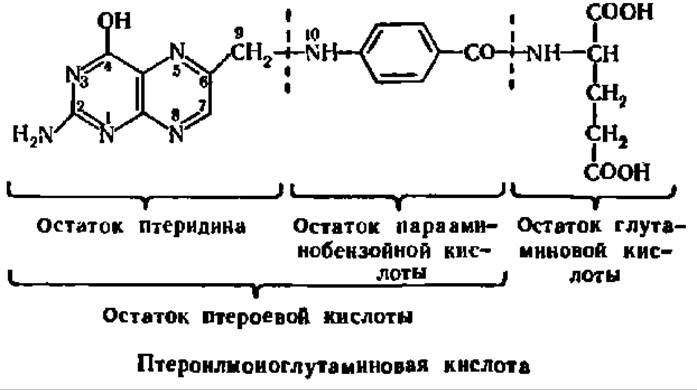
The remaining folic acids differ from pteroylmonoglutamic acid by the presence of a greater or lesser number (from 3 to 6) of glutamic acid residues attached to the terminal glutamic acid residue in the form of a y-glutamyl peptide.
Folic acid forms yellow, needle-shaped crystals containing two moles of crystallization water per mole of acid. They are stable in air and cannot be characterized by a melting point because they decompose at 250°C. They are sparingly soluble in water (25 mg/L), glacial acetic acid, and alcohols, and insoluble in ether, acetone, and chloroform. Prolonged exposure to light destroys folic acid.
Experiments have established that if folic acid is lacking in the diet of animals (e.g., chicks), their growth is retarded and hematopoiesis is disrupted. Lactic acid bacteria are very sensitive to a deficiency of vitamin Bc, for which it serves as an essential growth factor. Humans rarely suffer from Bc avitaminosis, as folic acid is synthesized by the microflora of the gastrointestinal tract and is always supplied to the body in sufficient quantities; however, if this avitaminosis develops in a human, it can manifest as anemia, accompanied by multiple functional Disorders of the digestive organs.
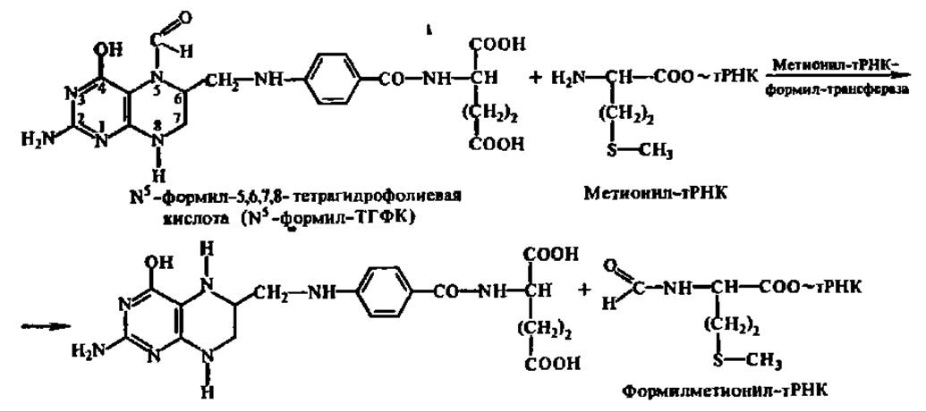
Folic acid, acting as a coenzyme for a number of enzymes, transfers single-carbon fragments during the biosynthesis of many compounds: the methyl group in the biosynthesis of methionine and thymine, the oxymethylene group (—CH2OH) in the biosynthesis of Serine, and the formyl group

Since these compounds play a leading role in protein and NUCLEIC ACID METABOLISM (a deficiency of methionine and serine limits Protein Biosynthesis, the absence of thymine and its corresponding nucleotide hinders DNA biosynthesis, and a shortage of purine bases impairs the de novo formation of DNA and all types of RNA), the physiological disorders observed in Bc avitaminosis are entirely understandable.
Folic acid transfers the aforementioned fragments while in a reduced state, in the form of 5,6,7,8-tetrahydrofolic acid. The attachment of fragments occurs at the N atom located at position 5, with the participation of a trifunctional enzyme—formyl-methenyl-methylenetetrahydrofolate synthetase (with an enzymatic molecular weight from 150,000 to 225,000 depending on the source). An example is the transfer of the formyl group during the biosynthesis of formylmethionyl-tRNA (protein biosynthesis begins with its attachment to the ribosome—see p. 290) in accordance with the equation
During the transfer of a methyl radical, THFA interacts with vitamin B12.
Human sources of folic acid include many foods, such as spinach, cauliflower, animal liver, and bread. The concentration of folic acid is particularly high in brewer's and baker's yeast.
Vitamin BT (carnitine). In 1948, G. Fraenkel and co-workers discovered a specific vitamin essential for normal growth and moulting in insects. The characteristic effect of this vitamin was identified in the yellow mealworm (Tenebrio molitor), which is why the corresponding index was used in its name.
By its chemical nature, vitamin BT proved to be ß-hydroxy-γ-trimethylaminobutyric acid:

When absent from the diet of certain insects, growth is arrested and death occurs during moulting. Although precise studies on the mechanism of carnitine action are lacking, it is believed to participate in methyl group transfer in insects. Such a function is also plausible in vertebrates, where it plays an active role in transporting acyl radicals across cell membranes—ultimately contributing to the oxidation and synthesis reactions of higher fatty acids.
Choline. This compound was isolated from Bile more than a century ago; however, it has only recently been recognized as having vitamin-like functions:

Choline chloride consists of colorless, hygroscopic crystals that are readily soluble in water and alcohols.
Although choline is synthesized within the animal body, under certain conditions a deficiency arises, leading to symptoms of choline deficiency. These manifest as Fatty liver degeneration, hemorrhages in the kidneys and other organs, a reduction in prothrombin synthesis, and alterations in conditioned reflex activity.
The mechanism of choline action is well understood. On the one hand, it acts as a partner to relevant acceptors in methyl group transfer reactions during the biosynthesis of key compounds such as methionine, purine and pyrimidine bases, etc. On the other hand, choline serves as a structural component of the active group of a biocatalyst that accelerates phospholipid synthesis (see p. 409). Additionally, it is a component of acetylcholine, which is involved in Nerve Impulse transmission.
Choline is abundant in chicken egg yolks, animal liver and kidneys, cabbage, and fish products.
Vitamin C (ascorbic acid). The earliest accounts of a specific organic substance whose presence in food prevents scurvy date back to 1885, when V. V. Pashutin rejected the prevailing view that scurvy was an infectious disease and proposed avitaminosis as its cause. In 1920, the anti-scurvy factor was named vitamin C; two years later, it was isolated in pure form, and by 1927 its chemical nature was fully elucidated and designated as ascorbic acid. The synthesis of vitamin C was achieved in 1932.
Ascorbic acid can be viewed as a derivative of the carbohydrate L-gulose; hence it is also referred to as 2,3-dehydro-L-gulono-γ-lactone.

The acidic properties of ascorbic acid—which, as seen from the formula, lacks a free carboxyl group—depend on the dissociation of the hydrogen from the hydroxyl group located at the 3rd carbon atom.
Vitamin C forms colorless crystals (m.p. = 192°C) with a sour taste, highly soluble in water and alcohol, but insoluble in benzene, chloroform, ether, and other fat solvents. In an oxygen-free environment, ascorbic acid crystals can be stored for years, but in the presence of oxygen or in solution—especially alkaline ones—vitamin C breaks down rapidly. Iron and copper ions further accelerate this degradation.
Ascorbic acid readily donates two H atoms, converting into dehydroascorbic acid, and vice versa. This critical property underlies the mechanism of action of ascorbic acid in the body: it acts as a component of redox systems, thereby ensuring the normal progression of vital processes in tissues.
The oxidation of L-ascorbic acid to L-dehydroascorbic acid is accompanied by the loss of two protons and two electrons:

Intermediate products, particularly the monodehydroascorbic acid free radical, are exceptionally reactive and interact with many other oxidoreductase coenzymes, such as Glutathione, NADH, FAD, Cytochromes, etc. The direct oxidation of ascorbic acid is accelerated by ascorbate oxidase (see p. 414).
Ascorbic acid is widely distributed in nature, present in virtually all tissues and organs of animals, plants, and microorganisms. Most frequently, it exists in its oxidized form, and in plants as bound ascorbic acid—so-called ascorbigen—which is formed through its interaction with 3-hydroxymethylindole. Ascorbbigen exhibits a slightly weaker physiological effect, but is more resistant to various physicochemical factors.
In humans, primates, and guinea pigs, insufficient Dietary intake of vitamin C leads to scurvy, a specific deficiency disease. The condition manifests as increased permeability and fragility of blood vessels, resulting in spontaneous hemorrhages and characteristic changes in bones and Teeth: teeth quickly decay, loosen, and fall out. These phenomena are rooted in impaired Synthesis of the adhesive intercellular protein Collagen, caused by hindered post-translational modification (see Chapter VII)—specifically, the inhibition of the oxidation of Proline and Lysine radicals into hydroxyproline and hydroxylysine radicals, respectively. As a result, non-fibrillar collagen is synthesized, leading to pathological changes in vascular walls and supporting tissues. Biosynthesis of ascorbic acid also fails to occur in certain birds (the family Passeridae) and several species of bats.
When evaluating the mechanism of action of ascorbic acid, great importance is currently attributed to its potential role in protecting active protein HS-groups from oxidation, including proteins with biocatalytic activity. This function is carried out by the reduced form of ascorbic acid. Naturally, any disruption of this process severely impacts the state of the organism. It has also been recently demonstrated that ascorbic acid serves as the active group of an enzyme
that accelerates the hydrolysis of certain thioglycosides, such as sinigrin found in mustard and horseradish seeds:
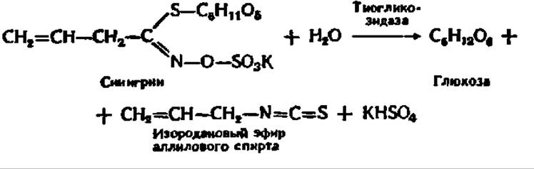
Ascorbic acid also acts as a paired donor in certain monooxygenase reactions (see Chapter X).
A wide variety of plant-derived foods serve as sources of vitamin C for humans. Rose hips, black currants, sea buckthorn, mountain ash, red peppers, lemons, and cabbage are particularly rich in this vitamin.
Vitamin P (rutin). In 1936, A. Szent-Györgyi and co-workers isolated a substance from lemon peel that improved capillary function, naming it vitamin P (from the Latin permeare meaning to penetrate). Vitamin P, which is nowadays conventionally referred to as rutin, represents a family of chemically related substances. All of them share a flavone backbone:

Currently, over a dozen compounds with P-vitamin activity are known, collectively termed bioflavonoids. They differ in the degree of hydroxylation of the benzene rings forming the flavone core, as well as in the various glycosidic groups attached to the 3rd carbon atom of the pyran ring. As an example, let us examine the structural formula of rutin, which contains a disaccharide residue—rutinose (C12H21O9):

Chemically pure preparations of P-group vitamins are yellow or orange crystalline substances that are sparingly soluble in water.
A dietary deficiency of vitamin P in humans and animals leads to increased capillary permeability, manifested by sudden hemorrhages after tissue compression, limb pain, general weakness, and rapid fatigue.
It is hypothesized that P-group vitamins participate in oxidation-reduction reactions, thereby ensuring the normal course of biological oxidation processes in the body. The actions of vitamins P and C are interrelated: in the presence of each other, they exhibit a significantly higher therapeutic effect than individually. Apparently, these vitamins function together in redox processes, forming a paired link within the respective system.
The dietary sources of vitamin P for humans are the same foods that are rich in vitamin C, such as black currants and lemons. In addition, significant amounts of vitamin P are found in lingonberries, blueberries, cranberries, plums, cherries, grapes, and other fruits, as well as in buckwheat and peppers.
Vitamin H (biotin). The vitamin properties of biotin were discovered back in the 1920s, but it was not until 1936 that 1.1 mg of a crystalline preparation, named biotin, was successfully isolated from 250 kg of egg yolk. Its structure was established in 1942, and chemical synthesis was achieved a year later:

The heterocyclic portion of the molecule consists of imidazole (A) and thiophene (B) rings, while the side chain is represented by a valeric acid residue.
Colorless, needle-like crystals of biotin (melting point = 220 °C) are readily soluble in water, sparingly soluble in alcohols, and practically insoluble in diethyl ether. Biotin is stable against molecular oxygen and H2SО4, but is degraded by the action of H2О2, bromine water, HCl, HNО3, and alkalis.
The essential role of biotin in normal vital activity is reflected in its very name (from the Greek bios meaning life). A deficiency of this vitamin in humans leads to a number of pathological changes: skin inflammation, hair loss, and excessive secretion by the skin's Sebaceous Glands (seborrhea). Protection against the development of seborrhea (from the Latin sebum meaning tallow and rheo meaning to flow) served as the basis for designating biotin as the anti-seborrheic vitamin.
The Mechanism of biotin action is likely multifaceted. It is believed that its primary role is serving as a coenzyme that is part of enzymes accelerating carboxylation reactions.
For instance, the carboxylation of acetyl-coenzyme A is of immense importance for the normal progression of biochemical processes, as it initiates the cycle of higher FATTY ACID BIOSYNTHESIS reactions (see p. 397). Biotin participates not only in CO2 fixation but also mediates transcarboxylation reactions—that is, the transfer of a carboxyl group from one compound to another:

The utilization of biotin as a coenzyme in carboxylation and transcarboxylation reactions is crucial for the Synthesis of purine bases, the conversion of pyruvic acid into oxaloacetic acid and subsequently into aspartic acid, as well as for various other metabolic reactions.
Dietary sources of vitamin H for humans include beef liver and kidneys, chicken eggs, milk, tomatoes, soybeans, carrots, potatoes, peas, and oatmeal. Biotin can also be supplied to the body by symbiotic microbes; for example, ruminants are entirely provided with biotin through the activity of their symbionts.
Vitamin U. In 1942, G. Cheney, based on experiments with chicks in which ulcers were induced by feeding them the alkaloid cinchophen, hypothesized that the accelerated healing of ulcers upon adding fresh greens, unboiled milk, and raw liver to the feed was due to the presence of an unknown factor, which he named vitamin U (from the Latin ulcus meaning ulcer). In 1954, R. Macrorie and co-workers identified natural vitamin U, isolated from cabbage juice as a crystalline bromide, with synthetic S-methylmethionine sulfonium bromide. It became clear that vitamin U is S-methylmethionine:

In the USSR, V. N. Bukin and co-workers synthesized S-methylmethionine sulfonium chloride in 1969, which has found widespread application as an anti-ulcer agent. It is a white crystalline powder with a slightly sweet-salty taste and a faint cabbage-like odor, highly soluble in water and water-alcohol mixtures, but insoluble in ethanol, glycerin, and diethyl ether.
The therapeutic mechanism of S-methylmethionine sulfonium chloride is attributed to the fact that it is a high-energy (macroergic) compound (see Chapter V) and an active donor of methyl groups for chemical processes associated with the regeneration of the mucous membranes of the digestive tract.
Sources of vitamin U for humans include asparagus, cabbage, tomatoes, celery, and green tea.
Last update: 06/08/2026
Editorial and Educational Adaptation: This material has been compiled based on the primary/original source text. The project team performed an editorial review, corrected technical inaccuracies, structured sections, and adapted the content for an educational format.
What was processed:
- elimination of formatting defects (OCR errors, structural breaks, corrupted characters);
- editorial organization of content;
- standardization of terminology in accordance with academic sources;
- verification of factual statements against the original source text.
All mentions of the author, publication year, and origin of the primary text have been preserved in accordance with the source.