Nephrology for the Family Physician - O.I. Bakaliuk 2003
Structural and functional organization of the urinary system
The Urinary System consists of two Kidneys, two Ureters, the Urinary Bladder, and the Urethra.
The kidneys are paired Organs of a specific shape. They feature anterior and posterior surfaces, medial and lateral borders, and superior and inferior poles. At the midpoint of the medial border lies the renal hilum (hilus renalis), which opens into a cavity known as the renal sinus (sinus renalis). Blood Vessels, Lymphatic vessels, and nerves enter and exit the Kidney through the hilum.
The kidneys are located retroperitoneally at the level of the 12th thoracic, 1st, and 2nd lumbar vertebrae on either side of the spine, and are surrounded by a thick layer of adipose tissue. The right kidney is positioned slightly lower than the left, and the superior poles of the kidneys are closer to each other than the inferior poles (Fig. 1).
Both kidneys weigh about 300 g, with the left kidney being heavier than the right. The ratio of the combined kidney mass to total body weight is 1:240. The kidneys have limited vertical mobility and are maintained in their normal position by the surrounding Connective Tissue (renal fascia) and adipose capsules, as well as the tone of the anterior abdominal wall Muscles. The Role of blood vessels in the fixation of the kidneys is relatively minor.
Class="center">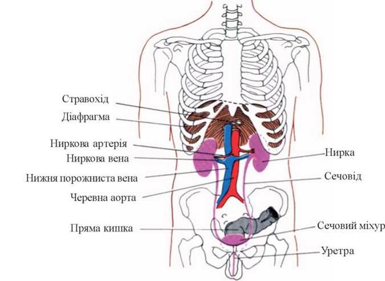
Fig. 1. Topography of the kidneys.
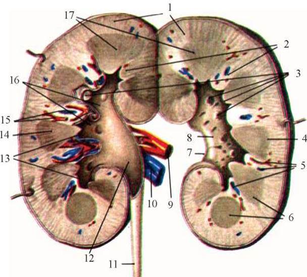
Fig. 2. Frontal section of the right kidney (schematic): 1 - renal cortex, 2 - renal columns, 3 - renal papillae, 4 - Base of the pyramid, 5, 15 - blood vessels, 6, 14 - renal pyramids, 7 - renal hilum, 8 - renal sinus, 9 - renal artery, 10 - renal vein, 11 - Ureter, 12 - renal pelvis, 13 - minor renal calyces, 16 - major renal calyces, 17 - renal medulla.
A Cytology/practical/54.html">Longitudinal section of the kidney from the lateral to the medial border reveals that the renal hilum leads into a centrally located cavity—the renal sinus. This cavity houses a significant portion of the renal pelvis, the Major and minor calyces, blood vessels, lymphatic vessels, and nerves, along with the apexes of the renal pyramids, which appear as papillae (papillae renales). The surface of each papilla features small openings, the papillary foramina (foramina papillaria), through which urine flows out (Fig. 2). The medullary substance (constituting 25–30% of total kidney mass) differs from the cortical substance in its lighter, pale-red coloration.
The Morphology and function of the nephron have been described in detail (G. Mazhdrakov et al., 1980; Yu.V. Natochin, 1994; V.V. Serov, 1995; A. Bohle et al., 1989); therefore, we will limit our Structure/133.html">Discussion to the core data essential for understanding the Structure and function of the kidneys.
The structural unit of the kidney is the nephron. The total number of nephrons in both kidneys reaches 2–2.5 million, with 4/5 of them located in the cortical substance and 1/5 at the corticomedullary junction. The nephron structure comprises the renal corpuscle (glomerulus and its capsule) and the renal tubule. The SCHEMATIC STRUCTURE OF a nephron is shown in Figure 3.
Two Types of nephrons are found in the human kidney: cortical (superficial) and juxtamedullary (deep). Juxtamedullary nephrons feature a Loop of Henle that extends down to the renal papilla. It is believed that the presence of these two nephron types is related to The Need for exceptionally fine Regulation of Blood sodium levels as The basis of the body's electrolyte Homeostasis (W. Cupples et al., 1996).
The glomerular tuft consists of approximately 50 capillary loops (Fig. 4).
The appearance of a renal glomerulus under an Electron microscope is shown in Figure 5.
The Capillary Wall acts as a glomerular filter, consisting of the epithelium, endothelium, and the intervening basement membrane (BM).

Fig. 3. Schematic STRUCTURE OF THE nephron.
The endothelium of the glomerular capillaries contains pores measuring 100–150 nm, which serve as the primary pathway for ultrafiltration.
The capillary BM (with a thickness of 250–400 nm) consists of three layers: the lamina densa, lamina rara externa, and lamina rara interna. The middle layer, the lamina densa, serves as the basement membrane proper. The BM contains highly specific Proteins (types IV and V Collagen, Laminin, entactin, nephritogenoside, Fibronectin, and negatively charged heparan sulfate Proteoglycans).
Type IV collagen plays a crucial role in ensuring proper BM function.
The BM contains 6 different variants of collagen chains of this type, designated from a1 to a6. Each chain is encoded by its own Gene, and these genes are arranged in pairs on three different Chromosomes. Genetic Defects in The structure of two of these six type IV collagen chains are associated with the clinical syndromes of Alport and Goodpasture (see below). The BM also contains pores (with an average size of 2.9 nm), the dimensions of which can vary depending on the hydrostatic pressure within the glomerular capillaries. The BM is not continuous; it is interrupted in areas where endothelial Cell nuclei are located, and mesangial Cells are "inserted" into these gaps within the BM. The protein Components of the BM also establish a specific electrostatic charge, a reduction of which is critical for The Development of proteinuria (PU).
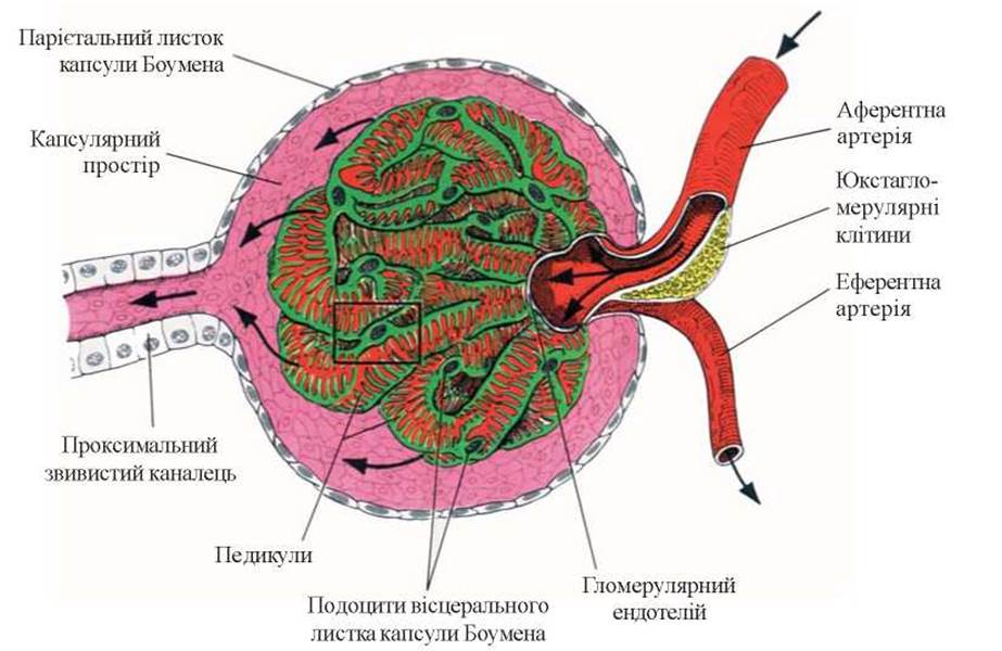
Fig. 3. Schematic structure of the glomerulus.
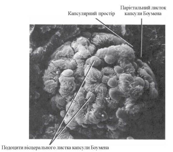
Fig. 3. Renal glomerulus. Scanning Electron Microscopy, x 1230 (R.G. Kessel et al., 1979).
Epithelial cells (podocytes) resemble cushion-like structures with cellular processes (or an octopus) that externally cover the capillary (Fig. 6).
The minor FOOT processes of podocytes (pedicels) branch out perpendicularly from the major processes and intertwine closely, forming the so-called slit Diaphragm (pecular diaphragma) along with fine fibrillar structures. Featuring a filamentous structure, the slit diaphragm thus creates a unique system of pores (5–12 nm in diameter), which also plays a critically important role in Glomerular Filtration.
The appearance of podocytes under an electron microscope is shown in Figure 7.
The glomerular capillary loops are suspended like the mesentery of the intestine at the vascular pole by the mesangium—a connective tissue structure comprising mesangial cells and the mesangial matrix. Recent studies have highlighted the vital role of the mesangium in maintaining normal nephron function and its impairment in pathology.
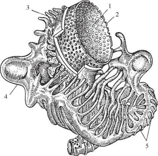
Fig. 6. Glomerular filter. Schematic diagram of the capillary wall structure (adapted from M.Ya. Ratner et al., 1987): 1 - capillary lumen; 2 - endothelium; 3 - basement membrane; 4 - podocyte; 5 - minor foot processes of podocytes (pedicels).
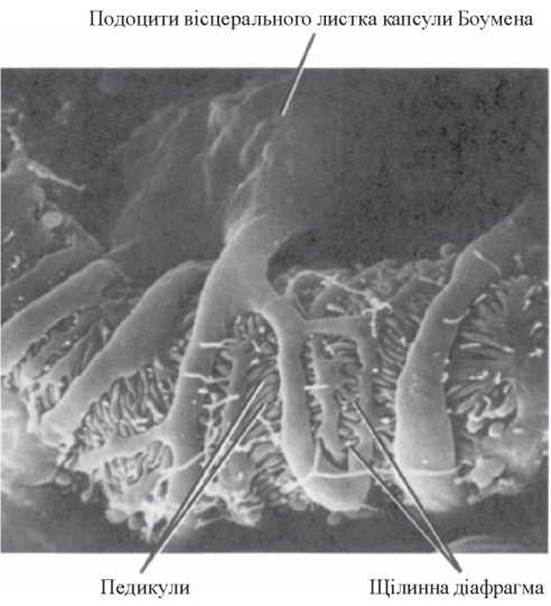
Fig. 7. Podocyte. Scanning electron micrograph, x 7800 (R.G. Kessel et al., 1979).
Two groups of mesangial cells are distinguished: intrinsic glomerular (contractile) cells and transient (resident) mononuclear leukocytes.
Contractile mesangial cells, which make up the bulk of the mesangial cell population, share many properties with vascular smooth Muscle cells and play a key role in regulating capillary blood flow and ultrafiltration. These cells contain Actomyosin and receptors for angiotensin II (AII). Under certain conditions, they are capable of exhibiting amyloidoclastic activity, producing renin, and contracting in response to vasopressin, parathyroid hormone, Prostaglandins (PG) I2 and E2, and histamine, which leads to a reduction in the total glomerular filtration surface area (J.I. Kreisberg et al., 1995). Simultaneously, these cells synthesize types I, III, IV, and V collagen, fibronectin, and glycosaminoglycans, and secrete neutral proteases (including collagenase) and specific matrix-degrading components. Thus, contractile mesangial cells ensure the turnover of the mesangial matrix and its physiological apoptosis.
Transient mesangial cells account for 3–7% of the total population; both in vivo and in vitro, they phagocytose circulating immune complexes (CICs), latex particles, Bacteria, and aggregated gamma globulin.
The glomerular mesangium has its own hemircirculation, which is relatively independent of capillary blood flow (A.F. Michael et al., 1990). This suggests that even a small number of mesangial mononuclear phagocytes, by releasing BIOLOGICALLY ACTIVE SUBSTANCES typically produced by circulating tissue monocytes-macrophages, can influence the state of the entire mesangial region. Consequently, the presence of a small number of mesangial cells in normal glomeruli creates conditions for the local execution of immune reactions, resulting in thickening and sclerosis of the capillary wall or complete obliteration of its lumen.
The primary components of the mesangial matrix also include types IV and V collagen, laminin, and fibronectin. Functionally, the matrix acts as a channel ("drainage system") for removing degradation products of protein molecules, microorganisms, or their fragments.
The glomerular capsule consists of the basement membrane and epithelium. The basement membrane of the capsule serves as the Water/144.html">Origin of the tubular basement membrane, while the capsular epithelium is functionally similar to the epithelium of the proximal tubule. The capsular epithelium also possesses contractile properties and is therefore capable of altering the volume of the capsule.
The tubular System of the nephron is divided into the proximal convoluted segment, the loop of Henle, the distal segment, and the collecting ducts. The endothelium of each of these segments features a specific ultrastructure that facilitates maximum performance of their Functions, such as reabsorption and secretion.
The appearance of renal tubules under an electron microscope is presented in Figure 8.
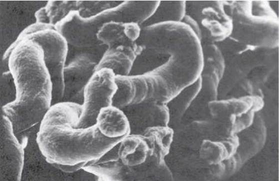
Fig. 8. Renal tubules. Scanning electron micrograph, X 500 (Courtesy of Fisher Scientific Company and S.T.E.M. Laboratories, Inc., Copyright, 1975).
At the site of contact between the convoluted part of the distal tubule and the vascular pole of the glomerulus, the epithelium of this region undergoes structural modification. As proposed by W. Zimmerman (1933), this portion of the tubule was named the macula densa. The cells of this region establish extremely close contact with the Structural elements of the juxtaglomerular apparatus (Fig. 9).
The juxtaglomerular apparatus includes:
- epithelioid Cells of the walls of the afferent and efferent arterioles of the glomerulus;
- Goormaghtigh cells (lacis cells), located in the space between the afferent and efferent arterioles of the glomerulus;
- glomerular mesangial cells.
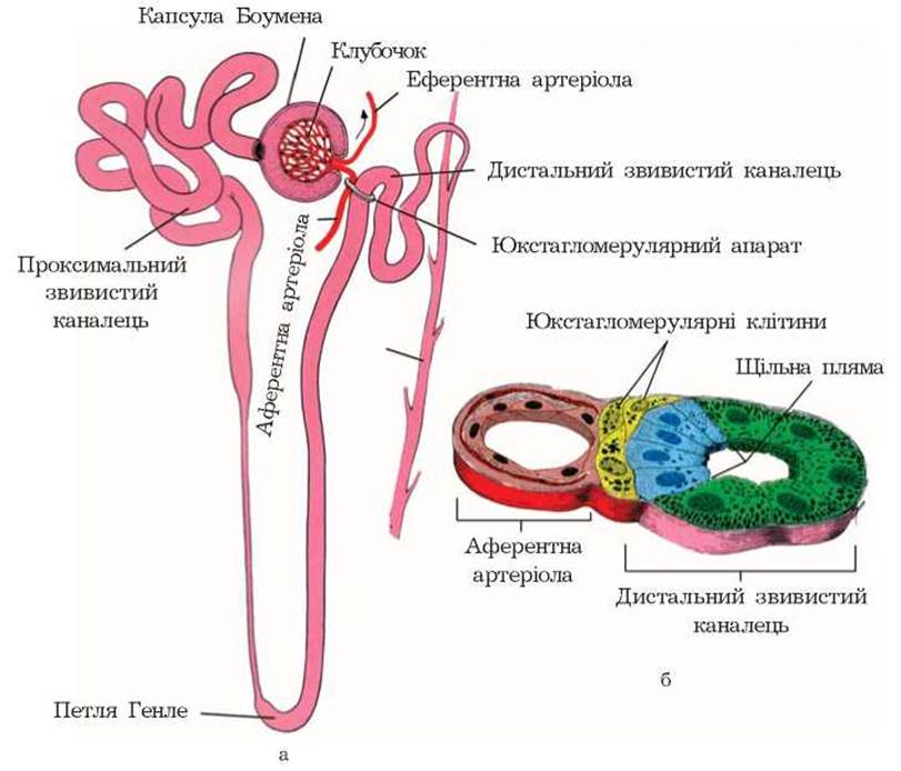
Fig. 9. Diagram of the structure of the renal juxtaglomerular apparatus: a - Location; b - cross-section.
The tight contact between the macula densa and Other components of the juxtaglomerular apparatus allows The chemical composition of urine within the distal tubule lumen to influence glomerular blood flow and intraglomerular pressure, while conversely mediating the hormonal effects of the juxtaglomerular apparatus on macula densa cell function.
At the level of the individual nephron, this renal structure acts as a local regulatory mechanism. Through the glomerulo-tubular shunt, it continuously compares the glomerular filtration rate (GFR) and the COMPOSITION OF THE plasma filtered in the glomerulus with the composition of the tubular urine. Functionally, the juxtaglomerular apparatus resembles a steam boiler water gauge Glass, with the key difference that the indicator is not fluid level, but rather the concentration of sodium ions in plasma and urine. When a mismatch occurs between the compared parameters, the juxtaglomerular apparatus can automatically—via renin release and local synthesis of Ang II—reduce the lumen of the afferent glomerular arteriole, thereby adjusting the filtration load to match the reabsorptive capacity of the tubules.
The collecting ducts ensure passive Water Transport and the secretion of hydrogen ions (H+).
The Endocrine System of the kidneys includes the prostaglandin system, the kallikrein-kinin system, and apudocytes. Prostaglandins are produced by medullary interstitial cells and collecting duct nephrocytes, while the components of the kallikrein-kinin system are synthesized by nephrocytes of the distal tubules. Renal apudocytes (oncocytes and M-cells) are part of a highly active Cellular Organization known as the APUD system. The latter is localized in various organs with or without hormonal activity (Pineal Gland, pituitary, thyroid, Parathyroid glands, Adrenal Glands, Liver, Pancreas, Esophagus, Trachea, Bronchi, taste buds of the Tongue, etc.). Renal apudocytes produce biogenic amines that function both as Hormones and Neurotransmitters/neuromodulators (dopamine, melatonin, serotonin).
The performance of vital renal functions is impossible without adequate blood supply. The kidneys are among the most heavily perfused organs: 1 minute sees up to 25% of Cardiac Output passing through the kidneys (ranging from 12% to 30%), which averages 1200 mL/min for a 70 kg human. The intensity of renal blood flow exceeds resting muscle blood flow by 100-fold or more. Another characteristic feature of Renal Circulation is its ability to autoregulate and maintain GFR across a fairly wide range, practically independently of systemic ARTERIAL BLOOD PRESSURE (BP).
There are two distinct circulations in the kidneys: cortical and juxtamedullary (Fig. 10).
Under physiological conditions, 85–95% of blood passes through the cortical (major) circulation and 3–10% through the juxtamedullary (minor) circulation. These two circuits are separated by efferent arterioles (vasa efferentia), which pose a significant resistance to blood flow. As a result, a relatively high pressure is maintained within the glomerular capillary bed (about 70 mmHg in the efferent arteriole, 45–60 mmHg in renal capillaries, 15 mmHg in the renal capsule and proximal convoluted tubules, 10 mmHg in the loop of Henle, and 10 mmHg in the interstitial fluid), whereas pressure in the peritubular bed remains low (6 mmHg in distal tubules, 2–0 mmHg in collecting ducts, 13 mmHg in peritubular capillaries, and 45 mmHg in the efferent renal vein) (Fig. 11).
The low pressure in the peritubular capillary bed and the slowed blood flow therein create favorable conditions for the reabsorption of various substances.
A small fraction (2–3%) of the blood reaching the kidney may pass through arteriovenous anastomoses (shunts). Under certain conditions (neuro-reflex and hormonal alterations), the volume of blood shunted can increase significantly, diverting blood directly into the pyramids while bypassing the cortex, which may lead to cortical ischemia and necrosis.
The Lymphatic system of the kidneys is functionally subordinate to the reabsorptive function of the tubules. The lymphatic vessels maintain closer contact with the Arterial System than with the Venous system. Lymphatics are absent in the glomeruli and pyramids; overall, the anatomical layout of the renal lymphatic system mirrors the architecture of its Vascular System to a certain extent. Initial lymphatic capillary networks surround the glomerular capsules and tubules.
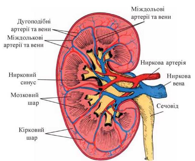
Fig. 10. Diagram of renal blood supply (after G. Mazhdrakov et al., 1980).
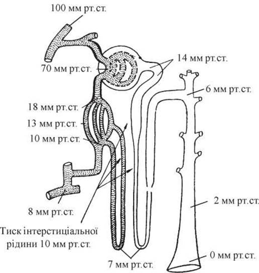
Fig. 11. Blood pressure in various segments of the nephron (after A.C. Guyton, 1971).
The Nervous system of the kidney is also quite complex in structure. Compared to other visceral organs, a greater number of nerve fibers penetrate the kidneys. Originating from the celiac plexus at the level of the 12th thoracic to 2nd lumbar vertebrae, they form the renal plexus (plexus radialis) at the renal hilum, which includes superior and inferior aortico-renal ganglia as well as small renal ganglia. The renal plexus is closely interconnected with the aortic and celiac plexuses and the lumbar sympathetic ganglia. The thoracolumbar Sympathetic trunk supplies the kidney with A large number of vasoconstrictor fibers, whereas parasympathetic fibers are far fewer in number (the Vagus nerve sends only a sparse number of branches to the kidney). Thus, the situation-appropriate vasomotor status of the kidney is primarily maintained through alterations in the vasoconstrictor activity of adrenergic nerves.
Adrenergic innervation predominates in the cortex; minor amounts of norepinephrine are found in the outer medullary layer, though even less is present in the inner medulla. Adrenergic nerves accompany interlobular and arcuate Arteries as well as afferent arterioles, enabling vasoconstriction via the contraction of vascular smooth muscle cells. These nerves are absent in the glomeruli, efferent arterioles, and tubules.
Cholinergic fibers are predominantly located in the region of the efferent arterioles of juxtamedullary nephrons and the sphincters of the vasa recta. Thus, medullar circulation and the effective control of intraglomerular pressure in juxtamedullary nephrons are exclusively associated with these vasodilatory fibers.
Renal hemodynamics are primarily controlled by alpha-adrenoceptors, whereas renin release is regulated by beta-adrenoceptors. Beta-adrenergic stimulation induces renal vasodilation, whereas alpha-adrenergic blockade causes vasoconstriction. Stimulation of both alpha- and beta-adrenoceptors is accompanied by the release of vasopressin and Erythropoietin. Intense stimulation of renal sympathetic nerves leads to a decrease in blood flow and urine filtration, whereas moderate stimulation results in decreased blood flow coupled with increased urine filtration. It is worth noting that a completely denervated kidney is capable of maintaining parameters such as GFR and renal plasma flow at appropriate levels, which aligns with METABOLISM/2.html">THE CONCEPT OF a high degree of autonomy in renal hemodynamics.
The kidneys play an active role in regulating the constancy of the internal environment by influencing the volume, ionic concentration, and Osmotic Pressure of extracellular fluid, acid-base balance (ABB), the metabolism of proteins, Lipids, CARBOHYDRATES, and vitamin D3, as well as erythropoiesis and systemic and Regional Blood Flow.
Modern understanding of Urine Formation mechanisms encompasses glomerular ultrafiltration, tubular reabsorption and secretion, and the renal Synthesis of specific compounds.
The initial stage of urine formation is the ultrafiltration of Blood Plasma. Currently, the glomerular filter is viewed as a selective system featuring barriers of increasing density. It comprises the capillary endothelium, the dense layer of the glomerular basement membrane (GBM), and the slit diaphragms of podocytes. In The Mechanism of ultrafiltration, special significance is attributed to the physicochemical state of the GBM (the magnitude of its negative charge) and the state of renal capillary blood flow, which under normal conditions establishes an additional (functional) barrier composed of high-molecular-weight Plasma Proteins—the so-called concentration-polarization layer. Reciprocal isometric Changes in the colloid viscosity of the outer GBM layer, the endothelial glycocalyx, and podocytes—which determine filtration pore size—along with steric conformity between the shape of the filtered substance and the filtration pores, alterations in GBM charge magnitude (congenital or acquired), and disruptions in intraglomerular pressure determine the glomerular filtration rate and filtrate composition under both normal and pathological conditions. The ultrafiltrate is virtually protein-free and has an electrolyte, glucose, urea, and creatinine concentration comparable to that of plasma. It contains only trace amounts of low-molecular-weight proteins (12–21 kDa), such as beta-2-microglobulin, Lysozyme, and retinol-binding protein. The total albumin concentration in the ultrafiltrate is 1 mg/L, compared to 40–50 g/L in blood plasma; an intact glomerular membrane allows no more than 0.1% of blood albumins to pass into the ultrafiltrate. The GFR volume averages 180 L/24 h, while the total protein content in the filtrate is 36 g/24 h. Incidentally, the total daily fluid filtration across all capillary membranes in the body, excluding glomerular membranes, amounts to only 4 L, with the filtered fluid returning to the central circulation via lymphatic vessels. We should also note that the glomerular filtration rate is a relatively stable value and remains virtually independent of systemic BP fluctuations within physiologically permissible limits. This stability is ensured by well-developed autoregulatory mechanisms, among which the juxtaglomerular apparatus plays a leading role, as previously mentioned. At the same time, the glomerulus (its vascular and mesangial components) is subject to the Influence of other locally and systemically produced bioactive substances, such as vasopressin, bradykinin, prostaglandins, norepinephrine, and acetylcholine.
The decisive factor driving glomerular ultrafiltration is the pressure gradient between the hydrostatic pressure generated by cardiac action and opposing forces. The latter include the colloid osmotic pressure of blood plasma (16–26 mmHg) and the hydrostatic pressure of fluid directly within Bowman's capsule (8–15 mmHg).
Thus, the net pressure gradient establishing the effective filtration pressure ranges between 8–12 mmHg, while the total GFR volume depends on the number of functioning glomeruli, the state of glomerular blood flow, and the permeability coefficient of the capillary wall.
The lumen of Bowman's capsule leads directly into the proximal tubule (which is 13–14 mm long within a total tubule length of 50 mm). This tubular segment is lined with a single layer of cuboidal epithelium whose cells feature a basally positioned Nucleus, numerous Mitochondria, and ribonucleic acid granules. The luminal surface of the cells is covered with microvilli that significantly expand the absorptive surface area. The microvillar zone is rich in alkaline phosphatase, ATPases, and 5'-nucleotidase, while the abundant mitochondria contain dehydrogenases, diaphorases, Hydrolases, and other oxidative Enzymes.
The ultrastructure and enzyme Chemistry of the cells in the principal tubular segment are adapted to perform the massive task of reabsorbing proteins, water, glucose, Amino Acids, electrolytes, and Vitamins. It is considered proven that all protein filtered in the glomeruli is reabsorbed in the proximal tubule (via an energy-dependent vacuolar-lysosomal reabsorption system). Consequently, various infections, intoxications, or genetically determined disorders play a critical role in precipitating acute or chronic blockade of this system, resulting in proteinuria and the development of Acute Kidney Injury (AKI).
The structure of the epithelium in the thin segment of the loop of Henle is adapted to minimize the filtration load of water and facilitate its "passive" reabsorption. The narrowest part of the tubule within the loop of Henle is lined with flattened, star-shaped epithelial cells. These cells are virtually devoid of oxidative enzymes, contain a limited number of mitochondria, and have intercellular spaces filled with supporting connective tissue. This specific epithelial structure limits reabsorption in this nephron segment and is closely linked to the operation of the countercurrent multiplier Mechanism of urine concentration (Fig. 12).
The operating principle of this mechanism was established back in the 1950s (H. Wirz et al., 1951), and our understanding of it has changed little since then. The kidney's ability to concentrate urine is intimately related to the Structural Features of the nephron, the arrangement of the loop of Henle, collecting ducts, and blood vessels.
The loop of Henle consists of two parallel limbs with opposite directions of urine flow. The Blood vessels of the medulla—the vasa recta—mirror the bends of the loop, and BLOOD FLOW IN the descending and ascending segments is likewise countercurrent. Functionally, this entire nephron architecture acts as a countercurrent multiplier system, which facilitates the generation of high solute concentrations through a small concentration gradient in each limb as fluid moves along the entire system. In juxtamedullary nephrons, located at the corticosomedullary junction, both limbs of the loop of Henle and their surrounding vascular branches descend deep into the medullary zones. Here they make a hairpin turn and HEAD back toward the cortex. Due to intensive sodium reabsorption in the loop of Henle and slowed blood flow in the vasa recta, a high osmotic sodium concentration is established in the medullary interstitium. This value progressively increases from the cortex toward the tip of the papilla.
Thus, the mechanism of urine concentration unfolds as follows: upon passing through the proximal tubule, an isotonic filtrate of reduced volume enters the loop of Henle. In this segment, intense sodium reabsorption occurs largely independently of water reabsorption, as the tubular wall is relatively impermeable to water. Consequently, the tubular fluid becomes diluted while the medullary interstitium accumulates a high sodium concentration. This diluted urine loses excess fluid in the distal tubule and becomes once again isosmotic to plasma. The reduced volume of isosmotic urine then enters the medullary collecting system, which is surrounded by a high osmotic pressure driven by elevated interstitial sodium. In the collecting ducts, antidiuretic hormone stimulates water reabsorption along the concentration gradient. The vasa recta traversing the medulla function as countercurrent exchangers—absorbing sodium on their descent toward the papillae and releasing it before returning to the cortex. Consequently, a high sodium content is maintained deep within the medulla, driving water reabsorption from the collecting system, which is The Essence of urine concentration. The energy required for the kidneys to perform this osmotic work is quite substantial; for instance, excreting 1 L of urine over 24 hours requires approximately 848 calories.
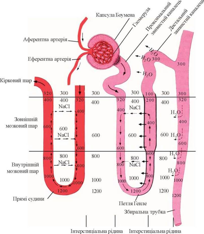
Fig. 12. Diagram of the countercurrent multiplier mechanism of urine concentration.
The distal tubule is lined with cuboidal epithelium featuring apically positioned nuclei, numerous cigar-shaped mitochondria, ribonucleic acid granules, and small vacuoles. The latter resemble Hydrochloric acid vesicles found in the Glandular Epithelium of the gastric mucosa. The distal tubular epithelium is rich in amino acids, basic and acidic ribonucleic acid proteins, Polysaccharides, and reactive SH-groups; it is also characterized by a high content of hydrolytic, glycolytic, and Krebs cycle enzymes. All these structures support the robust biochemical activity required to maintain high-level Ion Exchange, ammonium synthesis, and other vital processes. The Structural and functional specialization of the distal tubules, coupled with their heightened vulnerability to Hypoxia, explains why they are among the first to suffer damage (up to necrosis) under The Influence of toxic acidic urine products.
Overall, the total length of the tubules reaches 100 km, while the surface area of the tubular epithelium spans 50–60 m2.
In the region of the collecting ducts, the cuboidal epithelium gradually transitions into a columnar epithelium (comprising light and dark cells with a well-developed basal labyrinth and high water permeability), containing relatively few cellular Organelles. Dark cells are associated with hydrogen ion secretion. Nephrocytes in this nephron segment secrete prostaglandins, and their cell membranes can alter permeability in response to antidiuretic hormone.
Thus, the specific architecture of the tubular epithelium and the high enzymatic activity of intracellular structures ensure the selective reabsorption or secretion of various elements to preserve homeostasis (Fig. 13).
It is worth noting that The excretion of certain metabolic end products and exogenous organic substances occurs primarily via tubular secretion. For instance, cells of the proximal nephron secrete Bile acids, uric acid, cAMP, adrenaline, acetylcholine, histamine, serotonin, iodopyracet, indomethacin, penicillin, furosemide, and ethacrynic acid into the tubular lumen.
Kidneys play a paramount role in regulating water-electrolyte balance, particularly sodium and potassium homeostasis. Sodium and potassium are the primary electrolytes whose distribution between intracellular and extracellular fluids is crucial for maintaining the normal osmotic concentration of the extracellular fluid. The concentration of sodium in plasma and extracellular fluid is 130–150 mmol/L, and in intracellular fluid 11–13 mmol/L; for potassium, these values are 4.0–4.5 mmol/L and 140–150 mmol/L, respectively. Normally, the total osmotic pressure of intra- and extracellular fluids is equal, transmembrane fluid movement is absent, and fluid redistribution between body compartments occurs only when extracellular sodium concentration changes. Normal extracellular sodium levels are regulated by the thirst mechanism—which drives fluid intake—and antidiuretic hormone secretion, which governs fluid excretion.
The kidneys maintain sodium balance by controlling urinary sodium excretion. These regulatory mechanisms are exceptionally complex and multifaceted. Freely filtered at the glomerulus, sodium is sequentially reabsorbed across all segments of the renal tubules According to the body's physiological needs. Therefore, sodium balance is typically evaluated through the ratio of glomerular filtration rate (GFR) to tubular reabsorption (glomerulotubular balance, James A. Schrier, 1999). Literature data (M. Shapiro et al., 1986; H.J. Reineck et al., 1993) indicate that despite The Importance of GFR, tubular reabsorption remains the primary driver of sodium homeostasis. This process is modulated by aldosterone, natriuretic Peptides (atrial natriuretic peptide and its analogs), physical factors operating in the proximal tubule (hydrostatic and oncotic pressure gradients across peritubular capillaries and the tubular lumen, magnitude and velocity of peritubular blood flow, capillary wall permeability), intrarenal blood flow redistribution, corticosteroids, estrogens, Growth Hormone, Insulin, progesterone, the kallikrein-kinin system, and prostaglandins (M. Cogan, 1990; T. Ferris, 1993). Among extrarenal factors, aldosterone—acting via the renin-angiotensin-aldosterone system (RAAS)—plays the most critical role in sodium reabsorption, even though quantitatively it regulates only about 2% of the total reabsorbed sodium. This is because its primary Site of Action is the principal cells of the cortical collecting ducts, a nephron segment reached only after roughly 90% of sodium has already been reabsorbed; thus, this site serves as the ultimate fine-tuning station for homeostatic sodium balance.
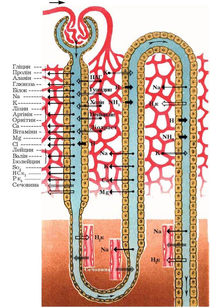
Fig. 13. Diagram of solute transport localization along the nephron. Arrows pointing into the tubular lumen denote secretion; arrows pointing outward denote reabsorption (Yu.V. Natochin, 1974).
A normal kidney filters approximately 23 mol of Cl- per day. Only 1% of this amount is excreted in the urine, while 99% of the filtered chloride ions are reabsorbed along the tubules: 50–60% in the proximal tubules, 5–10% in the distal segments, 20–30% in the thick ascending limb of the loop of Henle, and 2–5% in the collecting ducts. Chloride reabsorption mechanisms are heterogeneous, supported by the existence of specialized "chloride-secreting cells" that act as modulators of the reabsorption process.
Extracellular potassium levels are also tightly regulated, primarily by aldosterone. Potassium distribution is mainly maintained by Na+, K+-ATPase, a universal transport enzyme embedded in all cell membranes. Rising plasma potassium concentrations stimulate aldosterone secretion, whereas declining levels suppress it, forming a crucial feedback loop. By the end of the thick ascending limb of the loop of Henle, an average of 90% of potassium has been reabsorbed, while the final decision to retain or excrete potassium rests with the collecting ducts: when dietary potassium is high, these structures secrete it; when low, secretion ceases. Additionally, extrarenal factors—such as blood insulin levels and the beta-adrenergic system—participate in potassium homeostasis. The exact mechanism by which insulin drives potassium into cells is not fully understood, though it is hypothesized that insulin may activate membrane Na+, H+ pumps. Conversely, hyperkalemia stimulates insulin release, whereas hypokalemia inhibits it. Catecholamines promote cellular potassium uptake via beta-adrenergic receptor activation, whereas alpha-adrenergic stimulation yields the opposite effect. Finally, acidosis must be mentioned as a state that triggers potassium efflux from cells; a 0.1 shift in blood pH alters serum potassium concentration by 0.6 mmol/L.
The regulation of Calcium and phosphorus metabolism is even more complex, reflecting the vital role of calcium in physiological homeostasis (V.V. Kolomiets, 1990; V.O. Bobrov et al., 1994). This ion actively participates in nearly all intracellular processes, generation of cell membrane electrical potentials, activation of numerous intracellular enzymes, formation of novel biological structures, Fertilization, secretion, neuromuscular transmission, Muscle contraction, membrane permeability, and Intermediary Metabolism. Furthermore, calcium concentration dictates the contractile force of cardiomyocytes, vascular and bronchial tone, motility of the Uterus, gastrointestinal tract, and ureters, as well as the secretion of insulin, Pituitary Hormones, and other Endocrine glands, alongside platelet aggregation (V.A. Bobrov et al., 1992; R.D. Bukowsky et al., 1991; J.T. Repke et al., 1991; K.B. Knight et al., 1992). Consequently, blood calcium concentration—specifically ionized calcium (V.N. Titov et al., 1992), which dictates its physiological activity—represents one of the most stable parameters of electrolyte homeostasis.
Of the three distinct calcium fractions present in blood serum (ionized, comprising 44% of total calcium; complexed/chelated, 10%; and protein-bound, 46%), only ionized calcium and its chelated compounds can cross the glomerular filtration barrier into the primary urine. Notably, serum ionized calcium is not only a primary regulated variable in Calcium Homeostasis but also acts as a feedback factor regulating the secretion of calcium-controlling hormones.
The most crucial Hormonal regulators of blood calcium concentration are parathyroid hormone (PTH), Calcitonin, and 1,25(OH)2D3.
Parathyroid hormone (the hormone of the parathyroid glands) enhances calcium reabsorption in the distal tubules while suppressing the tubular reabsorption of sodium, phosphates, and several amino acids; it also inhibits hydrogen ion secretion and sodium-hydrogen exchange, leading to bicarbonaturia. Alongside locally synthesized tumor necrosis factor-alpha and interleukin-1, PTH promotes bone calcium resorption by activating osteoclasts, without significantly affecting intestinal calcium absorption. As renal disease progresses, prominent marrow fibrosis develops, eventually revealing the histological picture of osteitis fibrosa. Nevertheless, PTH simultaneously stimulates osteoclast activity (Sh. Massry, 1999). Thus, skeletal manifestations of excess PTH in patients with Chronic Kidney Disease (CKD) include increased numbers of osteoclasts and osteoblasts, accelerated osteoclastic bone resorption, enlarged Haversian canals, endosteal fibrosis, and accumulation of fibrous Bone tissue and osteoid.
In contrast to PTH, calcitonin (secreted by the parafollicular C-cells of The Thyroid Gland) lowers plasma calcium concentrations, thereby preventing hypercalcemia. Functioning as a physiological antagonist to PTH, calcitonin acts on specific outer Membrane Receptors to reduce the number and activity of osteoclasts in bone tissue, inhibiting PTH-induced osteocytic osteolysis. Calcitonin also exerts a phosphaturic effect by suppressing proximal tubular phosphate reabsorption.
Based on serum hormone levels, patients can generally be divided into two groups: the first exhibits normal alkaline phosphatase, high calcitonin, and relatively low PTH levels; the second displays high alkaline phosphatase and PTH levels alongside low calcitonin. These findings point to an inverse correlative feedback mechanism between PTH and calcitonin, likely mediated indirectly through blood ionized calcium concentrations (S.I. Ryabov et al., 2000).
Finally, a critical role in maintaining calcium homeostasis belongs to 1,25(OH)2D3, the most metabolically active form of vitamin D3. Normally, vitamin D3 is synthesized in the Skin or obtained from dietary sources. A fraction of this biologically inactive compound is converted within the microsomal and mitochondrial fractions of hepatic cells into 25-hydroxycholecalciferol (25(OH)D3), which is subsequently transformed into 1,25(OH)2D3 within the proximal convoluted tubules of the kidneys. The production of 1,25(OH)2D3 is co-stimulated by parathyroid hormone (via a cAMP-dependent pathway) and calcitonin (by upregulating 1-alpha-hydroxylase activity).
Vitamin D3 metabolites, which in healthy individuals are produced exclusively in the kidneys, primarily exert extrarenal effects: elevating blood calcium through enhanced bone resorption and intestinal absorption while reducing urinary calcium excretion. Vitamin D3 influences bone mineralization through multiple pathways—affecting collagen synthesis and maturation, and directly stimulating and/or elevating calcium and phosphorus levels in the extracellular fluid surrounding bone tissue (Sh. Massry, 1999).
It should be noted that along with data on the Biological Role of vitamin D3 in the regulation of Mineral Metabolism, reports have recently emerged regarding The Effect of its active metabolites on the status of immunocompetent cells (F.I. Rusnak et al., 1997). It has been proven, for example, that in infectious and allergic diseases, the expression of lymphocytic receptors for calcitriol is almost always detected, whereas among healthy individuals, such expression is determined in only 17 % of cases.
However, the role of The Kidneys in maintaining calcium homeostasis is not limited to this.
An important role in this process is played by the participation of the kidney in the metabolism of calcium-regulating hormones (G. Lehmann et al., 1987; J.H. Laragh et al., 1992). Under normal conditions, Parathyroid hormone and calcitonin are continuously filtered in the renal glomeruli, and the ultrafiltrate enters the lumen of the proximal tubules. There, these hormones are hydrolyzed into amino acids, and the latter are reabsorbed into the blood for their de novo synthesis. The active form of vitamin D3, like other Steroid Hormones, is also gradually inactivated in the kidneys, which creates the prerequisites for maintaining its constant blood level by changing The rate of the inactivation process. The situation changes dramatically in the event of renal failure (RF) (H. Saha et al., 1991; Y. Tsukatomo et al., 1991). A decrease in nephron mass leads to a reduction in the rate of hormone filtration, their inactivation, and subsequent accumulation in the blood, accompanied by a simultaneous decrease in the synthesis of 1,25(OH)2D3. As a result, severe disorders of calcium metabolism develop, leading to Osteomalacia, osteitis fibrosa, and osteosclerosis.
We should also note the adverse effect of acidosis on the calcium balance and the development of skeletal abnormalities. Acute acidosis causes a significant loss of acid-soluble calcium carbonate by the Skeleton. In stable RF with its inherent persistent hydrogen ion retention, the skeleton provides buffer reserves by depleting calcium carbonate stores.
Already at an early stage of Renal Dysfunction, as a result of a decrease in the filtration charge of phosphorus, hyperphosphatemia develops, which is accompanied by a reciprocal decrease in The amount of ionized calcium in the blood. The latter induces parathyroid hormone secretion and phosphaturia. Thus, at the cost of parathyroid hormone hypersecretion and increased bone resorption, the blood phosphorus level returns to normal. With a further decrease in GFR and an increase in blood phosphorus levels, parathyroid hormone secretion is re-induced.
The total amount of magnesium in the body is about 2000 mEq or 25 g. 60 % of this amount is located in the bones, 20 % in the muscles, and 20 % in other organs, Tissues, and extracellular fluid.
The kidneys filter about 2 g of magnesium per day, while its excretion is only 100-130 mg/day. It has been established that a reciprocal relationship exists between calcium and magnesium reabsorption—the administration of one of these electrolytes reduces the reabsorption of the other. The Use of mineralocorticoids, alcohol, and glucose increases magnesium excretion, with the ascending limb of the loop of Henle playing the leading role in this process; the role of the terminal nephron segments is minimal.
The main mechanism of renal acid-base balance (ABB) regulation is the tubular secretion of H+ ions, which ensures the reabsorption of bicarbonate and an equivalent amount of Na+. Maximum bicarbonate reabsorption (80-90 %) occurs in the proximal tubule; 5 % of bicarbonate is reabsorbed in the loop of Henle, and 3-4 % in the distal convoluted tubules. The secretion of H+ ions also plays an important role in the renal excretion of acidic products (phosphates, ammonia). The combination of these two processes—bicarbonate reabsorption and H+ secretion—ensures the constancy of blood pH (normally 7.35-7.43). A certain role in Blood pH Regulation belongs to aldosterone (facilitating H+ secretion by increasing the negative charge of the basement membrane and directly stimulating H+-ATPase), as well as parathyroid hormone, as mentioned above. It should be noted that the renal response to changes in blood ABB occurs much slower than the corresponding response from the Respiratory system.
Below is Table 1, which shows the filtration, reabsorption, and excretion values for major inorganic and organic substances.
Our knowledge regarding the active role of the kidneys in maintaining the physiological state of the hemostatic system has also expanded significantly.
Normal aggregate properties of blood are a vital prerequisite for the kidneys to perform their functions. Under normal conditions, the kidneys produce and release A number of procoagulant factors into the blood and urine (VII, VIII, IX, X, thromboplastin), and also synthesize and excrete anticoagulant factors such as heparin, urokinase, and tissue plasminogen activator. The kidneys absorb and catabolize a portion of fibrinogen and factor XIII, which participates in the synthesis of glomerular capillary basement membrane collagen. In pathological conditions involving the deposition of immune complexes and/or their formation *in situ* in the glomeruli, and the development of inflammation with enhanced transudation of procoagulant proteins, the functional contribution of the kidneys to hemostasis is lost on the one hand, while a process of local activation of the Blood Coagulation SYSTEM with intravascular hypercoagulation arises on the other. Under certain conditions, such local hypercoagulation can progress into a disseminated process.
The role of the kidneys in regulating hematopoiesis is also important. Hypochromic normocytic anemia is a classic symptom of renal dysfunction. Its Pathogenesis is complex and includes various links—enhanced hemolysis, a hypercatabolic state, and microhemorrhages (G.D. Shostka, 1997)—among which the impairment of renal erythropoietin production is assigned the leading role.
Erythropoietin is a glycosylated polypeptide with a Molecular Weight of 34,000, comprising 165 amino acids. Its carbohydrate moiety is responsible for preserving the biological activity of the hormone in the body, whereas the asialo form is rapidly degraded by hepatocytes. The primary stimulus for its production is a decrease in pO2 directly within the capillaries of the renal parenchyma, more precisely at the venous end of the capillary. For this reason, erythropoietin hyperproduction is associated not only with renal pathology, but also with Disorders of the blood, cardiovascular, and respiratory systems.
The First stage of the cascade reaction leading to enhanced erythropoietin synthesis is increased calcium influx into the prostaglandin-producing cells of renal structures—a direct stimulating effect of PGE2 on erythropoiesis in the Bone Marrow has been revealed. Erythropoietin production is also stimulated by Adrenal Cortex Hormones, thyreoidin, and androgens.
Receptor-stimulators of erythropoietin production (sensor cells) must always maintain close and constant contact with the blood. Such a zone in the kidneys is the cortex and the outer part of the medulla, where autoregulation mechanisms ensure a constant blood supply across a fairly wide range of blood pressure (from 70 mm Hg to 200 mm Hg). According to recent data, these receptor-stimulators are located in the endothelial cells of the peritubular capillaries of the proximal tubules in the cortical and medullary layers. Erythropoietin is most likely produced right there. Under conditions of pronounced hypoxia, erythropoietin can also be produced and inactivated by hepatocytes and Kupffer cells.
In the bone marrow, erythropoietin promotes the transition of unipotent erythroid committed progenitors of erythropoiesis into the erythron, the initial cell of which is the proerythroblast and the final cell is the erythrocyte.
Several directions of erythropoietin action at THE CELLULAR LEVEL are distinguished: stimulation of proliferation and maturation processes of early intermediate erythropoietin-sensitive cells, shortening of the intermitotic period, acceleration of the maturation of normoblasts and bone marrow reticulocytes, omission of one or more mitotic divisions, and a reduction in the proportion of so-called "ineffective" erythropoiesis.
Another important cause of anemia in renal pathology is hemolysis resulting from the accelerated destruction of erythrocytes in morphologically altered (fibrin deposition, dystrophic changes in the endothelium) arterioles and capillaries. Passing through such altered microvessels, erythrocytes become traumatized, lose their shape, and rapidly hemolyze.
We should also point out the altered iron metabolism in RF—its reduced intake when patients are on a low-protein diet, the toxic effect on heme synthesis, the metabolism of aluminum porphyrin from dialysis water, and parathyroid hormone (G.D. Shostka, 1999; A. Lach et al., 1995).
In recent years, data have appeared regarding the NEGATIVE IMPACT OF elevated serum levels of middle-molecule peptides (MMPs) on erythropoiesis (R.Z. Ismagilov et al., 1998; D.S. Rao et al., 1993).
Erythrocytosis occurs relatively rarely in kidney diseases. This state of hematopoiesis is characteristic of Renal Tumors, in particular hypernephroid carcinoma, renal cysts, Hydronephrosis, and Polycystic Kidney Disease. The explanation for this is found in the increased production of erythropoietin in these forms of pathology.
Urine transport during The final stage of excretion (from the kidney to the bladder) is also an active process (G. Mazhdrakov et al., 1980). The movement of urine is ensured by the contraction of muscle fibers located in the walls of the Urinary Tract, and this active process begins at the level of the renal pelvis.
Four small muscles are distinguished in the musculature of the calyces and pelvis: *m. levator fornicis*, *m. sphincter fornicis*, *m. longitudinalis calycis*, and *m. sphincter calycis*. The activity of these muscles is strictly coordinated, which ensures normal urine passage. During the so-called diastolic phase, *m. levator fornicis* and *m. sphincter fornicis* relax, while the upper part of *m. longitudinalis calycis* is in a contracted state, and *m. sphincter calycis* separates the caliceal cavity from the pelvis. As a result, a negative pressure is created in the peripapillary space, which facilitates the "suction" of urine from the kidney. This is followed by caliceal systole, during which *m. levator fornicis* and *m. sphincter fornicis* contract while *m. longitudinalis calycis* and *m. sphincter calycis* relax simultaneously. Caliceal filling (diastole) lasts 30–60 s, and emptying (systole) lasts 1–3 s.
Through this mechanism, urine is injected from the kidney into the pelvis in small portions. While the relaxed pelvis is filling, the most distal part of its neck remains closed. Subsequently, with the simultaneous opening of the pelvic neck and contraction of the muscles in its wall, a specific portion of urine enters the proximal section of the ureter.
A urine-filled, pointed "spindle" is formed in the ureter. This "spindle" moves along the ureter at a speed of 2.5–3 cm/s. Only one "spindle" passes through the ureter at a time, which is ultimately propelled into the urinary bladder during the final stage. When necessary (increased diuresis), the length and radius of the "spindles" increase and the intervals between their formation decrease; during very high diuresis (e.g., diabetes insipidus), physiological hypotonia of the ureters occurs.
As the urinary bladder fills, urine is voluntarily eliminated to the outside through the urethra.
Last update: 08/08/2026
Editorial and Educational Adaptation: This material has been compiled based on the primary/original source text. The project team performed an editorial review, corrected technical inaccuracies, structured sections, and adapted the content for an educational format.
What was processed:
- elimination of formatting defects (OCR errors, structural breaks, corrupted characters);
- editorial organization of content;
- standardization of terminology in accordance with academic sources;
- verification of factual statements against the original source text.
All mentions of the author, publication year, and origin of the primary text have been preserved in accordance with the source.