FUNDAMENTALS OF ENZYME STRUCTURE AND KINETICS IN BIOLOGICAL SYSTEMS - O. A. Naumenko - 2017
3 Bioenergetics
3.1 METABOLISM, its Functions, and Regulation
3.1.1 Definition of Metabolism and its Functions in Living Systems, REGULATION OF METABOLISM
Metabolism is the totality of metabolic processes within an Organism. Through metabolism, the organism interacts with its environment, acquiring energy for vital life processes (Energy Metabolism) and building blocks for The formation of subcellular structures, Cells, Organs, Tissues, and the organism as a whole (plastic or anabolic metabolism).
Despite the immense diversity of living organisms on our planet, a clear unity of metabolism is evident. Overall, the general metabolism of all classes of Organic compounds follows the same fundamental principles, and the Metabolic pathways of many organic compounds are identical across All living organisms.
Two Main Pathways of metabolism are distinguished in living systems:
- anabolism, or plastic metabolism — the synthesis of compounds, which requires an input of energy;
- Catabolism, or energy metabolism — The breakdown of molecules with the release of energy, supplying The Cell with high-energy compounds.
These processes run concurrently and simultaneously in living systems; The regulation of metabolism at all Levels of Organization, from the cell to the whole organism, is carried out by protein Enzymes.
Depending on how they regulate metabolic pathways, all Enzymes can be divided into 3 major groups:
1) anabolic (regulating biosynthetic processes);
2) catabolic (catalyzing energy-yielding reactions);
3) amphibolic (regulating both processes).
Conclusion: thus, the regulation of metabolism within an organism essentially boils down to the Introduction/15.html">Regulation of enzyme Activity.
Functions of metabolism:
- supplying the organism with energy derived from the breakdown of energy-rich nutrients or through The conversion of solar energy;
- converting dietary molecules into precursors utilized by the cell for The Biosynthesis of its own macromolecules;
- assembly of macromolecular (Biopolymers) and supramolecular structures of the living organism, i.e., plastic and energetic maintenance of its Structure;
- Synthesis and degradation of Biomolecules that perform specific functions in the body (Membrane Lipids, intracellular messengers, and pigments).
Metabolism proceeds via the following MAIN TYPES OF Chemical Reactions:
- oxidation-reduction reactions;
- synthesis of compounds utilizing ATP energy;
- isomerizations;
- addition or removal of functional groups.
Since these reactions are enzymatic in nature, the types of chemical reactions correspond to the Classification of Enzymes.
An enzymatic chain of chemical reactions in which the products of one reaction become the substrate for the next is called a metabolic pathway (MP).
Some Metabolic pathways are linear, others are branched, and still others are cyclic.
A linear MP features a single starting material and a single end product, such as Glycolysis or the Electron Transport Chain.
A branched MP involves either generating multiple end products from a single precursor or converting several starting substances into a single product, such as The Tricarboxylic Acid Cycle or the urea biosynthesis cycle.
A cyclic MP occurs when one of the substances entering the metabolic pathway is regenerated through a series of sequential reactions.
Based on their carbon assimilation strategy, all Living organisms are divided into two groups:
- autotrophic (self-feeding), which assimilate atmospheric CO2 during Photosynthesis and use it to build all their organic compounds (photosynthetic Bacteria, green plants);
- heterotrophic (feeding on others), which derive carbon from complex organic compounds (cells of higher animals and most microorganisms)—meaning they feed on the metabolic byproducts of other organisms.
In the biosphere, autotrophs and heterotrophs drive the global Carbon and Oxygen cycles.
With respect to oxygen requirements, heterotrophs are classified into:
- aerobes (requiring oxygen);
- anaerobes, which do not require oxygen to oxidize nutrients;
- facultative anaerobes, which can thrive in both oxygen-rich and oxygen-depleted environments;
- obligate anaerobes, which live exclusively in oxygen-free environments.
As for nitrogen, organisms require it for the synthesis of Amino Acids and NUCLEOTIDES.
Metabolic Regulation is achieved by modulating enzyme activity. Changes in enzyme levels occur through Gene induction or repression, as well as via intracellular proteolytic degradation.
Enzymes whose levels remain relatively constant in a living organism are called constitutive, whereas those whose concentrations fluctuate depending on the metabolic state are termed adaptive or inducible.
Another key mechanism of metabolic regulation is the alteration of membrane permeability. Changes in membrane function lead to shifts in the rates of metabolite and gas fluxes, Electrochemical Potential, Nerve Impulse transmission, and receptor signaling.
In The Human Body, metabolic coordination is carried out by the nervous and endocrine systems.
3.1.2 Phases of Metabolism
Metabolism comprises a combination of opposing Catabolic and anabolic processes, which possess several distinct characteristics (Table 3.1)
Class="center">Table 3.1 - Features of Catabolic and Anabolic Processes
Catabolism |
Anabolism |
1. Breakdown (degradation) |
1. Biosynthesis |
2. Oxidation |
2. Reduction |
3. Energy release |
3. Energy consumption |
4. Various starting compounds yield identical end products |
4. Identical starting compounds yield various end products. |
Although catabolic and anabolic pathways differ in many respects, they are closely interrelated. At different stages of life, either anabolic or catabolic processes predominate.
3.2.1.1 Metabolic cycles. Some metabolic pathways are characterized by the regeneration of one of the starting metabolites at the end of the process, which closes the pathway into a circular, cyclic process. Such closed metabolic pathways are referred to as metabolic cycles (H —> H). Examples of cyclic metabolic pathways include the tricarboxylic acid cycle, The Glyoxylate cycle, the Ornithine Urea Cycle, the Cori cycle, and The Calvin Cycle, among others.
The existence of metabolic cycles has a profound biological significance, as they carry out specific biochemical reactions with extreme economy, repeatedly utilizing the same amount of the starting compound, which is regenerated with each turn of the cycle and re-enters the process.
Individual reactions within a metabolic pathway (cycle) are called stages, links, or steps of metabolism.
Metabolic pathways can intersect. The intersection point of metabolic pathways is called a metabolic branch point (or node), and the corresponding metabolite through which different metabolic pathways pass is referred to as a branch-point (or key) metabolite (D). For instance, glucose-6-phosphate, acetyl-CoA, and others serve as branch-point (key) metabolites, allowing metabolism to switch from one pathway to another.
Very often, the Formation of the same metabolite can occur via two or more different pathways.
3.1.2.2 Primary and secondary metabolic pathways. A metabolic pathway with a greater metabolic capacity is called the primary (or major) metabolic pathway (A —> B —> C —> D), whereas other pathways with lower metabolic capacity are classified as secondary (or alternative) metabolic pathways (A —> M —> N —> D).
Side pathways of metabolism are defined as those that branch off from a common metabolite of the primary pathway but lead to the formation of a different (secondary) end product (metabolite) (E —> O —> P).
Regulatory (rate-limiting) steps. The intensity of metabolic flux, or the metabolic capacity of a given pathway, depends on its bottleneck—the so-called "rate-limiting step," which is the step possessing the lowest metabolic capacity.
To increase (enhance) metabolic flux, there is no need to alter every step of the metabolic pathway; modifying just one or a few is sufficient. As a rule, regulatory steps represent irreversible reactions. Most often, the regulatory step is the first enzymatic step of a metabolic pathway. Examples include the hexokinase reaction in The Glycolytic Pathway and the glucose-6-phosphate dehydrogenase reaction in the oxidative Pentose Phosphate Pathway. Through these rate-limiting steps, Nature has engineered the remarkable efficiency of metabolic regulatory systems.
Depending on The sequence of metabolic processes occurring in the organism, the sequence of metabolic events is divided into three stages.
1. Digestion AND ABSORPTION (resorption). During DIGESTION IN THE gastrointestinal tract, high-molecular-weight compounds undergo Hydrolysis to form simpler ones. This also includes the bacterial action of the gastrointestinal flora on dietary substances, which is of particular importance in ruminants. Subsequently, nutrients are absorbed through the intestinal mucosa and transported across membranes into cells.
2. Intermediary Metabolism — the totality of intracellular Chemical Reactions Involving synthesis and degradation, resulting in the formation of intermediate and End products of Metabolism.
3. Excretion of metabolic end-products.
3.1.3 Metabolic pathways and Stages of Nutrient energy extraction
Depending on The amount of energy released during the Breakdown and Oxidation of nutrients, H. Krebs identified three MAIN STAGES OF nutrient energy extraction.
In the first (preparatory) stage, large (polymeric) nutrient molecules are broken down into small units (monomers) within the gastrointestinal tract. Polysaccharides are broken down into Monosaccharides (primarily hexoses such as glucose, fructose, and galactose). Fats are split into glycerol and Fatty acids, and Proteins into amino acids. The end-products of this First stage comprise approximately 25–30 different chemical compounds (amino acids, monosaccharides, glycerol, and fatty acids). Only about 0.6–1% of the total nutrient energy is released at this point, which is solely dissipated as heat rather than utilized for other purposes. The Biological Significance of the chemical reactions in The first stage lies in preparing nutrients for subsequent energy release.
In the second stage (the initial phase of intermediary metabolism), the substances formed in the first stage undergo further breakdown—specifically, the degradation of polymers into intermediates. The outcome of these transformations may at first seem unexpected, since the numerous chemical compounds representing the end-products of the first stage are funneled into just three principal metabolites: 2-oxoglutarate, oxaloacetate, and acetyl-CoA, with acetyl-CoA being the most abundant. These secondary-stage metabolites play a central role in metabolism. This stage accounts for the release of 25–30% of the energy contained in the nutrients.
The Third Stage (the final phase of intermediary metabolism, characterized by degradation into CO2 and H2O with the participation of oxygen) includes the tricarboxylic acid cycle and The electron transport chain—the ultimate pathways for The oxidation of nutrients into CO2 and H2O. It is during this third stage that the remaining 70–75% of nutrient energy is released and captured within the phosphate bonds of ATP.
The tricarboxylic acid cycle and the electron transport chain, coupled with Oxidative Phosphorylation, serve as the common central highways of material and energy metabolism through which CARBOHYDRATES, lipids, and proteins are processed.
Review Questions for the topic
4. Define metabolism.
5. What are the primary functions of metabolism?
6. How is metabolic regulation achieved?
7. Catabolism and anabolism, and their interrelation.
8. The sequence of metabolic processes and stages of nutrient energy extraction.
9. WHAT IS A metabolic pathway?
10. What are the types of metabolic pathways?
11. What is a branched metabolic pathway?
12. What is the term for a common intermediate shared by different metabolic pathways?
13. What is The Role of acetyl-CoA in biochemical reactions?
3.2 Biological Oxidation (Tissue Respiration)
3.2.1 Definition of biological oxidation. Pathways of oxygen utilization
The transfer of a phosphate group to ADP yielding ATP is the primary reaction of Bioenergetics. The source of energy for this reaction is the electron flow driven by oxidation-reduction reactions.
In non-photosynthetic organisms, the source of electrons is reduced components (food); in photosynthetic organisms, electron Donors are chemical molecules that transition to an excited state under The Influence of light.
Biological oxidation, or tissue respiration, is a set of oxidation-reduction processes taking place in living cells, whose main function is to supply the organism with energy in a usable form and reducing equivalents for biosynthetic processes.
A substance is oxidized when it loses electrons, or simultaneously loses electrons and protons (hydrogen atoms, dehydrogenation), or gains oxygen (oxygenation). The opposite transformations are reduction processes.
The ability of molecules to donate electrons to another molecule is characterized by the oxidation-reduction potential (redox potential, E°′).
The redox potential is determined by measuring the electromotive force in volts. The standard used is the Redox Potential of the reaction at pH 7.0: Н2 ⇄ -2Н+ + 2 е-, which is equal to 0.42 V.
Redox potentials are related to The change in Free energy ΔG°' by the Nernst equation:
∆G°’ = -zF x ∆Е°’
z is the number of electrons transferred in the reaction; F is the Faraday constant, ∆Е°’ is the difference in redox potentials of the electron-donor and electron-acceptor pairs.
In the 18th century, A. Lavoisier established that the respiration process is associated with the oxidation of substances and drew attention to the similarity between the combustion of organic matter and animal respiration. Consequently, the scientific community long held the view that biological respiration is a slow form of combustion.
A fundamental feature of tissue respiration, unlike combustion, is that oxidative processes in the organism occur at a low Temperature without abrupt changes and without the appearance of a flame, since energy is released not all at once, but in portions (stepwise).
The second feature is that during biological oxidation, Water is not only an end product, as in combustion, but also a direct participant in oxidative processes, which proceed more intensively in tissues with a high water content.
The third feature of biological oxidation is that the reaction supplying energy in living organisms, with minor exceptions, is the oxidation of hydrogen by oxygen, which under normal conditions proceeds with an explosion.
To explain the features of biological oxidation, several theories have been proposed.
The first of these was the oxygen activation theory (A. N. Bach, C. Engler, and J. Wild), followed by V. I. Palladin and H. Wieland, who put forward the hydrogen activation theory. The discovery of Cytochromes (D. Keilin, 1925), which remove electrons from hydrogen, and the enzyme cytochrome oxidase (O. Warburg, 1928), which receives electrons from hydrogen, made it possible a few years later to formulate the modern theory of biological oxidation (or tissue respiration), known as the electron transport chain, which is largely based on the two preceding theories.
When studying oxidative processes in cells, it is advisable to follow the following scheme of oxygen utilization (Table 3.2):
Table 3.2 - Pathways of oxygen utilization in cells
Substrate oxidation |
||||
Dehydrogenation |
Oxygenation |
Free-radical oxidation |
||
-2Н |
-2Н |
+1/2 O2 |
+ O2 |
O'- |
to 1/2 O2 |
to O2 |
+1/2 O2 |
+ O2 |
НО'- NO' ONOO- O3 |
Н2O |
Н2O2 |
R-OH |
RO2 |
|
Tissue respiration |
Simple oxidation systems |
Monooxygenase pathway |
Dioxygenase pathway |
|
ATP |
detoxification |
detoxification |
Cleavage of aromatic rings |
|
Heat |
Heat |
|||
There are three main pathways for oxygen utilization:
1) substrate oxidation via dehydrogenation, involving the transfer of two hydrogen atoms to an oxygen atom to form H2O (the oxidation Energy is stored as ATP, accounting for over 90% of oxygen consumption) or to an oxygen molecule to form H2O2;
2) Addition of an oxygen atom to form a hydroxyl group or an oxygen molecule;
3) generation of oxygen free radicals, which serve both to protect the cell's internal environment from foreign molecules and to damage membranes during oxidative stress.
3.2.2 Stages of Tissue Respiration
In biochemistry and cell biology, tissue (cellular) respiration refers to the redox processes through which cells consume oxygen and release carbon dioxide.
In aerobically respiring cells, oxygen serves as the final electron and proton acceptor. Consequently, tissue respiration in aerobic cells proceeds in three main stages.
1. Formation of Acetyl-CoA from organic molecules (glucose, fatty acids, amino acids) through their oxidation.
2. Acetyl-CoA enters the tricarboxylic acid cycle, where its acetyl group is enzymatically oxidized to CO2, releasing HS-CoA. The energy released during oxidation is stored in reduced coenzyme carriers (NAD, FADH2).
3. Electrons are transferred to O2 as the final acceptor via a sequential chain of enzyme electron carriers, known as the Respiratory Chain or electron transport chain (ETC).
The transfer of electrons along the respiratory chain releases a large amount of energy, which is used to synthesize ATP via oxidative phosphorylation.
Tissue respiration is evaluated using the respiratory quotient (RQCO2):
RQCO2 = moles of CO2 produced / moles of O2 consumed.
This metric helps determine the type of fuel molecules utilized by the organism: Complete oxidation of carbohydrates yields a respiratory quotient of 1, proteins 0.8, fats 0.71, and a mixed diet yields an RQCO2 of 0.85.
The ETC is embedded in The inner mitochondrial membrane. Electrons are passed down the chain from more electronegative components to more electropositive oxygen: from NADH (-0.32 V) to oxygen (+0.82 V).
3.2.3 Structural Features of Mitochondria
The outer mitochondrial membrane is permeable to many molecules and ions (voltage-dependent anion channels) because it contains mitochondrial porins—proteins with a Molecular Weight of 30 to 35 kDa (atomic mass units, daltons (Da)).
Functional components of mitochondria:
1) the outer mitochondrial membrane (delimiting the internal space), permeable to O2 and various low-molecular-weight substances;
2) contains lipid and monoamine metabolism enzymes;
3) the intermembrane space (IMS) contains adenylate kinase and ADP phosphorylation enzymes not associated with the respiratory chains;
4) the inner mitochondrial membrane (IMM) contains proteins, 20–25% of which are enzyme proteins involved in the proton-Electron Transport Chain and Oxidative Phosphorylation; it is permeable only to small molecules (oxygen, urea) and contains specific transmembrane carriers;
5) the matrix contains Enzymes of the tricarboxylic acid cycle, β-Oxidation of Fatty acids, and enzymes for mitochondrial DNA, RNA, and Protein Synthesis.
Review Questions for the Topic
1. Pathways of oxygen consumption (biological oxidation).
2. THE CONCEPT OF redox potential.
3. The Nernst equation.
4. Pathways of oxygen utilization in oxidative processes.
5. The respiratory quotient.
6. Structural Features of the mitochondrial membrane.
7. What are the respiratory quotients for proteins and carbohydrates?
8. What is tissue respiration?
9. Name the three main stages of tissue respiration.
10. Where is the ETC located?
3.3 Respiratory Chains
3.3.1 Types of Electron Transfer in the Respiratory Chain
The mitochondrial respiratory chain consists of protein enzymes that sequentially transfer electrons.
There are Three types of electron transfer:
1) direct electron transfer, for example, through the REDUCTION OF Fe3+ to Fe2+;
2) transfer of hydrogen atoms (H+ and e-);
3) transfer of a hydrogen hydride ion (H)-, which contains two electrons. Electrons for the respiratory chain are generated by the action of
dehydrogenase enzymes—pyridine-dependent dehydrogenases and flavin enzymes—which collect electrons from catabolic substrates and accumulate them in universal electron acceptors, namely nicotinamide nucleotides (NAD+ or NADP) or flavin nucleotides (FMN+ or FAD+).
3.3.2 Components of the Respiratory Chain
The respiratory chain is part of the oxidative phosphorylation process. Its components catalyze the transfer of electrons from NADH+ and H+ or reduced ubiquinone (QH2) to molecular oxygen. Due to the large difference in redox potentials between the donors (NADH+ and H, QH2) and the acceptor (O2), the reaction is highly exergonic. Most of the energy released in the process is used to generate a proton gradient and, ultimately, to synthesize ATP via ATP synthase.
The respiratory chain includes three Protein Complexes (complexes I, III, and IV) embedded in the inner mitochondrial membrane, along with two mobile carrier molecules: ubiquinone (coenzyme Q) and cytochrome c. The enzyme succinate dehydrogenase (SDH), which strictly belongs to the citrate cycle, is considered complex II of the respiratory chain (Figure 3.1). ATP synthase is designated as complex V, although it does not directly participate in electron transport.
Figure 3.1 - STRUCTURE OF THE respiratory chain
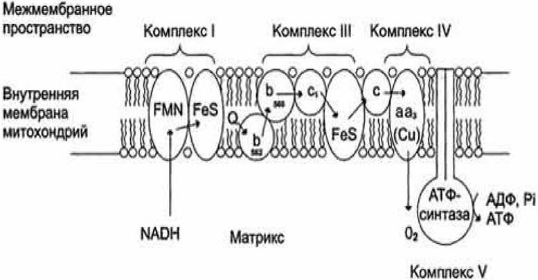
The complexes of the respiratory chain are composed of multiple Polypeptides and contain various protein-bound redox Coenzymes. These include flavins (FMN or FAD in complexes I and II), iron-sulfur centers (in complexes I, II, and III), and heme groups (in complexes II, III, and IV). The structure of the complexes is presented in Table 3.3.
Different types of Hemes covalently bound to proteins take part in electron transfer. Heme b types correspond to those found in Hemoglobins. In complex IV, a copper ion (CuB) and heme a3 interact directly with oxygen.
Table 3 - Complexes of the Mitochondrial Electron Transport chain
Name of enzymatic complexes |
Molecular weight (kDa) |
Number of subunits |
Prosthetic groups |
INADH dehydrogenase (NADH-CoQ oxidoreductase) |
850 |
42(14) |
FMN, FeS |
II succinate dehydrogenase |
140 |
5 |
FAD, FeS |
III ubiquinone:cytochrome c oxidoreductase |
250 |
11 |
Hemes, FeS |
cytochrome c complex |
13 |
1 |
Heme |
IV cytochrome c oxidase (cytochrome c oxidase) |
160 |
13 (3-4) |
Hemes, CuA CuB |
Note: cytochrome c is not a part of the complex; cytochrome c is a soluble protein that shuttles between complexes III and IV.
3.3.3 Types of Oxidizable Substrates
Oxidation substrates are molecules that undergo dehydrogenation (lose 2H) upon oxidation. There are three types of substrates:
1) Type 1 substrates (hydrocarbon-based) — succinate, acetyl-CoA. The average cleavage energy for an e- pair is about 150 kJ/mol. This is lower than the energy of e- in the NADH coenzyme. For this reason, NAD cannot participate in the dehydrogenation of these substrates;
2) Type 2 substrates (alcohol-based). Their dehydrogenation yields ketones. The average cleavage energy for an electron pair is about 200 kJ/mol, which allows NAD to participate in the dehydrogenation of Type II substrates;
3) Type 3 substrates (aldehyde-based). The cleavage energy for an e- pair is about 250 kJ/mol. Dehydrogenases of Type III substrates often contain multiple coenzymes, with a portion of the energy being stored prior to the electron transport chain.
3.3.4 Function of Respiratory Chain Enzymes
Electrons from second- and third-type donor substrates enter the respiratory chain via various pathways. During the oxidation of NADH+ and H+, complex I transfers electrons through FMN and FeS centers to ubiquinone. Electrons generated during the oxidation of succinate, acyl-CoA, and other first-type substrates are transferred to ubiquinone by complex II or another mitochondrial dehydrogenase via an enzyme-bound FADH2 or flavoprotein. In this process, the oxidized form of coenzyme Q is reduced to aromatic ubiquihydroquinone. The latter transfers electrons to complex III, which delivers them via two hemes b, one Fe/S center, and heme c1 to a small heme-containing protein, cytochrome c. Cytochrome c then transfers electrons to complex IV, cytochrome c oxidase. To carry out redox reactions, cytochrome c oxidase contains two copper-containing centers (CuA and CuB) and hemes a and a3, through which electrons finally reach oxygen. The reduction of O2 yields a strong basic anion O2-, which binds two protons and is converted into water. The electron flow is coupled with the proton gradient generated by complexes I, III, and IV.
Thus, complexes I and II catalyze the transfer of electrons from various donors: complex I from NADH and complex II from succinate; complex III transfers electrons from ubiquinone to cytochrome c, while complex IV transfers electrons from cytochrome c to oxygen.
Proton translocation by complexes I, III, and IV proceeds vectorially from the matrix into the intermembrane space. During electron transfer along the respiratory chain,
the concentration of H+ ions increases, i.e., the pH decreases. In intact mitochondria, essentially only ATP synthase enables the reverse movement of protons back into the matrix. This underlies the regulatory coupling of electron transport with ATP synthesis.
Due to its nonpolar side chain, ubiquinone moves freely within the membrane. The water-soluble cytochrome c is located on the outer side of the inner membrane.
The oxidation of NADH (NADH) by complex I occurs on the inner side of the membrane as well as in the matrix, which is also the site of The Citric Acid Cycle and β-oxidation—the most important sources of NADH. Furthermore, the reduction of O2 and the formation of ATP take place in the matrix. The synthesized ATP is transported via an antiport mechanism (against ADP) into the intermembrane space, from where it enters the Cytoplasm through porin proteins.
The change in free energy ∆G in reduction reactions depends solely on the difference in redox potentials between the donor and acceptor. The total reaction energy (over 200 kJ/mol) is broken down into smaller, more manageable "packets," the magnitude of which is determined by the redox potential differences of the respective intermediates. This provides the respiratory chain with a remarkably high energy yield of approximately 60%.
3.3.5 Complete and Shortened Respiratory Chains
Depending on the type of oxidation substrate (i.e., the cleavage energy of the e- pair), complete and shortened respiratory chains (Electron Transport Chains - ETC) are distinguished.
The ETC is a universal conveyor system for transferring protons and electrons from oxidation substrates to oxygen. Type II and III substrates enter the complete ETC, whereas Type I substrates enter the shortened ETC. Pyridine-dependent dehydrogenases are absent in the shortened ETC.
Complete respiratory ETC. Substrates of the second and third types transfer protons and electrons (i.e., undergo dehydrogenation) to the NAD coenzyme of pyridine-dependent dehydrogenases (the first ETC complex). The NAD coenzyme is not tightly bound to the dehydrogenase and, following substrate dehydrogenation, freely diffuses to the inner mitochondrial membrane.
NADH dehydrogenase is localized in the inner mitochondrial membrane and contains two prosthetic groups: FMN and FeS proteins. FMN participates in the transfer of two hydrogen atoms, after which FeS proteins accept 2e-, while the remaining 2H+ are pumped into the intermembrane space and "await" the moment when 2e- negatively charge the oxygen atom. In other words, proton and electron streams diverge at the NADH dehydrogenase stage.
Incomplete (shortened) respiratory ETC. This begins with first-type substrates. Their dehydrogenation occurs via a CoQ-ferment succinate dehydrogenase containing FAD and bound to the inner mitochondrial membrane. FAD attaches two hydrogen atoms from the substrate, then FeS accepts 2e-, two Protons are pumped into the matrix, and electrons are subsequently transferred to oxygen, just as in the complete ETC.
3.3.6 Properties of Coenzyme Q and Cytochromes
Two electrons are transferred to ubiquinone CoQ. Human tissues contain CoQ10, which possesses a side chain of 10 isoprene units. CoQ can move within the lipid phase of the membrane and transfer 1e- or 2e- to the cytochrome chains. Up to CoQ, electron transfer is two-electron; after CoQ, it is single-electron.
Each molecule of reduced CoQ transfers an electron to the cytochrome chain. (Consequently, two cytochrome chains must participate thereafter.) Cytochromes are arranged according to their redox potentials. These are heme-containing enzymes whose prosthetic groups consist of heme and heme-like structures. Heme derivatives of protoporphyrin IX—the heme of Hemoglobin and the heme of cytochrome b—are structurally similar.
Cytochromes differ in:
1) by the radicals in the porphyrin ring;
2) by the structure of the apoenzyme;
3) by the type of bond between the prosthetic group and the apoenzyme.
In cytochrome b, the iron atom is attached via its fifth and sixth coordination bonds to the nitrogen atoms of the Histidine imidazole rings of the apoenzyme. In cytochrome c, the fifth bond is linked to histidine, and the sixth to Methionine of the apoenzyme. During electron transfer, the valence of the iron atom changes from Fe3+ to Fe2+. HCN, H2S, and CO can bind to the sixth coordination bond of cytochrome a iron. In this case, the valence of iron (Fe) becomes fixed, and the flow of electrons ceases. This is the MECHANISM OF ACTION of Respiratory Chain Inhibitors.
The complex of cytochromes a + a3 is called cytochrome c oxidase. Cytochrome a3 contains copper atoms. Electrons are accepted by the subunits of cytochrome a and transferred to cytochrome a3, which passes them on to oxygen. This transfer is accompanied by a valence change of copper from Cu2+ to Cu1+. The oxygen atom acquires a negative charge and gains The ability to interact with protons to form H2O.
Review Questions for the Topic
1. Name the five main complexes of the respiratory chain.
2. Which complexes are embedded in the mitochondrial membrane?
3. Which complexes are located in the intermembrane space?
4. What are the structural features of the first complex?
5. How does the respiratory chain function?
6. What types of substrate oxidation are distinguished?
7. Which substrates belong to the first type?
8. What is the full respiratory chain?
9. What is the shortened respiratory chain?
10. Types of electron transfer.
11. What are cytochromes?
3.4 Oxidative Phosphorylation
3.4.1. Concept of Oxidative Phosphorylation
The synthesis of ATP from ADP and inorganic phosphate, coupled with the transfer of protons and electrons along the respiratory chain from substrates to oxygen, is called oxidative phosphorylation.
The oxidation energy sufficient for the formation of an ATP molecule is released in the respiratory chains at the following sites:
1) NADH dehydrogenase;
2) ubiquinone:cytochrome c oxidoreductase;
3) cytochrome c oxidase.
The energy is sufficient for the formation of a high-energy ATP bond (30.2 kJ/mol). The decrease in free energy accompanying the transfer of protons and electrons to oxygen As a result of a single dehydrogenation is approximately 220 kJ/mol. Meanwhile, the synthesis of ATP in the full respiratory chain can utilize 30.2 × 3 = 90.6 kJ/mol. Hence, the efficiency of the electron transport chain is about 40%. The remaining energy is dissipated as heat (maintaining body temperature).
There are three main hypotheses regarding oxidative phosphorylation.
The mechanochemical, or conformational hypothesis (Green & Boyer, 1960s). The transfer of protons and electrons induces a conformational change in enzyme proteins. They enter a high-energy state and subsequently return to their original conformation, releasing energy to drive ATP synthesis. This hypothesis has been partially validated: Eox —> Econformational shift —> EATP.
The chemical coupling hypothesis (Lipmann, Slater, Lehninger, 1930s–1940s). Coupling agents, such as substance X, participate in linking respiration and phosphorylation. Substance "X" accepts protons and electrons from the first enzyme at the coupling site and interacts with CH3PO4. Upon transferring protons and electrons to the second enzyme of the coupling site, the bond becomes high-energy. This high-energy group is then transferred to ADP, yielding ATP.
Mitchell's chemiosmotic hypothesis (1961). Peter Mitchell proposed that ATP synthesis is driven by a proton gradient across the inner mitochondrial membrane. In this process, Electron transport along the respiratory chain components of the inner mitochondrial membrane drives proton translocation across the inner mitochondrial membrane to its cytosolic side. As a result, an electrochemical gradient is established across the inner mitochondrial membrane.
According to modern understanding, respiration and phosphorylation are coupled via the electrochemical potential (proton-motive force) across the inner mitochondrial membrane.
To understand this mechanism, the following concepts are required:
a) the inner mitochondrial membrane is impermeable to H+ and OH-;
b) ATP synthase (Complex V) is embedded in the inner mitochondrial membrane, catalyzing the reversible reaction:
АТФ + Н2O ⇄ АДФ + Р;
c) ATP synthesis occurs as protons flow through ATP synthase from the intermembrane space (IMS) to the matrix.
The core Mechanism of Oxidative phosphorylation: powered by the energy of electron transfer within the electron transport chain (Eoxidation), protons are pumped across the membrane into the mitochondrial intermembrane space, generating an electrochemical potential (Epmf). As protons flow back through ATP synthase, the energy of the electrochemical potential is transformed into bond energy: ATP — a phosphoric acid residue.
Respiratory chain complexes: NADH dehydrogenase, ubiquinone:cytochrome c oxidoreductase, and cytochrome c oxidase pump protons into the intermembrane space. Protons are sourced from matrix H2O or generated via Conformational Changes in the enzymes. The matrix side of the membrane develops a negative charge (an excess of OH-), while the intermembrane space side becomes positive (due to H+). This results in an electrochemical potential comprising two components:
- a chemical component (due to the concentration gradient of H+ ions);
- an electrical component (due to the electrical potential difference ΔμH+ = Δφ + ΔpH). This value has been measured and is equal to -0.14 V.
ATP is synthesized during the reverse flux of protons through the ATP synthase channel (membrane discharge).
3.4.2 Structure of ATP Synthase
ATP synthase consists of the following components:
- F0 (oligomycin-sensitive) — a hydrophobic segment composed of 13 polypeptide chains, integrated into the inner mitochondrial membrane;
- F0 functions as a proton channel through which, under normal conditions, only protons can traverse the membrane;
- F1 (the coupling factor identified as essential for oxidative phosphorylation) — a peripheral membrane protein that catalyzes ATP Synthesis Coupled to proton translocation.
The mitochondrial F1 component consists of 9 subunits of five types: α, β, and γ. Each β subunit contains a single catalytic site for ATP synthesis. The catalytic HEAD of the F1 component forms a sphere 8 nm high and 10 nm wide, consisting of α and β subunits arranged like orange segments (Figure 3.1). The polypeptides forming the F1 stalk are arranged asymmetrically: one domain of the γ subunit forms the central shaft passing through F1, while the second domain of the γ subunit interacts with one of the three β subunits, designated as the "empty" β subunit.
Figure 3.1 - Structure of the ATP synthase enzyme
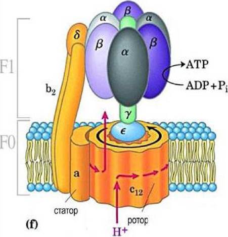
Although the Amino acid sequences of the three β subunits are identical, they differ in conformation; consequently, the γ subunit can bind to only one β subunit at a time. The Conformations of the β subunits are designated as β-ATP, β-ADP, and empty β, reflecting the nucleotides currently bound to them. This is critical for The Mechanism of action of the complex.
The F0 component forms the proton channel and consists of three types of subunits: a, b, and c. The c subunit is a small hydrophobic polypeptide comprising two transmembrane helices located almost entirely within the membrane, with a small loop protruding from the matrix side of the membrane.
3.4.3 Mechanism of ATP Synthesis
The mechanism of ATP synthesis was proposed by P. Boyer and involves three stages resulting in a conformational change of the β-subunit:
1) the β-subunit is in the β-ADP conformation and binds ADP and inorganic phosphate;
2) the subunit undergoes a conformational change that produces the β-ATP form, which tightly binds and stabilizes ATP;
3) the subunit shifts to the β-empty conformation, which has a low affinity for ATP, releasing the newly synthesized molecule from the enzyme surface.
The next cycle begins when the β-subunit transitions back to the β-ADP conformation and binds ADP and inorganic phosphate.
Conformational changes are driven by the movement of protons through the F0 component of ATP synthase. The flow of protons through the F0 channel causes the central stalk of the γ-subunit to rotate.
Thus, a full Rotation of the γ-subunit causes each β-subunit to pass through all possible conformational states, yielding 3 molecules of ATP per one complete head rotation.
3.4.4 Quantitative evaluation of Oxidative Phosphorylation
To quantitatively express oxidative phosphorylation, a coefficient is introduced, which represents The ratio of the number of inorganic phosphate molecules incorporated into ATP during respiration to each atom of oxygen consumed.
The question arises: how many protons are required for the synthesis of 1 molecule of ATP? Synthesizing 1 molecule of ATP requires 4H+, one of which is used to transport Pi, ATP, and ADP across the mitochondrial membrane. Consequently, the P/O ratio is 2.5 (10/4).
3.4.5 Respiratory Control
Respiratory control is the Regulation of the electron transfer rate along the respiratory chain by the ATP/ADP ratio. The lower this ratio (meaning ADP is prevalent), the more intensive respiration becomes (driving the reaction ADP + Pi → ATP).
Uncoupling of respiration and oxidative phosphorylation occurs when the permeability of the mitochondrial membrane to protons increases anywhere other than solely through the ATP synthase channel. As a result, no electrochemical potential is generated, and the energy of oxidation is dissipated as heat. Ionophores (such as 2,4-dinitrophenol, valinomycin, etc.) act in this manner. They transport protons back across the membrane, leveling out the pH and Membrane Potential gradients. Medicinal drugs (aminobarbital), microbial metabolites, excess THYROID Hormones (which induce the accumulation of Unsaturated fatty acids acting as ionophores), and other factors lead to the uncoupling of respiration and phosphorylation, resulting in hyperthermia.
The Thermoregulatory function of tissue respiration is based on the uncoupling of respiration and phosphorylation. Tissue respiration that occurs in mitochondria without the formation of high-energy compounds is referred to as free or non-phosphorylating oxidation.
A natural uncoupling agent is thermogenin, a proton channel located in the mitochondria of brown adipose tissue. Brown fat is found in newborns and hibernating animals, serving for heat production. Upon body cooling, norepinephrine activates hormone-sensitive lipase.
Due to active lipolysis, the body generates a large amount of free fatty acids, which undergo degradation via β-oxidation and enter the respiratory chain. Since fatty acids simultaneously open the thermogenin proton channel, their degradation is independent of ADP availability—meaning it proceeds at maximum velocity and generates energy in the form of heat.
3.4.6 Free Radical Oxidation. Antioxidant Defense
The containment of free radical oxidation processes is carried out through non-enzymatic and enzymatic mechanisms.
1. Non-enzymatic defense includes: metal-chelating complexes (ethylenediaminetetraacetic acid); in the aqueous phase — Vitamin C, urates; in the lipid phase — Vitamins A and E, thyroxine, Steroids.
2. Enzymatic defense includes the enzymes superoxide dismutase, catalase, and peroxidase (most commonly Glutathione peroxidase).
Antioxidant additives such as β-carotene, α-tocopherol, and butylated hydroxyanisole are added to food products. Vitamin complexes containing vitamins A, E, C and β-carotene, E, C are used to prevent radiation injury.
Review Questions for the Topic
1. Definition of oxidative phosphorylation.
2. What is the mechanism of oxidative phosphorylation?
3. Outline three hypotheses regarding the mechanism of oxidative phosphorylation.
4. Structure of ATP synthase.
5. Mechanism of ATP synthesis.
6. Quantitative assessment of oxidative phosphorylation.
7. Respiratory control and impairment of cellular respiration.
8. Respiratory inhibitors.
9. Uncoupling of respiration and oxidative phosphorylation.
10. What are the recognized Hypoenergetic States?
11. What is free radical oxidation?
12. What constitutes the antioxidant defense system of cells?
13. What is oxidative and reductive stress?
14. Which enzymes comprise the antioxidant defense system?
3.5 Tricarboxylic Acid Cycle
3.5.1 History of the Discovery of the Tricarboxylic Acid Cycle
The German-British biochemist Hans Adolf Krebs was born in Hildesheim, Germany, into the family of otolaryngologist Georg Krebs and Alma Krebs (née Davidson). He received his early education at the Andreanum Gymnasium in Hildesheim. In 1937, while studying intermediate Carbohydrate Metabolism, Krebs made his second major contribution to biochemistry. He described The Citric Acid cycle, or tricarboxylic acid cycle, which is now commonly referred to as the Krebs cycle. This cycle serves as the common final pathway for the degradation of carbohydrates, proteins, and fats into carbon dioxide and water, acting as the primary energy source for the majority of living organisms.
In 1953, Krebs was awarded the Nobel Prize in Physiology or Medicine “for his discovery of the citric acid cycle.” Krebs shared the prize with Fritz Lipmann. In his congratulatory speech, Erik Hammarsten of the Karolinska Institute remarked: “The Krebs cycle explains two simultaneously occurring processes: degradative reactions that release energy, and synthetic processes that consume this energy.” Figure 3.2 illustrates An Overview of this cycle.
Figure 3.2 - Tricarboxylic Acid Cycle

The complete “combustion” of both Fatty Acids and carbohydrates requires the oxidation of an acetyl group attached to coenzyme A down to СO2 and water. This oxidation occurs via a system of reactions known as the tricarboxylic acid cycle or the Krebs cycle.
3.5.2 Stages of the Tricarboxylic Acid Cycle
TCA cycle enzymes are localized within the mitochondrial matrix. This reaction system begins with the attachment of the acetyl group from acetyl-coenzyme A to oxaloacetate (the salt of oxaloacetic or ketosuccinic acid), forming the tricarboxylic acid salt, citrate. Subsequently, citrate undergoes a series of sequential transformations accompanied by two decarboxylation steps—i.e., the release of СO2—ultimately resulting in the regeneration of oxaloacetate.
Let us examine each stage of the tricarboxylic acid cycle in detail.
1. The cycle begins with the attachment of acetyl-CoA to oxaloacetate, yielding citric acid (citrate). Then, through a series of dehydrogenations (removal of hydrogen) and two decarboxylations (elimination of СO2), citric acid (a six-carbon compound) loses two carbon atoms and is reconverted into oxaloacetate (a four-carbon compound) within the Krebs cycle. In other words, as a result of one full turn of the cycle, a single molecule of acetyl-CoA is oxidized to СO2 and Н2O, and an oxaloacetate molecule is regenerated. The first reaction is catalyzed by the enzyme citrate synthase (Figure 3.3):
Figure 3.3 - First reaction of the tricarboxylic acid cycle

2. In the second reaction, the resulting citric acid undergoes dehydration to form cis-aconitic acid, which then adds a molecule of water to yield isocitric acid (isocitrate). These reversible Hydration-dehydration reactions are catalyzed by the enzyme aconitate hydratase (aconitase). This process results in the interchange of H and OH within the citrate molecule (Figure 3.4):
Figure 3.4 - The second reaction of the tricarboxylic acid cycle

3. Third reaction. Isocitric acid is dehydrogenated in the presence of an NAD-dependent isocitrate dehydrogenase. Concurrently with the isocitrate dehydrogenase reaction, isocitric acid undergoes decarboxylation (Figure 3.5):
Figure 3.5 - The third reaction of the tricarboxylic acid cycle

4. During the fourth reaction, Oxidative Decarboxylation of α-ketoglutaric acid takes place, yielding the high-energy compound succinyl-CoA. This reaction requires 5 coenzymes: TPP, lipoamide, HS-CoA, FAD, and NAD+ (Figure 3.6):
Figure 3.6 - The fourth reaction of the tricarboxylic acid cycle

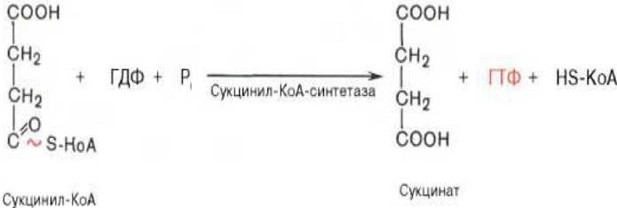
5. The fifth reaction is catalyzed by the enzyme succinyl-CoA synthetase. In this step, succinyl-CoA is converted into succinic acid (succinate) with the participation of GTP and inorganic phosphate. Simultaneously, a high-energy phosphate bond of GTP is formed at the expense of the high-energy thioester bond of succinyl-CoA (Figure 3.7):
Figure 3.7 - The fifth reaction of the tricarboxylic acid cycle
6. As a result of the sixth reaction, succinate is dehydrogenated to fumaric acid. The oxidation of succinate is catalyzed by succinate dehydrogenase, an enzyme in which the FAD coenzyme is tightly (covalently) bound to the protein (Figure 3.8):
Figure 3.8 - The sixth reaction of the tricarboxylic acid cycle

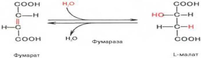
7. The seventh reaction is driven by the enzyme fumarate hydratase (fumarase). The resulting fumaric acid is hydrated, producing malic acid (malate) as the reaction product (Figure 3.9):
Figure 3.9 - The seventh reaction of the tricarboxylic acid cycle
8. During the eighth reaction of the tricarboxylic acid cycle, mitochondrial NAD-dependent malate dehydrogenase drives the oxidation of L-malate to oxaloacetate (Figure 3.10):
Figure 3.10 - The eighth reaction of the tricarboxylic acid cycle

As can be seen, one full turn of the cycle—consisting of eight enzymatic reactions—results in the complete oxidation ("burning") of a single molecule of acetyl-CoA. For the cycle to run continuously, a steady supply of acetyl-CoA is required, and the coenzymes (NAD+ and FAD) that have been reduced must be oxidized over and over again. This oxidation is carried out by the electron transport chain (respiratory chain) localized in the mitochondrial membrane.
3.5.3 Summary of the Krebs Cycle
As noted earlier, one molecule of NADH (equivalent to 3 ATP molecules) is generated during The oxidative decarboxylation of Pyruvate to acetyl-CoA.
The breakdown of one glucose molecule yields 2 pyruvate molecules, and upon their oxidation to 2 molecules of acetyl-CoA followed by 2 turns of the tricarboxylic acid cycle, 30 molecules of ATP are synthesized (consequently, the oxidation of one pyruvate molecule to CO2 and H2O yields 15 molecules of ATP).
To this amount, one must add 2 ATP molecules generated during aerobic glycolysis, as well as 6 ATP molecules synthesized through the oxidation of 2 extramitochondrial NADH molecules, which are formed during the oxidation of 2 glyceraldehyde-3-phosphate molecules in the dehydrogenase reaction of glycolysis.
Consequently, the breakdown of a single glucose molecule in tissues According to the equation C6H12О6 + 6O2 —> 6СO2 + 6Н2O results in the synthesis of 38 ATP molecules.
Undoubtedly, in energetic terms, the complete breakdown of glucose is a far more efficient process than anaerobic glycolysis.
It should be noted that the 2 NADH molecules formed during the conversion of glyceraldehyde-3-phosphate may subsequently yield only four ATP molecules upon oxidation rather than six. The reason is that extramitochondrial NADH molecules themselves are unable to cross the mitochondrial membrane.
During the steady-state functioning of the tricarboxylic acid cycle, none of the core cycle components are consumed. However, some of them are required for biosynthetic processes, such as the Synthesis of specific amino acids and nucleotides.
Nevertheless, the electrons they donate can be transferred into the mitochondrial biological oxidation chain via the so-called glycerophosphate shuttle mechanism.
Review questions for the topic
1. What is The history of the discovery of the tricarboxylic acid cycle?
2. What is the reaction sequence of the tricarboxylic acid cycle?
3. Provide a characterization of each of the eight stages of the Krebs cycle.
4. What is the biological Significance of the tricarboxylic acid cycle?
5. How is the tricarboxylic acid cycle regulated?
6. What is the shuttle Mechanism of enzyme Action?
7. How many ATP molecules are produced during the breakdown of 1 glucose molecule?
8. How are the enzyme Cofactors of the Krebs cycle reduced?
Last update: 06/08/2026
Editorial and Educational Adaptation: This material has been compiled based on the primary/original source text. The project team performed an editorial review, corrected technical inaccuracies, structured sections, and adapted the content for an educational format.
What was processed:
- elimination of formatting defects (OCR errors, structural breaks, corrupted characters);
- editorial organization of content;
- standardization of terminology in accordance with academic sources;
- verification of factual statements against the original source text.
All mentions of the author, publication year, and origin of the primary text have been preserved in accordance with the source.