BOTANY VOLUME 2 - PLANT PHYSIOLOGY - 2007
7. DEVELOPMENTAL PHYSIOLOGY
7.6. Control of Development by Phytohormones
As previously mentioned in this book, many developmental processes are regulated by phytohormones. Hormones are low-molecular-weight signaling molecules synthesized by all or many plants that elicit characteristic physiological responses at low concentrations (≤ 10-6 M). The sites of synthesis and action of a hormone are generally spatially separated. Phytohormones thus serve for intercellular regulation in Multicellular Organisms in much the same way as animal and human hormones, but, unlike the latter, phytohormones rarely regulate METABOLISM in already differentiated organisms (Examples include the control of guard Cells by Abscisic acid, see 7.6.4, and The regulation of germination by gibberellin, see 7.6.3); instead, phytohormones predominantly control growth and differentiation processes themselves, interacting in complex ways (which are not yet fully understood). Furthermore, the active hormone is frequently produced near or even directly at The Site of Action, and transport (if required at all) occurs over very short distances that can be bridged by diffusion. In such cases, phytohormones behave analogously to well-known animal paracrine or autocrine signaling substances: paracrine factors affect cells in the immediate vicinity of the synthesis site, whereas autocrine factors affect The Cell that produces them. Another difference from animal hormones lies in the low tissue- and organ-Specificity of phytohormones, which is why they often exhibit a broad spectrum of activity. It thus becomes clear that a phytohormone Functions merely as a stimulus. The Nature of the elicited process depends on the prevailing state of Cell Differentiation, i.e., on the set of active genes capable or incapable of activation.
The concentration of each phytohormone at its site of action is tightly regulated. It is the net result of synthesis, degradation, conjugation, storage, and influx or efflux. At the same time, the sensitivity of various Organs to specific phytohormones can vary greatly. Not only insufficient supply of phytohormones (as, for example, in Biosynthesis-deficient mutants), but also their excessive influx (often achievable only experimentally) leads to characteristic developmental defects.
Alongside the five groups of long-known phytohormones (Auxins, Cytokinins, Gibberellins, abscisic acid, and Ethylene), other classes of substances with phytohormone-like activity—brassinosteroids and jasmonates—have emerged in recent years; in addition, There are many physiologically active substances with more specific functions and a limited distribution within plants. Synthetic analogues of many phytohormones find application in ornamental and agricultural plant cultivation, as well as in PLANT CELL CULTURE.
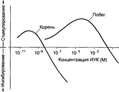
According to Thimann1, auxins (from Latin augere, to grow) are natural or synthetic compounds that, albeit at very different concentrations (Fig. 7.31), stimulate cell elongation growth and thereby SHOOT or ROOT elongation; at higher concentrations, they inhibit growth. Growth stimulation is particularly noticeable in bioassays using auxin-depleted Tissues, such as cereal coleoptile cylinders. Importantly, substances are classified as auxins not based on their chemical Structure, but on their characteristic action.
1 Auxins were independently investigated by Thimann (Germany), Went (the Netherlands), and Kholodny (Russia). The authors mention only the German discoverer. — Ed. note.
Class="center">Fig. 7.31. Shoot and root elongation growth as a function of indole-3-acetic acid (IAA) concentration in the medium (schematic). Experiments were performed on auxin-depleted organ segments.
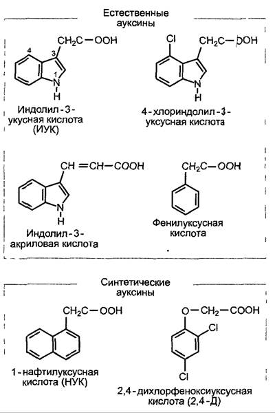
The most widespread auxin in plants is indole-3-acetic acid (β-indolylacetic acid, IAA, Fig. 7.32). This compound appears to occur in all pro- and eukaryotes, yet it serves as a signaling molecule only in higher plants. Other auxins, such as phenylacetic acid (in tobacco), indoleacrylic acid, and halogenated derivatives of indoleacetic acid (in legumes), are not of universal significance (Fig. 7.32). Among synthetic auxins, 2,4-dichlorophenoxyacetic acid (2,4-D), 1-naphthylacetic acid (NAA), and indole-3-butyric acid (IBA) are frequently used, from which IAA can be formed within the plant via β-oxidation. Common to all active auxins is the presence of a carboxyl group (which dissociates at physiological pH values) and a partial positive charge at a distance of 0.55 nm from the negative charge of the dissociated carboxyl group.
Fig. 7.32. Natural and synthetic auxins
The main sites of IAA production in higher plants are considered to be, on the one hand, embryonic tissues (Meristems, embryos) and photosynthesizing organs (especially growing leaves), although The Root System is also capable of IAA biosynthesis.
Elucidating the pathways of IAA biosynthesis has proved difficult and is still not considered complete. The amounts of IAA extractable from tissues are extremely low (e.g., 24 µg • kg-1 in maize coleoptiles, 69 µg • kg-1 in rosette leaves of Arabidopsis thaliana, approximately 350 µg • kg-1 in maize root tips). Correspondingly, The activity of IAA biosynthetic Enzymes is low. To study metabolism, isotope-labeled precursors are frequently added externally to tissues at concentrations far exceeding those of endogenously present auxin metabolites. This carries the risk of non-physiological Side Reactions.
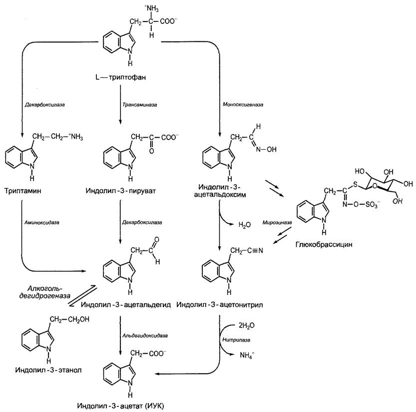
IAA is formed from L-Tryptophan1. Biosynthesis can proceed via various pathways depending on the plant and tissue (see Fig. 7.33), but very little is yet known about the enzymes and regulatory mechanisms.
1 This pathway is not the sole one. The possibility of IAA synthesis bypassing tryptophan, originating from earlier precursors, is also discussed. — Ed. note.
Fig. 7.33. Biosynthesis of indole-3-acetic acid (IAA) from L-tryptophan. The main pathway proceeds via indole-3-Pyruvate, while the tryptamine pathway is of secondary importance. Indole-3-ethanol is considered a temporary storage form of the IAA precursor indole-3-acetaldehyde. In Brassicaceae, indole-3-acetic acid is formed via indole-3-acetonitrile. The release of indole-3-acetonitrile from glucobrassicin, a glucosinolate widespread in Brassicaceae, potentially facilitates IAA formation. Within the cell, IAA is present almost entirely in the dissociated form as indole-3-acetate (pKa value for IAA = 4.8).
A small fraction of the plant's IAA supply can originate from epiphytic Bacteria and rhizosphere microorganisms (bacteria and Fungi), where microbial IAA production in the rhizosphere is based on tryptophan exuded by plant roots. During crown gall tumor formation (see Box 9.2)—which results from The transfer of several genes from the pathogenic soil bacterium Agrobacterium tumefaciens into the host cell's nuclear genome—two genes are also transferred whose products establish an additional, plant-cell-independent pathway of IAA biosynthesis from tryptophan via the intermediate indole-3-acetamide (see Box 9.2) in transformed cells. In crown gall tumors, the content of free or conjugated (see below) IAA is strongly elevated.
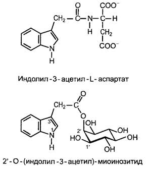
Regulation of tissue IAA supply is achieved not only through synthesis, but also via inactivation of unneeded hormone. Certain IAA inactivation products, following conjugation with sugars (especially glucose), are deposited in vacuoles. However, IAA can also be conjugated and stored as amino acid conjugates (thereby removing it from the site of action). Endogenously, IAA-amides with aspartate (Fig. 7.34) and glutamate occur predominantly. Externally applied IAA is converted by plant cells primarily into sugar conjugates (above all with glucose). High-molecular-weight storage forms may also occur (e.g., in seeds). IAA conjugates, which ensure the irreversible breakdown of excess hormone, can also serve to maintain Homeostasis, as well as for temporary IAA storage and as a transport form of the phytohormone. For instance, during cereal germination, IAA is transported as 2'-O-(indolyl-3-acetyl)-myo-Inositol (Fig. 7.34) to the coleoptile tips, where the phytohormone is hydrolytically released. In cell cultures, conjugates with sugars are rapidly formed from auxin added to the medium (IAA or more stable synthetic auxins, see Fig. 7.32).1 These likely serve for the long-term supply of auxin to the tissue, i.e., they represent auxin storage forms.
1 The formation of conjugates of synthetic auxin analogues is a subject of debate. Not all analogues serve as substrates for IAA-glycosidases. For instance, 2,4-D is hardly metabolized at all. — Ed. note.
Fig. 7.34. Structural examples of indole-3-acetic acid conjugates
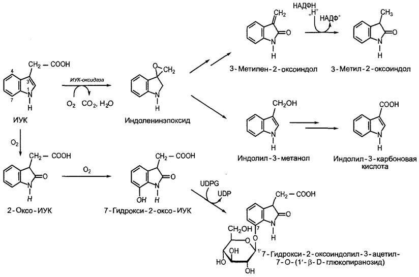
The breakdown of IAA (Fig. 7.35) proceeds via oxidative pathways, with the specific sequence of reactions varying somewhat depending on the plant species. Catabolism down to 3-methylene-2-oxindole, 3-methyl-2-oxindole, and indole-3-carboxylic acid is widespread and catalyzed by a relatively unspecific peroxidase; this enzyme, in turn, is activated by monophenols (such as Tyrosine or p-hydroxybenzoic acid) and Mn2+, and inactivated by diphenols (such as caffeic acid) (IAA oxidase). In some species (Pinus sylvestris, Vicia faba, Zea mays), the attachment of an acetyl side chain converts IAA into 7-hydroxy-2-oxo-IAA, which is then stored as the highly Water-soluble O-β-D-glucopyranoside; this compound occurs in large quantities, for instance, in maize endosperm. IAA catabolites are physiologically inactive and no longer conform to the 0.55 nm rule (see above).
Fig. 7.35. Oxidative catabolism of indole-3-acetic acid. The reaction sequence initiated by IAA oxidase is widespread among plants, whereas the 2-oxo-IAA pathway occurs, for example, in Pinus sylvestris, Zea mays, and Vicia faba
7.6.1.3. Transport of Indole-3-Acetic Acid
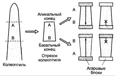
Over long distances, IAA can be transported within the assimilate stream of the phloem (see 6.8). In addition, There is a directed parenchymal transport of auxin known as polar auxin transport. In various isolated shoot segments (coleoptiles, shoot axes, leaf petioles, or pedicels), exogenously applied IAA is transported at a rate of 2 — 14 mm • h-1 in a polar, basipetal direction—that is, independently of the orientation of the specimen, meaning the EFFECT OF GRAVITY can be ruled out (Fig. 7.36). This polar basipetal auxin transport is active (i.e., metabolism-dependent) and can be inhibited by specific inhibitors (such as 1-N-naphthylphthalamic acid or 2,3,5-triiodobenzoic acid), in contrast to passive acropetal (shoot apex-directed) transport, which occurs via simple diffusion.
Fig. 7.36. Demonstration of basipetal polar transport of IAA in coleoptile segments. Regardless of segment orientation (normal or inverted), IAA diffusing from Agar blocks is transported through the tissue exclusively from the apical end to the basal end (arrow) and can be detected in the receiver block. IAA applied via the basal end penetrates the tissue slightly by diffusion but undergoes no Active Transport. Experiments utilize radioactively labeled IAA (e.g., using 14C) for high-sensitivity detection. Radioactivity is absent in the agar blocks shown in white
In roots, polar IAA transport proceeds acropetally (toward the root tip) within the central cylinder, while a fraction of the IAA is transported basipetally in the exodermis (from the tip toward the root base). These velocities are very close to those observed in shoots (4 — 10 mm • h-1). The Significance of polar auxin transport in axis formation during Embryogenesis was discussed previously (see 7.4.1).
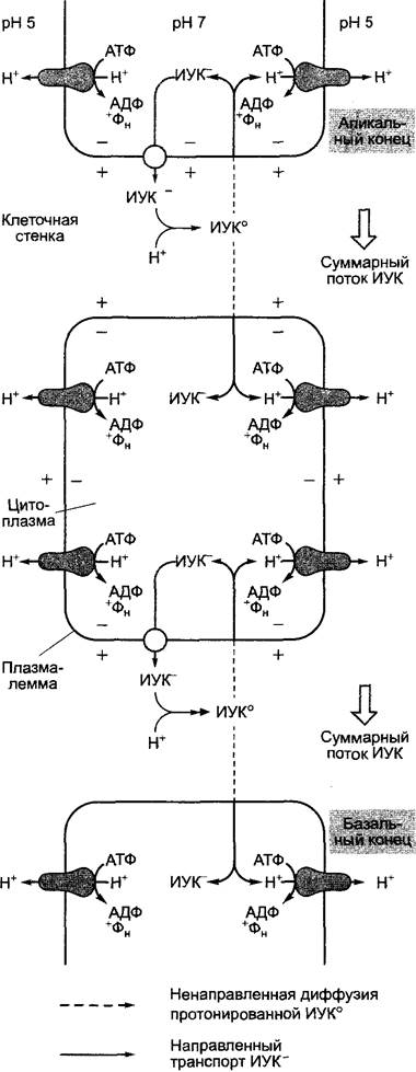
The Mechanism of polar auxin transport is not yet fully understood. According to the chemiosmotic model (Fig. 7.37), specific translocators (IAA carriers) are localized in The Plasma Membrane at the basal pole of the cell, where they export indole-3-acetate (IAA-). This process utilizes the energy of the electrochemical gradient maintained across the plasma membrane by a proton-pumping ATPase (see Figs. 6.4 and 6.5). Because of the low pH in the apoplast, a portion of the extruded indole-3-acetic acid (~50 %) remains undissociated. Undissociated IAA easily diffuses across cellular membranes, thereby re-entering the cell passively. This diffusion step is non-directional. According to this model, the overall directional process is maintained solely by the polar localization of IAA carriers, the direct identification of which is currently underway.1 An added complexity arises from the fact that the apoplast is generally subject to a mass flow of water driven by Transpiration, which operates counter to the vector of IAA transport (see 6.3.1.2; Fig. 6.32). Under certain conditions, the direction of polar transport within the plant can reverse—for example, in response to gravity, unilateral illumination (see 8.3.1.1), or during embryogenesis (see 7.4.1).
1 This information is outdated. In recent years, IAA carrier Proteins have been identified in Arabidopsis: AUX1 (mediating passive influx) and PIN1 (mediating active efflux). — Note by the Editor.
Fig. 7.37. Chemiosmotic model of polar IAA transport
The diverse Physiological effects of IAA include primarily the following:
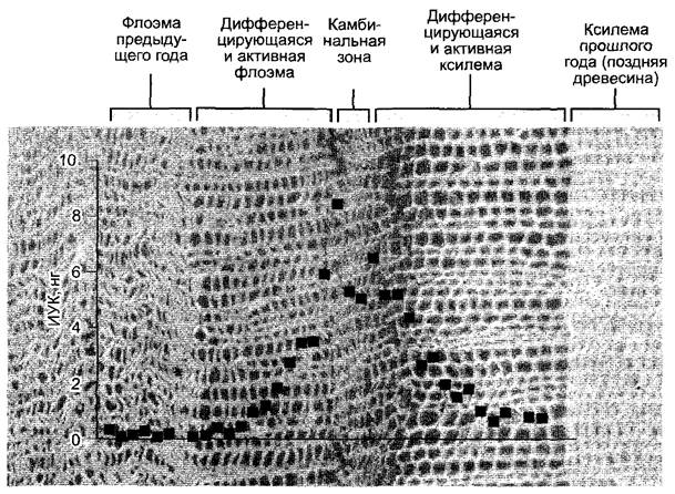
Stimulation of cambial activity to increase the number of xylem elements. Highly sensitive mass spectrometry techniques have demonstrated that the concentration of IAA in the cambial zone is higher than in the surrounding tissue (Fig. 7.38). It is hypothesized that this auxin gradient contributes to positional information that influences the developmental fate of cambial initials as they differentiate into phloem or xylem. Thus, IAA in this context acts more as a morphogen than as a classical phytohormone (see 7.3.3, 7.4).
Fig. 7.38. Radial concentration gradient of indole-3-acetic acid in the cambial zone of Pinus sylvestris, showing the corresponding phytohormone content per tissue disc (1 cm2 area, 30 µm thickness) tangentially excised from a tissue block using a freezing microtome. Further studies have revealed that phloem or xylem differentiation is governed less by the absolute concentration of IAA in the cambium than by the radial distribution of the phytohormone. The radial gradient of IAA is less steep toward the xylem differentiation side than toward the phloem side. The transverse section used as a Background for the graph allows visual assessment of IAA content across the respective tissue layers
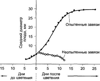
Stimulation of Seed and Fruit formation and development. The initial supply of active IAA is delivered by pollen. Later, it is produced by developing ovules and released into the surrounding Tissues of the Ovary, where it primarily stimulates cell growth. The first phase of ovary growth (prior to anthesis) is generally characterized by robust growth via Cell Division accompanied by relatively modest cell elongation. In many species (e.g., tomato and currant), cell division ceases after flowering, and subsequent growth is driven entirely by cell elongation—a process triggered only if pollination has occurred (Fig. 7.39). These cells can enlarge to the point of being visible to the naked eye (e.g., in watermelon, Citrullus vulgaris).
Fig. 7.39. Ovary growth in Cucumis anguria. In unpollinated flowers, post-flowering growth decelerates (with the decline caused by tissue desiccation), whereas pollinated Ovaries exhibit a typical sigmoid growth curve
If pollination fails to occur, flowers typically abscise; if it succeeds, petals and stamens wither, and fruit development is initiated. For the initial phase of fruit growth ("fruit set"), Fertilization is generally not strictly required; pollination alone—frequently even with foreign pollen incapable of effecting fertilization—is often sufficient. Pollen, being very rich in auxin, acts by releasing IAA. Consequently, pollination can often be mimicked by applying IAA (or other auxins) to the stigma. In most fruits, pollination triggers only fruit set rather than subsequent fruit growth, which is initiated only after fertilization has taken place and is once again regulated by auxin. Here, developing ovules serve as the source of auxin. As a result, in many fruit types (e.g., grapes, apples, pears, tomatoes, currants), the final size of the fruit is generally proportional to the number of developing seeds. In certain species (e.g., tomatoes, currants, tobacco, figs), fruit set and growth can be induced without prior pollination (parthenocarpy) through the application of IAA (or synthetic auxins) to the stigmas, resulting in seedless fruits. This technique is applied in greenhouse tomato production to achieve synchronized fruit set and harvest.
In spontaneously occurring parthenocarpic and consequently seedless fruits (e.g., certain varieties of tomatoes, cucumbers, figs, oranges, bananas, and pineapples), development proceeds either entirely without pollination or following Pollination and Fertilization coupled with subsequent embryo abortion. In these plants, the auxin synthesis in the ovules or other ovary parts required for nutrient influx and fruit growth apparently operates independently of, or requires only minor, external correlative influences.
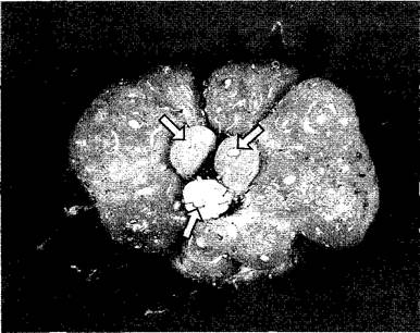
The stimulatory effect of IAA can be clearly demonstrated experimentally in strawberries. If the achenes developing after pollination are prematurely removed, the localized expansion of the fleshy receptacle beneath them is suppressed (Fig. 7.40); if all achenes are removed, receptacle growth ceases entirely, but it can be fully restarted by applying an auxin solution to the receptacle in place of the removed achenes. The linkage between fruit growth and successful fertilization followed by seed development is biologically vital: a substantial influx of nutrients for continued fruit growth occurs only when biologically justified. As with other growth processes, auxins are not the sole hormones driving fruit development. Evidence suggests that developing seeds, alongside auxins, also release gibberellins into surrounding tissues, which play a role in controlling fruit development. In some species, Treatment with gibberellins (rather than auxins) induces parthenocarpy (e.g., in Prunus species). Finally, fruits undergoing active cell division during growth exhibit the highest concentrations of cytokinins (e.g., apple, tomato, banana).
Fig. 7.40. Development of the strawberry aggregate fruit (etaerio of achenes). The receptacle does not become fleshy in areas where fruit development is not initiated due to pollinated failure. In the center of the strawberry shown, only three achenes are developing (arrows). The receptacle expands only in the immediate vicinity of these fruits. In this case, the achenes release the growth factor indole-3-acetic acid, which strongly stimulates the growth of receptacle cells
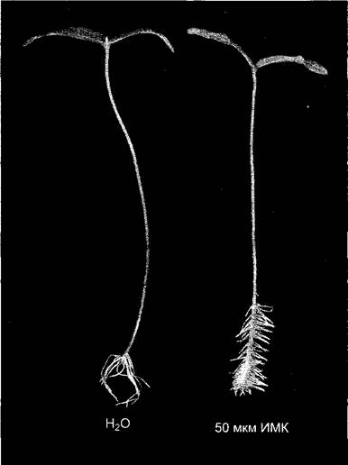
Stimulation of lateral and adventitious root initiation — rhizogenesis (Fig. 7.41). As with The Effect of IAA on cambial activity, this process demonstrates that auxin can stimulate cell division.
Fig. 7.41. Auxin-stimulated formation of adventitious roots in mung bean cuttings. Cuttings were placed for 7 days in a 50 µM solution of indole-3-butyric acid (right) or water (left). Upon cellular uptake, indole-3-butyric acid (IBA) is converted into indole-3-acetic acid — this is the underlying principle of IBA action. Control plants form sparse, long adventitious roots, whereas those treated with IBA produce numerous short roots. This occurs because the additional IAA generated by the tissue from IBA increases the overall available IAA concentration, thereby stimulating the initiation of new adventitious roots, although their elongation growth is inhibited
Induction of regeneration in cell culture. The process taking place with the participation of cytokinin is described in more detail in Section 7.6.2.3.
Inhibition of lateral bud outgrowth by auxin produced by the apical bud (apical dominance) (see 7.5). Cytokinin acts as an antagonist to auxin and stimulates the outgrowth of lateral buds.
Inhibition of leaf, flower, and fruit abscission. As long as a sufficient amount of IAA is transported from the leaf blade, flower, or developing fruit
via the petiole or flower stalk, respectively, the Differentiation of the abscission layer at the Base of the petiole (abscission zone) is suppressed. When auxin supply is insufficient (e.g., after the completion of leaf development, in the absence of pollination or fertilization), abscisic acid (see 7.6.4) and especially ethylene (see 7.6.5) induce the differentiation of the abscission layer, resulting in the shedding of the respective organ.
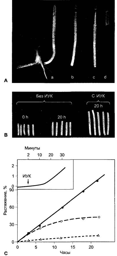
Auxin-stimulated extension growth has been investigated in particularly great detail. The application of auxin to intact plants has virtually no effect on shoot growth and generally exerts an inhibitory effect on roots. This is explained by the optimal auxin supply already present in intact tissues. However, in several pea varieties differing in height, a direct correlation between IAA concentration and elongation growth rate was demonstrated. In other cases, no such relationship was found. Conversely, in auxin-depleted shoot and coleoptile segments, the application of IAA induces a strong, phytohormone-concentration-dependent extension growth (Fig. 7.42). Growth begins after a certain latent period (lag phase) lasting about 10 minutes and continues in the presence of osmotica taken up by the cells (e.g., sucrose, KCl) for periods ranging from several hours to a day or more.
Fig. 7.42. Stimulation of coleoptile extension growth by IAA: A — pretreatment of corn coleoptile segments. The coleoptile tip supplies the organ with IAA. Therefore, the coleoptile segments (B) are depleted of IAA and show only weak extension growth, which is attributable to residual endogenous IAA. In the presence of IAA in the incubation medium, the extension growth of the coleoptile segments is significantly stimulated; C — time course of IAA-induced extension growth of oat coleoptile segments (10 µmol IAA, pH 6) in the absence (o) or presence (•) of 2% sucrose in the incubation medium (d — sucrose without IAA). Auxin-stimulated extension growth begins after a lag phase of approximately 8 – 10 min (see graph at top left)
The driving forces for cell growth can be described by The water potential equation (see Equation 6.15), neglecting the gravitational potential, which plays no significant role in measurements at the single-cell level:

A cell in pure water takes up water from its surroundings until the internal pressure $p$ (turgor) generated as a result of elastic wall tension is counterbalanced by the osmotic pressure $-\Pi$ ($p = \Pi,  = 0$). Thus, equilibrium between the cell and an aqueous solution with a negative water potential is established when
  and consequently
  . Growth is always associated with an irreversible increase in volume, i.e., water uptake, and occurs when
the turgor pressure generated by osmotic water influx exceeds the maximum possible elastic deformation of The Cell wall, leading to irreversible (plastic) extension of the cell wall. Therefore, a specific growth rate is the result of turgor-driven plastic extension of the cell wall, water influx, and the uptake of osmotica (primarily KCl) or the intracellular generation of osmotica (primarily CARBOHYDRATES) to maintain the osmotic potential. As a rule, growth is only possible in cells with a primary wall (see 2.2.12) and is coupled with the continuous de novo Synthesis of cell wall components (Cellulose and matrix constituents); this also controls the threshold potential for plastic deformation, preventing rupture of the cell wall.
During isodiametric growth, these processes proceed uniformly across the entire cell surface; during elongation growth, the cell stretches along an axis that is presumably determined by the orientation of cellulose microfibrils. In cells prior to the onset of growth, these microfibrils, embedded in a matrix, lack any pronounced preferential direction (random texture) (see Fig. 2.72, B). The arrangement of cellulose microfibrils is evidently governed by the Cytoskeleton (Fig. 7.43).
Fig. 7.43. Highly simplified model of IAA-stimulated cell elongation. Activation of proton transport into the apoplast leads to the loosening of Hydrogen Bonds, particularly between cellulose and hemicellulose, thereby increasing the plasticity and extensibility of the cell wall (1). Acidification simultaneously activates (presumably) cell wall enzymes required for the de- and repolymerization of matrix polymer components. The reorganization of cortical microtubules within the cell, and consequently of newly synthesized cellulose fibrils (see Fig. 2.72) in the cell wall, establishes the longitudinal axis of cell expansion (double arrow) (2). Finally, under The Influence of IAA, new cell wall material is intensively synthesized (3)
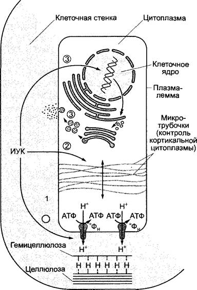
An increase in the growth rate induced, for example, by auxin causes an elevation in the driving force for water influx ("suction force") and can therefore be mediated through an increase in the osmotic potential and/or plastic deformation of the cell wall. All evidence indicates that auxin application enhances the plastic deformation of the primary wall. The molecular mechanisms remain poorly understood (Fig. 7.43).
According to the acid growth hypothesis, externally applied IAA induces marked acidification of the apoplast in auxin-depleted coleoptile or shoot segments (in coleoptiles from pH = 5.5 to pH ≤ 4.5), which is attributed to a massive efflux of protons from the cells. This acidification, which can be demonstrated unequivocally by experiments, accounts for the lag phase of auxin action and, according to the hypothesis, triggers the following processes.
✵ Hydrogen bonds between cell wall components are weakened. These bonds exist primarily between cel-
lulose microfibrils and appressed hemicellulose molecules (xyloglucans in dicots, see Fig. 2.64). Indeed, a cell wall protein has recently been described that becomes active in an acidic environment and catalyzes the disruption of hydrogen bonds between cellulose and xyloglucan. The addition of this protein, designated expansin1, very clearly enhances the pH-dependent plastic deformation of isolated cell walls.
1 From English expansion. — Editor's note.
✵ Enzymes that cleave (and re-form) covalent bonds of cell wall polymers are activated, allowing cell wall components to slide relative to one another under the influence of turgor pressure. Such enzymes include xyloglucan endotransglucosylase (XET), which breaks covalent bonds in hemicellulose polymers and reconstructs them. Through the de- and repolymerization of interconnected matrix components (structural model; see Fig. 2.68), not only is the plastic deformation of the cell wall increased, but the incorporation of newly secreted structural elements is also facilitated.
The mechanism behind auxin-induced apoplast acidification remains unclear. It likely involves a P-type H+-ATPase (see Figs. 6.4 and 6.5) that translocates hydrogen ions. This enzyme is activated by IAA via a yet unknown mechanism, and/or its Abundance in the Plasmalemma is increased by IAA. A highly efficient activation mechanism for this ATPase was discovered only recently (see Fig. 9.15): the H+-ATPase is the target of fusicoccin, a toxin produced by the phytopathogenic fungus Fusicoccum amygdali, which strongly stimulates cell expansion by elongation in coleoptiles and also affects other processes mediated by the H+-ATPase (such as stomatal movement, see 8.3.2.5). It is unlikely that auxin affects the H+-ATPase through the same mechanism.
The acid growth hypothesis is supported by the following evidence:
✵ acidic buffers stimulate cell expansion by elongation just like IAA, but without a lag phase;
✵ neutral buffers abolish IAA-stimulated elongation growth by neutralizing the H+ ions secreted by the cell;
✵ Inhibitors of the H+-ATPase suppress auxin-induced elongation growth.
Elongation growth triggered by acidic buffers is short-lived, whereas the effect of auxin (see Fig. 7.42) is significantly more sustained. This demonstrates that the acid growth hypothesis accounts for only one aspect of auxin action.
According to current understanding, alongside stimulating H+ ion transport into the apoplast, auxin is involved in the following processes:
✵ stimulating the synthesis and transport of cell wall components;
✵ inducing the reorganization of microtubules located in the peripheral (cortical) Cytoplasm, which causes cellulose microfibrils—synthesized by cellulose synthase—to orient predominantly perpendicular to the longitudinal axis of cell elongation (thereby reducing resistance to stretching in this direction).
Ultimately, the cell's capacity for longitudinal growth is lost because cell stretching shifts the cellulose microfibrils, making them run increasingly parallel to the longitudinal axis. Secondary wall formation then begins via the apposition of cellulose layers containing parallel fibers. Plastic deformation thus ceases, leaving the cell with only elastic properties that permit merely limited, reversible stretching.
7.6.1.5. MOLECULAR MECHANISMS OF Auxin Action
Auxin-regulated physiological processes proceed via changes in Gene Expression, distinguishing between early-response genes (primary or directly regulated genes) and late-response genes (secondary or indirectly regulated genes). The latter are believed to be controlled by
products of primary auxin-responsive genes, some of which function as Transcription factors. In the promoters of primary auxin-responsive genes, whose activity increases sharply just 5 – 10 min after auxin application, it is often possible to identify several auxin-responsive elements (AREs); these are sequence regions about -25 – 30 bp long, which consist respect
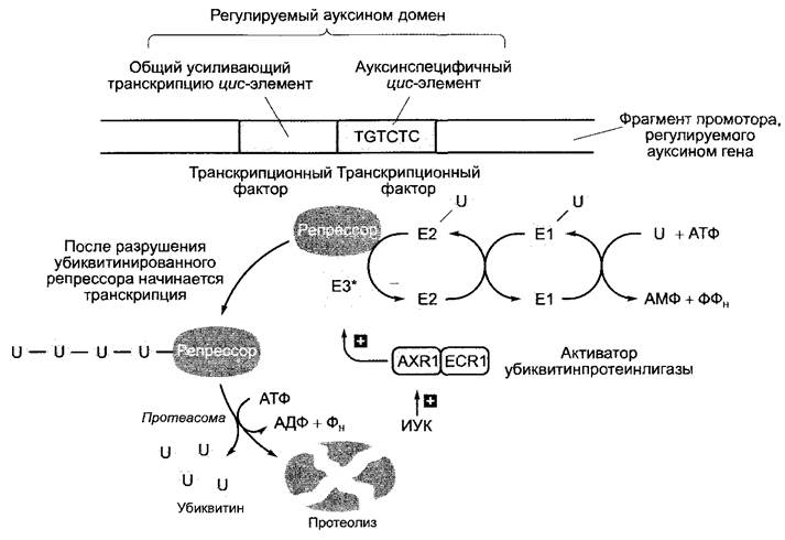
ively of an auxin-specific and a general transcription-activating element. According to recent, albeit still partly hypothetical models, IAA induces ubiquitination (see 7.3.1.3) and thereby the proteolytic degradation of a repressor protein; once this repressor is removed, transcription of directly auxin-regulated genes begins (Fig. 7.44). The activating protein AXR1 is involved in ubiquitination; a mutation in the corresponding gene leads to a loss of the cells' ability to respond to auxin (AXR, English auxin resistant — the axr mutant phenotype).
Fig. 7.44. Hypothetical model of gene activation by indole-3-acetic acid (IAA). IAA activates the heterodimeric protein complex of AXR1 and ECR1 localized in the Cell Nucleus, which in turn activates ubiquitin-protein ligase (E3*). This ligase promotes the ubiquitination of specific repressor proteins that prevent the transcription of auxin-regulated genes, thereby inducing the destruction of the repressors by the proteasome (see 7.3.1.3). The breakdown of ubiquitinated repressors initiates the Transcription of the corresponding genes. The ubiquitin-conjugation system consists mainly of a ubiquitin-activating enzyme (E1), which transfers ubiquitin to a ubiquitin-conjugating protein (E2); from there, ubiquitin is transferred to a protein substrate with the assistance of a substrate-specific ubiquitin-protein ligase (E3). This produces polyubiquitinated proteins that are rapidly degraded by the proteasome, releasing ubiquitin, provided they carry 4 or more ubiquitin molecules. The cell contains several E3-type enzymes; these are often heterooligomeric Protein Complexes that require activation (E3*) by a heterodimeric activator. This activator bears similarity to the E1 enzyme: one component corresponds to the N-terminal part and the second to the C-terminal part of the E1 enzyme. In the case of auxin-regulated genes, this role is played by the AXR1/ECR1 complex. Impairment of AXR1 function, for example due to a mutation in the AXR1 gene, results in the loss of The ability to respond to auxin. The protein was named after the axr1 mutant (auxin resistant), and ECR1 (E1-C-terminus related): the protein was discovered based on this similarity
Cytokinins are N6-substituted Purines (Fig. 7.45) that were discovered based on their stimulating effect on cell division (cytokinesis).
Fig. 7.45. Examples of natural and synthetic cytokinins. Natural cytokinins occur in the cell not only as free bases, as shown, but also as ribosides and ribosyl-5'-monophosphates (see Fig. 7.46)

7.6.2.1. Diversity of cytokinins
In tissue culture experiments using tobacco pith on media of defined composition, it was found that the addition of indole-3-acetic acid alone stimulates only cell expansion, but is insufficient to activate cell divisions. As a result of a systematic search for a cell division-stimulating factor, strong activity was first detected in autoclaved DNA preparations. The active compound was subsequently identified as N6-furfurylaminopurine (Fig. 7.45). It is formed during the autoclaving of DNA AS A result of Hydrolysis, phosphate Cleavage, and the migration of deoxyribose from its initial position (1' −> 9) (cf. Fig. 1.4) to a position involving dehydration (5' −> 6). This substance, named kinetin, does not occur in plants, but natural cytokinins are also N6-substituted adenine derivatives. The most important of these are N6-isopentenyladenine (IPA) and trans-zeatin (tZ); they occur in the plant as free bases, ribosides, or ribosyl-5'-monophosphates. Only the free bases act as active cytokinins, primarily trans-zeatin, which is also the predominant cytokinin in most tissues. In physiological studies, for example with cell cultures, synthetic cytokinins (see Fig. 7.45) are preferred due to their higher stability.
We mentioned earlier that N6-substituted adenine, such as IPA, occurs as a rare base in certain tRNAs (see Fig. 1.10). It is possible that in tissues with a high RNA content, cytokinins are generated during tRNA breakdown, though the physiological significance of this process remains unclear. For instance, zeatin also occurs as a rare base in certain tRNAs, but exclusively as the cis-isomer, whereas the free form is exclusively trans-zeatin. The initial hypothesis that cytokinins stimulate mRNA and Protein metabolism through their incorporation into tRNA was not confirmed; it subsequently became clear that N6-substituted adenine arises in tRNA solely through the subsequent prenylation of adenine (see 1.2.4).
N6-substituted adenines, already found in bacteria, also occur in fungi. They may have physiological significance in phytopathogens (e.g., Agrobac-
terium tumefaciens, see Box 9.2) and symbiotic bacteria (e.g., Phyllobacterium rubiacearum) or in mycorrhizal fungi (see 9.2.3). Thus, shoot fasciation caused by Rhodococcus fascians is due to cytokinins secreted by this actinomycete.
The physiological action of cytokinins and the compounds themselves have been detected across all groups of land plants, starting from mosses; however, they have been best studied in higher plants.
7.6.2.2. Metabolism and Transport
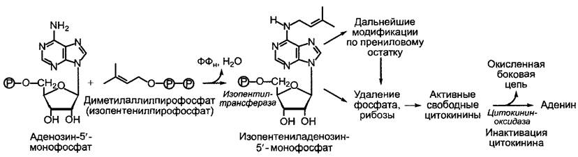
Disregarding the potential release of cytokinins during tRNA degradation—whose contribution to supplying the tissue with cytokinins is nonetheless doubtful—cytokinins are formed by transferring a dimethylallyl moiety from dimethylallyl pyrophosphate1 to adenosine 3'-monophosphate. This is followed by further transformations, which include the hydroxylation and subsequent saturation of side chains on the one hand, and the possible removal of the phosphate moiety and ribose upon each such combination on the other (Fig. 7.46). Along with isopentenyladenine, zeatin, and dihydrozeatin, plants contain their respective ribosides and ribotides; however, as already mentioned, only free bases exhibit cytokinin activity.
1 In equilibrium with its isomer, isopentenyl pyrophosphate, respectively. — Ed. note.
Fig. 7.46. Main reactions of cytokinin metabolism
Root apices are considered the primary site of cytokinin biosynthesis. From the roots, cytokinins are distributed throughout the plant via the xylem stream; the main transport form is trans-zeatin riboside (tZR). In grapevine bleeding sap, for example, 5–100 µg · L-1 of cytokinin has been detected. There is no polar transport of cytokinins in tissues; consequently, diffusion represents the sole mechanism for short-distance transport.
However, alongside the roots, very young leaves and developing seeds are also considered sites of cytokinin production1. The auxin of the apical bud apparently suppresses cytokinin biosynthesis or the import of cytokinins into those axillary buds whose development is inhibited due to apical dominance. Following the removal of the apical bud, and before the growth of young shoots begins, the cytokinin content in the shoot increases sharply, especially in the axillary buds. If the apical bud is replaced with an agar block containing auxin, cytokinin accumulation is suppressed and apical dominance is maintained.
1 Primarily the root pole of the embryo. — Ed. note.
Cytokinins can be converted into various sugar conjugates (glucosides), which may serve as storage, transport, or inactivating forms. A widespread inactivation mechanism is the oxidative Cleavage of the prenyl residue in cytokinins at the base (the cytokinin oxidase reaction): IPA yields adenine and 3-methyl-2-butenal, while trans-zeatin yields adenine and 3-hydroxymethyl-2-butenal.
Like all phytohormones, cytokinins also influence numerous physiological processes and interact in the process with other phytohormones.
The property of cytokinins to stimulate cell division was already mentioned above. This property forms The basis of the most important bioassay for cytokinin activity: the tobacco pith callus assay. On specific nutrient media, the biomass gain of sterilely cultured callus tissues is proportional to the cytokinin concentration. Admittedly, The stimulation of cell division is tied to the presence of auxin in the nutrient media. Both cytokinins and auxins are required for the progression of the Cell Cycle, specifically for the initiation of DNA Replication and mitosis (see 7.3.2, Fig. 7.19, A).
Auxin- and cytokinin-induced growth in cell cultures depends not so much on the absolute concentrations of both phytohormones as on their ratio. If the auxin concentration is increased relative to the cytokinin concentration, root regeneration (rhizogenesis) takes place; conversely, if the cytokinin concentration is increased relative to the auxin concentration, shoots regenerate (Fig. 7.47). When regenerating plants from cell culture, shoot formation is typically induced first, followed by the rooting of shoots on a medium promoting rhizogenesis. The auxin-to-cytokinin ratio during embryogenesis likely exerts a decisive influence on organ formation in the developing embryo.
Fig. 7.47. Dependence of growth and Organogenesis of a tobacco stem pith tissue fragment (explants) on the IAA and kinetin content in the nutrient agar medium. Left: state at the beginning of the experiment; right: after several weeks of cultivation. Organogenesis is determined primarily by The ratio of the concentrations of the two growth substances

Tumors in differentiated plants are also characterized by a disrupted auxin/cytokinin ratio and, frequently, by strongly elevated absolute concentrations of both phytohormones. This holds true for the previously mentioned crown gall tumors induced by Agrobacterium tumefaciens (see Box 9.2). Among the bacterial genes integrated into the plant nuclear genome during tumor formation is the ipt gene, which encodes isopentenyltransferase (IPT)—an enzyme that catalyzes the initial step of cytokinin biosynthesis just as it occurs in the plant (Fig. 7.46). Crown gall tumors are thus auxin- and cytokinin-independent, allowing them to be propagated indefinitely on nutrient media without the addition of these phytohormones. If the ipt gene in the bacteria is mutated, an excess of auxin arises: instead of a tumor, a root teratoma forms on the plant. If one of the two (or both) auxin genes is deleted, a cytokinin excess arises, leading to the formation of a shoot teratoma, entirely analogous to experimental regeneration achieved by shifting the auxin/cytokinin ratio in cell culture (Fig. 7.47). In principle, a teratoma is understood to be a tumor in which differentiated tissues and/or organs can be recognized.
Genetically induced tumors occur in various interspecific hybrids, primarily within the genera Nicotiana and Brassica. These tumors are non-infectious and are attributed to the combination of two incompletely compatible genomes whose cooperative action leads to disruptions in the normal developmental program. Since genetically induced tumors in in vitro culture are also auxin- and cytokinin-independent and contain large amounts of these phytohormones, a hormonally driven failure of cell cycle control is evidently at play here.
Cytokinins act as auxin antagonists in the disruption of apical dominance. Presumably, the formation of "witches' brooms"—that is, the outgrowing of numerous lateral buds (for example, in chrysanthemums, petunias, willows, and larches following infection with Rhodococcus fascians, formerly known as Corynebacterium fascians)—is likewise stimulated by the aforementioned bacterial cytokinin synthesis. The species name fascians derives from fasciation: the formation of ribbon-like, flattened shoots that represent multiple simultaneously growing lateral shoots. This is a symptom of Rhodococcus fascians infection (Lat. fascis, bundle), which is also linked to apical dominance.
Cytokinins stimulate:
• cell expansion during leaf development;
• chloroplast development in angiosperms (which, upon cytokinin addition,
proceeds to a large extent in the dark as well);
• bud induction on the caulonema of leafy mosses (see 11.2), from which the gametophyte subsequently differentiates.
Delaying senescence processes, especially in leaves, is a very important function of cytokinins. Senescence refers (by definition) to a developmental process that, unless halted or reversed, inevitably leads to the death of the entire Organism or the decay of its organs.
Based on the overall pattern of plant Aging, a distinction is generally made between monocarpic species, which flower and fruit only once, and polycarpic species, which produce flowers and fruits repeatedly.
All annual and biennial species are monocarpic, along with a limited number of perennials that may grow vegetatively for several years before perishing after flowering and fruiting (such as agave, bamboo, or Corypha umbraculifera, which can live for over 300 years). In these monocarpic species (unlike polycarpic ones), Senescence and Death are closely tied to the formation of reproductive organs. If the formation of flowers is prevented in annuals or biennials, such as sugar beets, their lifespan can be extended by many years.
This correlative link between aging and reproduction is not explained—or at least not solely explained—by the idea that developing flowers and, above all, fruits with their high nutritional demands simply drain vital resources from the rest of the plant. For instance, in dioecious spinach, the flowering of male plants triggers the exact same leaf yellowing as flowering and fruiting do in female plants. Therefore, it is more likely that senescence and death are driven by other interactions between the reproductive organs and the rest of the plant. It is possible that aging factors are released by flowers and fruits, or that the high demand for cytokinins originating from the roots creates a deficiency in the plant's other parts.
In polycarpic species, normal death apparently does not result from inevitable, programmed meristem senescence, but rather from the increasing difficulty of supplying them with water, mineral salts, nutrients, and BIOLOGICALLY ACTIVE SUBSTANCES. Apical meristems can often be kept alive almost indefinitely through continuous, unbroken propagation via cuttings (as seen in Lombardy poplars and many crops, such as strawberries, bananas, and roses) or in vitro culture. Thus, death here is governed solely by correlative factors.
Many trees can attain a very great age. According to reliable tree-ring counts, the age of poplars and elms, for instance, can reach up to 600 years, oaks up to 1,000 years, lindens 800 to 1,000 years, Sabina tibetica over 1,200 years, Fitzroya cupressoides in Chile over 2,000 years, the giant sequoia (Sequoiadendron giganteum) up to 4,000 years, and Pinus longaeva (= P. aristata) over 4,800 years. Many common Central European trees live for several centuries, and even unassuming plants like Vaccinium myrtillus can reach an age of 28 years. Using molecular biology techniques, the age of a long-lived clone of the alpine sedge Carex curvula was estimated at approximately 2,000 years. This sedge clone has consequently survived vastly different climatic eras in the exact same Location. It should be noted that long-lived plants undergo constant cellular renewal: in trees, for example, not only in the apical meristem but primarily in the cambium. The lifespan of individual plant cells—such as wood ray parenchyma cells in trees or parenchyma cells in succulent cacti—rarely exceeds 100 years unless some form of cellular "rejuvenation" occurs via division and growth. However, most cells do not reach such an advanced age. A slow but inexorable aging process occurs even during dormancy, which seeds and spores achieve through significant dehydration. Although metabolism almost completely ceases, experience shows that germination capacity rarely persists beyond 100 to 200 years. Extremely long-lived seeds are found among legumes, mallows, and the sacred lotus (Nelumbo nucifera); for the latter, lifespans of up to 1,000 years are sometimes cited. Similarly, the seeds of many weed species (such as Spergula arvensis and Chenopodium album) can remain viable for centuries in the complete absence of Respiration. Claims regarding the germinative capacity of so-called "mummy wheat" from Egyptian tombs are false, as wheat grains retain viability for a maximum of 10 years. Seeds of tropical plants, which are unadapted to surviving unfavorable climatic periods, often remain viable for even less than a year.
Individual organs of perennial plants—leaves, flowers, and fruits—have a significantly shorter lifespan than the plant as a whole. In woody plants, hemicryptophytes, and geophytes (see 4.2.4), all above-ground parts regularly die back in autumn.
Leaves exhibit either sequential or synchronous senescence. In the former case, only the oldest leaves age and die, whereas in the latter case—such as the autumn leaf fall of deciduous trees—all leaves undergo senescence simultaneously. Leaf senescence is a highly organized process in which mineral elements, primarily phosphorus, nitrogen, and sulfur, are broken down from Organic compounds, converted into suitable transport forms, and translocated via the phloem to storage tissues or organs (phloem transport, see 6.8.3).
Senescence is characterized by a decline in respiration and Photosynthesis rates, a slowdown in all anabolic metabolic processes (especially RNA and Protein Synthesis), and an acceleration of catabolic processes, such as the breakdown of chlorophyll, RNA, and proteins. Degradation products actively flow into senescing leaves, where synthesis is blocked; these leaves thus become net exporters of Amino Acids and ions mobilized through the phloem. In deciduous plants, the primary sink tissues in autumn are the storage parenchyma of the trunk and roots, whereas in sequentially senescing leaves, the sink role is fulfilled by younger, still-developing leaves.
The autumn breakdown of chlorophyll in deciduous plants occurs very rapidly: in Western Europe, the "wave of yellowing" sweeps from polar regions southward at a speed of 60 to 70 km per day, lasting at any given location for only 2 to 3 days. In the tropics, leaf discoloration and leaf drop at the onset of the dry season also take place over just a few days. The rapid degradation of chlorophyll into colorless products is physiologically necessary because pigmented intermediate products could exert phototoxic effects. According to various estimates, roughly 300 million metric tons of chlorophyll decompose annually on land, supplemented by another 900 million metric tons in the oceans resulting from the death of short-lived Algae. In addition, around 200 million metric tons of carotenoids break down into colorless products each year. Chlorophyll typically disappears a few days earlier than carotenoids, which is why leaves frequently transition from green to yellow. Furthermore, certain species synthesize anthocyanins1, creating a vibrant "golden autumn" display.
1 Imparts a red coloration to the leaves. — Ed. note.
Sequential leaf senescence appears to be driven by the progressive accumulation of ballast ions and metabolic waste, whereas synchronous senescence is photoperiodically regulated (see 1.1.22) and accelerated by low temperatures. In both cases, aging is hormonally controlled: it takes place against the backdrop of rising levels of senescence-promoting phytohormones (abscisic acid, see 7.6.4, and especially ethylene, see 7.6.5) alongside a decrease in cytokinins, auxins, and gibberellins.
In some plants (such as Rumex, Tropaeolum, and Taraxacum), senescence can be halted primarily by gibberellins (see 7.6.3), whereas in the leaves of woody plants, auxins also play a role (see 7.6.1).
Cytokinins are the key phytohormones that inhibit leaf senescence. This can be demonstrated most clearly using excised leaves, which age at an accelerated rate in the absence of natural cytokinin sources (primarily root-derived cytokinins), particularly in the dark. Applying cytokinin significantly delays this aging process. For instance, if cytokinin is applied to only one half of a leaf, senescence is delayed exclusively at the application site, while the untreated half undergoes accelerated correlative aging.
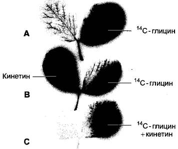
Through The Use of various combinations of cytokinins and phloem-mobile metabolites (such as The amino acid Glycine), it has been demonstrated that nutrients move within the plant from regions of low cytokinin concentration to regions of high cytokinin concentration (the "attractant effect" of cytokinins), while significantly fewer nutrients are exported from well-supplied tissues (the "retention effect" of cytokinins) (Fig. 7.48). Evidently, cytokinin concentration regulates, among other things, source-sink relations1 within the plant and thereby directs The pathway of phloem transport. Tissues well-supplied with cytokinins become functional sinks, importing nutrients from their cytokinin-depleted surroundings. At THE MOLECULAR LEVEL, cytokinin induces the accumulation of invertase within the cell wall. This leads to enhanced sucrose cleavage, which on one hand improves hexose supply to cytokinin-rich tissues and, on the other, promotes intensified sucrose unloading from the phloem (see 6.8.4). These mechanisms are crucial for establishing metabolite sinks with the aid of cytokinins.
1 The so-called "source-sink relations" (Eng.). — Ed. note.
Fig. 7.48. Attractant and retention effects of cytokinins in delaying leaf senescence: A – C — Autoradiograms of pinnate leaves of Vicia faba following the application of glycine labeled with the radioactive carbon isotope 14C; B, C — additionally treated with the cytokinin kinetin. The autoradiograms show the distribution of radioactivity within the leaves. Lines indicate the sites of substance application. A — Untreated control with kinetin; radioactive 14C-glycine is distributed throughout the treated leaflet and exported via the petiole. Only a minimal amount of radioactivity is present in the leaflet untreated with 14C-glycine; B — Attractant effect of cytokinin: massive accumulation of radioactivity occurs in the kinetin-treated leaflets; C — Retention effect of cytokinin: when kinetin and 14C-glycine are applied to the exact same spot on the leaf, no export of radioactivity occurs to other leaflets or through the leaf petiole.
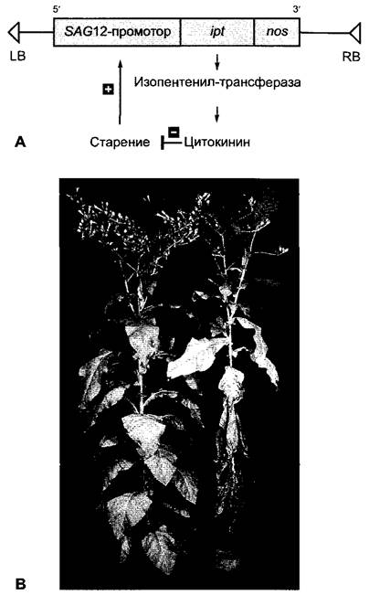
The significance of cytokinins in the aging of intact plants can also be illustrated through an elegant experiment using transgenic tobacco plants (for methodology, see Box 7.3) (Fig. 7.49). These plants expressed the previously mentioned ipt gene from Agrobacterium tumefaciens under the control of a promoter from a senescence-activated tobacco gene, SAG12 (senescence-activated gene). The onset of aging triggers the activation of this promoter, leading to The production of isopentenyl transferase in the Transgenic Plants and, consequently, enhanced cytokinin synthesis. As a result, the aging process is slowed down, causing promoter activity to subside once more. As illustrated in Fig. 7.49, this self-regulating system indeed leads to a substantial delay in sequential leaf senescence in tobacco. Only time will tell whether this approach holds potential for engineering genetically improved crop plants with enhanced yields.
Fig. 7.49. Delaying leaf senescence through regulated cytokinin synthesis in transgenic tobacco plants: A — Diagram of the genetic construct. Introduced into tobacco plants via Agrobacterium tumefaciens (Box 9.2), the chimeric gene consists of the senescence-activated promoter of the tobacco SAG12 gene, the coding region of the isopentenyl transferase (ipt) gene from A. tumefaciens, and the non-coding transcription termination region from the nopaline synthase (nos) gene of A. tumefaciens at the 3' end of the ipt gene. LB, RB — Left and right borders of the T-region of the A. tumefaciens Ti plasmid, respectively; B — In plants transformed with the chimeric gene (left), leaf senescence is markedly delayed compared to non-transformed control tobacco plants (right).
"Green islands" are occasionally observed on senapsed (or already shed) leaves: localized zones characterized by a visibly delayed aging process. This phenomenon is caused by the local release of cytokinins by parasitic bacteria or fungi (such as Erysiphe graminis or Uromyces phaseoli) or by parasitic insect larvae (such as Stigmella argentipedella). In the case of larvae, the labial glands are likely the sites of cytokinin biosynthesis. These parasites create a localized nutrient sink for themselves, effectively securing a "free meal."
A more practical and therefore widely studied aging process is fruit ripening, which shares common features with leaf senescence but also involves distinct specific processes and is discussed later (see 7.6.5.2).
7.6.2.4. Molecular mechanisms of cytokinin action
The exact mode of action of cytokinins is understood only in broad outlines, but many of their physiological effects are likely mediated by the Introduction/30.html">Regulation of Gene Expression. Specific genes responsive to cytokinins have been identified during Cell cycle regulation (see Fig. 7.19, A). The nitrate reductase gene is also under transcriptional control by cytokinins. Cytokinin receptors, much like ethylene receptors (see 7.6.5.3, Fig. 7.63), belong to the family of two-component receptor Kinases and are localized in the plasma membrane. Arabidopsis thaliana possesses two receptors structurally similar to each other and to ethylene receptors, designated CKI1 and CRE1 (named after the mutant phenotypes: cytokinin-insensitive and cytokinin-resistant, respectively)1. The binding of cytokinin to these receptors stimulates their phosphorylation at a conserved Histidine residue typical of two-component receptor kinases. The phosphate group is subsequently transferred to an aspartate residue (as in the ethylene receptor) and then to cytoplasmic Proteins of the AHP family (Arabidopsis histidine-phosphorelay proteins). Phosphorylated AHP proteins translocate into the cell nucleus and activate (via phosphorylation) a group of resident transcription factors, which in their phosphorylated form bind to the promoters of various target genes, thereby initiating their transcription. Consequently, at the Second Stage of signal Transduction (downstream of the AHP proteins), the signaling pathways originating from ethylene versus cytokinin receptors diverge.
1 Alternatively derived from "cytokinin receptor." — Ed. note.
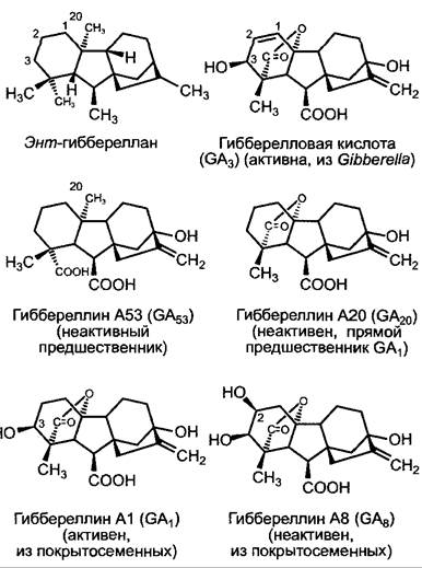
The extensive group of gibberellins consists of Diterpenes (see 6.16.2); a common structural feature of these hormones is the tetracyclic *ent*-gibberellane Skeleton (Fig. 7.50). To date, more than 100 structures have been described, but only a few of them are physiologically active. They differ in their action, specifically in stimulating internode elongation—particularly in dwarf varieties with impaired gibberellin biosynthesis or, correspondingly, in rosette plants, which are utilized for highly sensitive bioassays (see 7.6.3.3).
7.6.3.1. Diversity of Gibberellins
Gibberellins were initially discovered as a pathogenic factor produced by the rice-infecting fungus *Gibberella fujikuroi* (imperfect form *Fusarium moniliforme*). Infected plants exhibit excessive elongation and are prone to lodging due to insufficiently developed mechanical tissues (hence the disease
is known in Japan as "bakanae," meaning "foolish seedling disease"). This factor was named gibberellic acid. However, numerous related compounds were soon discovered, prompting the Introduction of a simple nomenclature system: gibberellin + A (for acid) + a number. Currently, gibberellins A1 to A116 (GA1–GA116) are known. Gibberellic acid from the bakanae pathogen is designated as GA3 under this nomenclature. Alongside *Gibberella fujikuroi*, gibberellins are also synthesized by the fungus *Sphaceloma manihoticola*, which induces giant growth in cassava plants. While gibberellin synthesis is less widespread among lower plants, it has been detected in all higher plants. The composition of gibberellins can vary significantly both among plant species and across different Organs of the same plant. As a rule, several gibberellins occur simultaneously within a plant organism (e.g., 14 in rice, 24 in immature apple seeds), though most of these are either precursors or catabolites of active gibberellins. The most important physiologically active gibberellins in angiosperms are gibberellin A1 (GA1) and gibberellin A4 (GA4); gibberellic acid (GA3) is rare in higher plants (for instance, in barley, where it plays a crucial role in mobilizing endosperm reserves, see 7.6.3.3). Many gibberellin-regulated processes can be triggered using GA3; consequently, this gibberellin, produced in large quantities from *Gibberella* culture filtrates, is most frequently used in experimental work.
Fig. 7.50. Structures of the gibberellin skeleton (*ent*-gibberellane) and some commonly occurring gibberellins. The prefix "*ent*-" derives from *enantio* (optical) and denotes a structure in which the chiral centers of the molecule are inverted. Thus, *ent*-gibberellane is the mirror image of gibberellane. The introduction of this seemingly overly complex nomenclature became necessary after The structure of kaurene—an intermediate in gibberellin biosynthesis—was found to be the mirror image of kaurene already described in the context of another synthesis, i.e., *ent*-kaurene.
7.6.3.2. Metabolism and Transport
Gibberellin biosynthesis is a multi-step process taking place across three stages and in three different cellular compartments, yet it involves only a few enzyme types (Fig. 7.51).
Fig. 7.51. Compartmentation of gibberellin biosynthesis. Double arrows represent multi-step reaction sequences in which four C5 units are combined to form geranylgeranyl pyrophosphate. For further explanations, see the text.
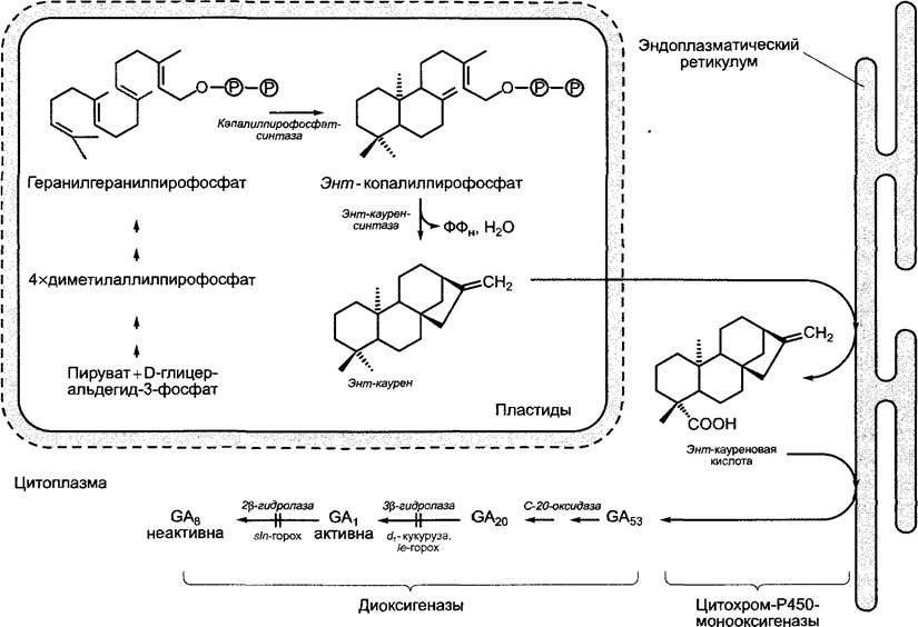
1. Formation of *ent*-kaurene from the universal diterpene precursor geranylgeranyl pyrophosphate (see 6.16.2). This reaction proceeds in two steps via the intermediate *ent*-copalyl pyrophosphate and is catalyzed by two enzymes—copalyl pyrophosphate synthase and *ent*-kaurene synthase—which belong to the terpene cyclases and are localized in Plastids. This step of gibberellin biosynthesis is inhibited by compounds such as chlorcholinchloride (= Cycocel, CCC) (Fig. 7.52). Cycocel is of practical importance in cereal cultivation (primarily wheat) and is used as a stem growth retardant to reduce lodging (lodging refers to the breakage of stems by wind or precipitation, which hinders mechanical harvesting).
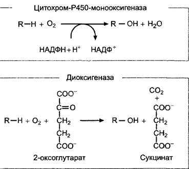
2. The water-insoluble pure hydrocarbon *ent*-kaurene leaves the plastids by an as-yet-unknown mechanism and is sequentially oxidized at The Endoplasmic reticulum to *ent*-kaurenoic acid, and subsequently to gibberellin A53 (GA53). This entire sequence of reactions is catalyzed by Enzymes of the cytochrome P450 monooxygenase group containing heme-bound iron (general reaction scheme, Fig. 7.53). An inhibitor of this biosynthesis step is ancymidol (see Fig. 7.52), which can also be used to cultivate plants with shortened internodes.
Fig. 7.52. Inhibitors of gibberellin biosynthesis.
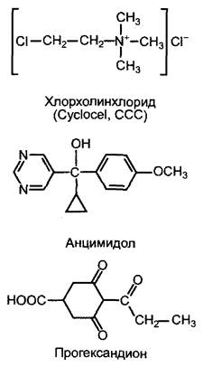
3. The formation of active gibberellins (most commonly GA1 from GA53) and their subsequent inactivation (not necessarily in the same cell) take place in the cytoplasm and are catalyzed by non-heme iron-containing Dioxygenases that oxidize 2-oxoglutarate as a co-substrate (for the general reaction scheme, see Fig. 7.53). In this process, carbon atom C-20 is first oxidized and released as CO2, after which the 19-carbon skeleton characteristic of active gibberellins, featuring a lactone ring (GA20), spontaneously forms. Activation occurs via 3β-hydroxylation of GA20 by the enzyme GA20 3β-hydroxylase, yielding GA1. Through the action of GA1 2β-hydroxylase, the active gibberellin A1 is converted into GA8, which is entirely inactive. The activities of 3β- and 2β-hydroxylases are thus critical in determining the level of active phytohormone within the cell. The transcription of the genes encoding these two enzymes is under strict control. Recently, specific inhibitors of 3β-hydroxylase have been developed, namely substituted cyclohexanediones (e.g., prohexadione, see Fig. 7.52). They induce very efficient internode shortening that can be reversed solely by GA1, but not by GA20 or GA8. Cyclohexanediones act as Competitive Inhibitors with respect to the co-substrate 2-oxoglutarate, preventing its binding to the catalytic center of the enzyme.
Fig. 7.53. General equations for Reactions Catalyzed by mono- and dioxygenases. Both enzyme types participate in gibberellin biosynthesis and in numerous other metabolic pathways.
Maize *d1* mutants (dwarf) and pea *le* mutants (used by Gregor Mendel in his crossing experiments, from "length") also provide evidence that a single specific gibberellin emerges during biosynthesis. GA1 serves as the active gibberellin in both maize and pea: both mutants lack 3β-hydroxylase function, and their dwarf phenotype can be rescued exclusively by GA1, but not by GA20 or GA8 (although, as already noted, the fungal gibberellin GA3 can be used instead of GA1). In pea *sln* mutants (slender), which exhibit excessive longitudinal growth, 2β-hydroxylase is impaired, preventing the inactivation of the active gibberellin.
In addition to the primary biosynthetic pathway discussed here, alternative pathways exist—for example, in gymnosperms—leading to gibberellins substituted at various positions. However, the general pathway of gibberellin biosynthesis is believed to be identical across all plants.
The genes for numerous enzymes involved in gibberellin biosynthesis have already been cloned, allowing their expression within the plant to be studied in greater detail and thereby providing insight into the sites of biosynthesis. Biosynthesis evidently occurs in many rapidly growing tissues (shoot meristems, growing leaves, shoot growth zones, root tips) and during early seed development; in these cases, the sites of synthesis and action cannot be spatially separated.
Within the shoot, gibberellins undergo non-polar transport; in roots, a weak acropetal polarity from the tip to the base is sometimes observed, with a transport velocity of 5–30 mm·h-1. Over short distances (e.g., in cereal grains, see 7.6.3.3), transport apparently occurs via simple diffusion. Gibberellins have been detected in both phloem and xylem sap; they are distributed throughout the plant via the mass flow of nutrients.
7.6.3.3. Action of Gibberellins
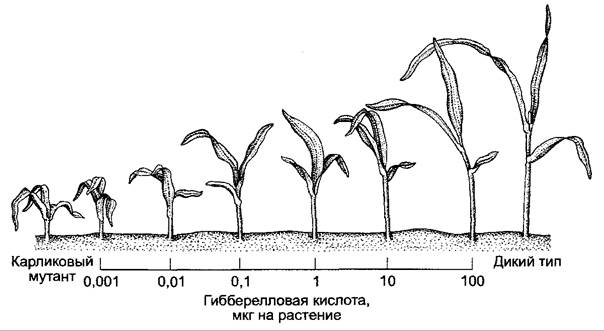
Gibberellins control a wide range of physiological processes: during vegetative development, they regulate shoot axis elongation (internode extension); during seed germination, they break dormancy (see 7.7.1.2) and mobilize reserve substances, particularly in cereal grains. During reproductive development, gibberellins can influence floral induction, flower Sex Determination, and fruit set. In this regard, their effects partly overlap with those of auxins. For instance, both induce parthenocarpy in apples and tomatoes; however, in many other processes, gibberellins and auxins exert opposing effects. Thus, gibberellins stimulate potato tuber sprouting (which auxins inhibit), inhibit lateral root initiation (which auxins stimulate), and stimulate root growth (whereas auxins stimulate at very low concentrations and inhibit at high ones); gibberellins have no effect on coleoptile elongation, whereas auxins do not stimulate internode extension. These differences in the regulation of internode and coleoptile growth form the basis for highly sensitive and specific gibberellin bioassays utilizing dwarf varieties (such as maize *d1* mutants) in which gibberellin synthesis is reduced or absent (Fig. 7.54).
Fig. 7.54. Growth response of dwarf maize seedlings (*dwarf1*) to a single application of various amounts of gibberellic acid (GA3 applied as an aqueous solution to the first leaf axil). Left: dwarf plant without GA3 application; right: wild-type plant of the same age (control).
Stimulation of internode elongation by gibberellins is due to the promotion of both cell growth and cell division. The molecular mechanisms remain largely unknown. However, gibberellin induces the formation of xyloglucan endotransglucosylase (XET — see 7.6.1.4) and thereby lowers, much like auxin, the threshold potential for plastic deformation of the cell wall, as this enzyme partially hydrolyzes the hemicellulose network of primary cell walls. This is thought to provide expansin with improved access to the hydrogen bonds between hemicellulose and cellulose, which are subsequently disrupted by its action. Unlike auxin, however, gibberellin action does not result in apoplast acidification. The difference between the effects of gibberellin and auxin is also evident from the fact that the contributions of both phytohormones to growth stimulation are additive.
In the induction of flowering or the stimulation of floral organ initiation, particularly in rosette plants, the application of gibberellin can frequently replace the effect of an external factor: for example, the effect of low temperatures (see 7.7.1.2, 7.7.1.3) or the inductive photoperiod in long-day plants that do not require cold treatment, such as Hyoscyamus niger or Spinacia oleracea (see 7.7.2.2). These external factors exert their effects by increasing endogenous gibberellin levels. In spinach, for instance, under conditions that induce Flower Formation and branching under long-day regimes (see 7.7.2.2), the levels of all intermediates from GA53 to active GA1 and its inactivation product GA8 steadily increase day by day (see Fig. 7.50). Among other processes, light stimulates the transcription of gibberellin 20-oxidase and gibberellin 3β-hydroxylase genes via the photoreceptor Phytochrome (see 7.7.2.4).
On sex expression in monoecious plants (see 11.2), such as cucumber, gibberellins and auxins exert opposing effects. While auxins stimulate the formation of female (pistillate) flowers and thus fruit set, gibberellins promote an increased formation of male (staminate) flowers. Consequently, inhibitors of gibberellin biosynthesis (see Fig. 7.52) stimulate the formation of pistillate flowers, much like auxins do, and are utilized in agricultural practice to promote fruit set in cucumbers. In maize, by contrast, the initiation of female inflorescence meristems occurs at a higher endogenous gibberellin level than the induction of male flowers. Gibberellin activates genes whose products block The Development of the androecium. This regulation
of sex is an example of how most developmental processes are governed not by a single phytohormone, but rather by the complex interplay of various phytohormones combined with external factors (see 7.7).
The Role of gibberellins in seed germination, particularly in Poaceae, has been studied most intensively. The majority of investigations have been carried out on barley.1 The Poaceae caryopsis is a dry fruit in which the seed coat and pericarp are fused together (see 11.2). These tissues surround the remaining seed tissues: the embryo and the triploid nutritive tissue (endosperm); the latter consists of the central starchy endosperm (whose cells die off at maturity) and an aleurone layer composed of living cells — either single-layered (e.g., in wheat) or multi-layered (e.g., three-layered in barley). The embryo adjoins the endosperm via its cotyledon, which is modified into a specialized absorbing organ termed the scutellum.2
1 Barley germination is of major economic importance in malting (a key component in beer brewing). — Editor's note.
2 The exact Nature of the scutellum in grasses remains a subject of debate. It has been suggested either that the scutellum is not a derivative of the cotyledon, or that it arises from the fusion of the cotyledon with other PARTS OF THE embryo. — Editor's note.
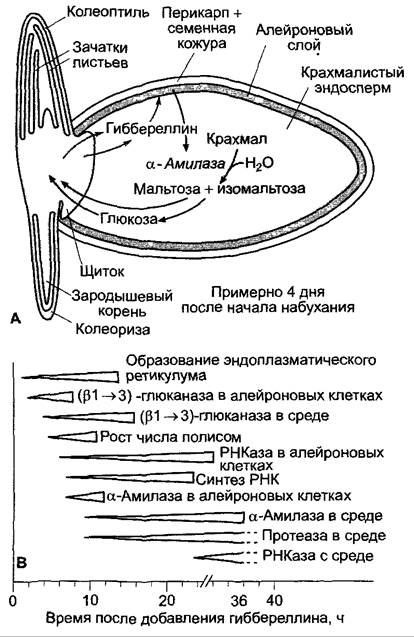
During germination, reserve starch is mobilized through hydrolytic breakdown (see 6.17.1.2). The enzymes required for this process (amylases) are partially secreted by the scutellum (β-amylases); however, the majority are produced in the aleurone layer in response to a signal from the embryo and are secreted into the starchy endosperm (α-amylases). The signal originating from the embryo consists of gibberellins (most likely GA3 in barley, and predominantly GA1 in wheat), which are released by the scutellum and diffuse into the endosperm. If the embryo is removed, α-amylase is not produced; however, the physiological action of the embryo can be functionally replaced by low concentrations of active gibberellin (e.g., GA3); an isolated aleurone layer, when incubated in GA3 solutions, synthesizes and secretes α-amylases on its own.
The effect of gibberellins on the aleurone layer is complex, and α-amylase secretion is preceded by a multitude of events (Fig. 7.55). In addition, many other hydrolytic enzymes are produced, which break down cross-linking glycans of the cell walls (glucanases), storage proteins (proteases), or Nucleic Acids (RNases). Gibberellin induces either both the Synthesis and Secretion of these enzymes (as in the case of α-amylase) or solely their secretion, while synthesis may proceed even in the absence of added gibberellin (certain glucanases, RNases). Enzyme production and secretion are consequently controlled independently of one another via gibberellin.
Fig. 7.55. Gibberellin-induced processes during caryopsis germination: A — state approximately 4 days after the onset of imbibition. Gibberellin release by the embryo begins roughly 12 h after THE START OF imbibition, and α-amylase secretion follows 8–10 h later. Starch hydrolysis begins near the scutellum and proceeds distally over several days; B — sequence of gibberellin-induced events in isolated barley aleurone layers
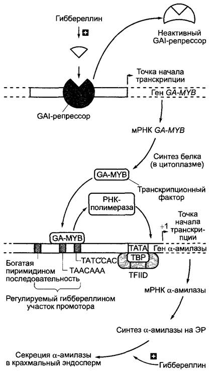
The molecular aspects of α-amylase production during gibberellin induction have been only partially elucidated (Fig. 7.56). Gibberellin stimulates the activation of α-amylase genes (enzymatic activity, represented here in a simplified manner as α-amylolytic, is provided by a family of Isoenzymes), thereby promoting the formation of α-amylase mRNA and the subsequent de novo biosynthesis of the enzyme protein. Both the transcription factor participating in this process and the cis-elements within the amylase promoters responsible for the physiological response to gibberellin have been identified (Fig. 7.56). The gibberellin receptor is apparently localized in the plasma membrane of aleurone cells.
Fig. 7.56. Model of the regulation of α-amylase production by gibberellin. An as-yet-unknown gibberellin signaling pathway stimulates the inactivation of a repressor protein encoded by the GAI gene1 (gibberellin-insensitive). This leads to the initiation of transcription of the GA-MYB gene, which encodes a specialized transcription factor of the MYB family. The GA-MYB transcription factor binds to sequence elements (likely a TAACAaa sequence) in the promoter of α-amylase genes, thereby triggering their transcription (see Fig. 7.10). Synthesized on the endoplasmic reticulum, α-amylase is secreted via the Golgi apparatus. The secretion process is also gibberellin-dependent and is regulated through a second, still poorly understood signaling pathway.
1 Data from Arabidopsis and cereals have been synthesized in this scheme. Specifically, the GAI gene originates from Arabidopsis. — Editor's note.
The phytohormone does not act alone in this process either. The action of gibberellin can be terminated by the simultaneous application of abscisic acid (see 7.6.4). An initially high level of abscisic acid in the caryopses upon the onset of germination drops rapidly before the gibberellin level rises. Therefore, abscisic acid is likely involved in the in vivo control of caryopsis germination processes.
The stimulation of seed germination by gibberellins is not restricted to cereals. In seeds or fruits of dicots, exogenously applied gibberellic acid not only accelerates germination but in many cases makes it possible even when the requisite external conditions are absent. For instance, the germination of hazelnut (Corylus avellana) typically requires a cold period (about 12 weeks at 5 °C). This stratification can be replaced by the application of gibberellic acid. Seeds whose germination requires light (light-dependent germination, see 7.7.2.2) can partially germinate in the dark if supplied with gibberellic acid.
Alongside phytohormones that exert predominantly stimulatory effects on metabolism and development, plants also contain biologically active substances with predominantly inhibitory effects. These include: abscisic acid (ABA), which was originally described as a factor promoting premature fruit abscission in cotton; abscisin II; and dormin as a factor ensuring bud dormancy. Research has demonstrated the structural identity of abscisin II and dormin.
7.6.4.1. Metabolism and Transport of Abscisic Acid
Abscisic acid is found in all lower and higher plants, including algae and fungi, as well as in cyanobacteria, but is absent in other bacteria and archaea. Large amounts of ABA are produced by the phytopathogenic fungus Cercospora rosicola (a pathogen of roses). In vascular plants, it is believed that all plastid-bearing cells are capable of ABA biosynthesis. It begins with the oxidative cleavage of 9-cis-neoxanthin, a violaxanthin derivative found in plastids and belonging to the xanthophylls. The cleavage product, xanthoxin, is converted in the cytoplasm via abscisic aldehyde into abscisic acid (Fig. 7.57). Consequently, ABA in vascular plants is an apocarotenoid.1 In fungi, ABA is formed via the cyclization of farnesyl pyrophosphate (see 6.16.2) and can thus be classified as a sesquiterpene. The older hypothesis postulating that this biosynthetic pathway should also occur in higher plants has not been confirmed. For example, maize Mutants with Impaired biosynthesis also synthesize significantly lower amounts of ABA (see 7.6.4.2).
1 The introduced terms are intended to emphasize the Specific features of biosynthesis. In Russian literature, ABA is characterized as a sesquiterpene, giving this term a purely structural significance. — Editor's note.
Fig. 7.57. Metabolism of abscisic acid (ABA). The phytohormone is formed in higher plants through the breakdown of the xanthophyll 9-cis-neoxanthin, and in fungi via the cyclization of farnesyl pyrophosphate. In the tomato mutants flacca (flc) and sitiens (sit), The conversion of abscisic aldehyde to ABA is impaired. Both mutants wilt very rapidly because, under water deficit, they are no longer able to close their Stomata. This defect can be rescued by spraying the plants with an ABA solution. In the viviparous mutants vp2 and vp5 of maize, disruptions occur in the Cytology/cytology/16.html">Early stages of carotenoid biosynthesis, leading to a deficiency in ABA and, consequently, to vivipary.
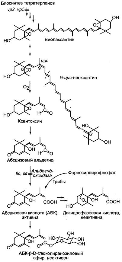
The phytohormone is inactivated by oxidation to dihydrophaseic acid or by Esterification with glucose.
ABA is found in all plant organs. The highest amounts of ABA are observed in autumn in dormant buds, as well as in seeds and fruits (primarily in fruit tissues). In certain situations, especially during drought, poorly watered tissues produce large amounts of ABA within a few hours. The biosynthesis of the phytohormone is triggered when turgor drops below a threshold value, rather than in response to a decrease in water potential (see equation 6.15). In leaves under water deficiency, the ABA content can increase more than 40-fold. ABA is also produced in the root system when turgor drops due to water shortage. The phytohormone is transported from the root via the xylem to the shoot; with the transpiration stream, it reaches the stomata and induces their closure (see 7.6.4.2;
8.3.2.5). The ABA produced and released in the leaf under water stress is transported via the phloem and thus reaches the root. There, ABA participates in increasing hydraulic conductivity, thereby enhancing the water-absorbing capacity of the root. Phloem and xylem transport of abscisic acid thus serve to coordinate the water status of the shoot and root. Over short distances, ABA is apparently transported by cell-to-cell diffusion; ABA released into the apoplast is distributed with the water flow (see Fig. 6.32). In the petioles of young leaves and internodes, the parenchymal transport of ABA is basipetally polarized and occurs at a rate of ~3 cm • h-1, i.e., twice as fast as IAA (see 7.6.1.3). Nothing is known about the transport system.
7.6.4.2. Action of Abscisic Acid
Abscisic acid was isolated from prematurely shed immature cotton bolls, where it is present in high concentrations. ABA stimulates organ abscission when applied to a leaf petiole or pedicel. This occurs only under certain conditions (e.g., when auxin supply is
suppressed in leaves due to removal of the leaf blade), but this does not represent the primary action of ABA. Organ abscission is explained rather by the release of ethylene induced by the treatment,1 which is why the name abscisic acid (from abscission) is poorly chosen, although it has become generally accepted.
1 ABA induces stress-induced leaf fall under water deficit. In the climate of Central Russia and Europe, mass leaf fall is regulated by ethylene, whereas seasonal leaf fall in arid regions is thought to be controlled by ABA. — Editor's note.
The physiological effects of ABA can be divided into two groups:
✵ Regulation of the dormant state of plant organs;
✵ regulation of the plant's water balance.
In addition, ABA often acts as an antagonist to other phytohormones. For example, externally applied ABA inhibits auxin-induced extension growth, and gibberellin-induced synthesis of α-amylase in the aleurone layer—they are also antagonists of ABA during the transition to dormancy or, respectively, upon emerging from it (see below). ABA stimulates leaf senescence, whereas cytokinins inhibit this process.
The accumulation of ABA in seeds or fruit flesh, due to its inhibitory effect on germination, is an important factor in seed dormancy. Seed dormancy depends on the Anatomical Structure of the seed coat, which in mature seeds, for example, impedes the access of water or oxygen. Germination occurs after its removal or mechanical damage. However, seed dormancy is often physiologically determined: in the absence of the seed coat, no further development takes place even under favorable conditions. On the other hand, in mutants with impaired ABA formation (e.g., in maize mutants vp2 and vp5, see Fig. 7.57), seed germination often occurs already on the mother plant (viviparity, hence the mutant designation viviparous). ABA has proven to be the embryonic factor of seed dormancy.
In experiments on Arabidopsis thaliana, it was shown that the embryo genotype, distinct from that of the mother plant, is responsible for ABA-dependent dormancy. Seed dormancy is thus determined by the ABA content of the embryo rather than the fruit tissues. In fleshy fruits such as berries, a presumably high level of ABA in the fruit flesh helps prevent premature seed germination. In some seeds (e.g., walnut, apple, rose), stratification (see 7.7.1.2) decreases The amount of ABA, thereby stimulating germination. Moreover, stratification often promotes the stimulation of gibberellin synthesis. In many cases, experimental seed dormancy induced by ABA can be interrupted by adding gibberellin. It has been shown that for the transition into dormancy or, respectively, the termination of embryo dormancy, the absolute content of both phytohormones is less important than the ratio of ABA and gibberellin concentrations.
During seed germination, ABA is not only responsible for embryo dormancy but also induces the formation of storage proteins in seeds. During embryogenesis, after the completion of cell divisions and before the onset of the cell elongation phase associated with the accumulation of storage substances in the embryo or endosperm tissue, a temporary sharp increase in abscisic acid concentration occurs in seeds. It is suggested that along with the induction of storage protein synthesis, other proteins (e.g., dehydrins) are also synthesized upon increased ABA concentration; they serve to structurally protect cells during the dormant phase of seed development when the water content in the tissue drops to 10% and below.
The onset of bud dormancy is usually (though not always) associated with an increase in ABA concentration and often with a decrease in cytokinin and gibberellin concentrations, whereas during vernalization (see 7.7.1.3), the ABA content decreases again while gibberellin and cytokinin concentrations simultaneously increase. If shoots are vernalized in the presence of exogenous abscisic acid, dormant buds, for example in ash trees, do not break dormancy.
ABA is of particular physiological importance for water balance regulation. Under water deficit, ABA stimulates stomatal closure, increases root hydraulic conductivity, and promotes root growth while simultaneously inhibiting shoot growth. The latter effect can be viewed as a long-term adaptation to chronic water scarcity (increasing the water-absorbing surface relative to the transpiring surface). The increase in hydraulic conductivity in root tissues in response to water deficit in the shoot occurs within a few hours and is likely regulated by ABA produced in the shoot and transported via the phloem to the root (mechanism unknown). Conversely, the control of stomatal aperture width by ABA occurs within minutes, allowing for an efficient, rapid, and reversible regulation of the balance between transpiration and turgor in the entire leaf (see Fig. 6.77) or even in localized areas within the leaf. With local water deficit, the time interval from the initiation of de novo ABA synthesis to the release of the phytohormone by mesophyll cells is short, so that the signaling substance rapidly reaches the target cells—the guard Cells of the stomata—with the transpiration stream.
Using highly sensitive Analytical Methods, ABA amounts were determined in isolated guard cells from well-watered (stomata open) or, respectively, water-stressed leaves (stomata closed) of Vicia faba: with closed stomata, the ABA content in guard cells is 20 to 25 times higher than with open stomata (Fig. 7.58). The significance of ABA for the regulation of stomatal transpiration is confirmed by experiments with ABA-deficient mutants, which wilt extremely rapidly and cannot close their stomata. Thus, the wilting tomato mutants flacca (flc) and sitiens (sit) contain about 10% of the ABA amount found in wild-type plants and can grow only in a water-vapor-saturated atmosphere. However, after ABA supplementation, the function of stomatal guard cells and water balance normalize.
Fig. 7.58. Abscisic acid content in guard cells of Vicia faba with open or, respectively, closed stomata. The analysis was performed on single isolated pairs of guard cells (A) using a highly sensitive immunological method, wherein in the shown experiments 1, 2, and 3 (B), 10, 20, or, respectively, 50 guard cell preparations from the epidermis of water-stressed plants W (stomata closed, dark bars with standard deviations) or, respectively, 100 guard cell preparations from the epidermis of well-watered plants (C — control, stomata open) were used for analysis; 1 femtogram (fg) = 10-15 g
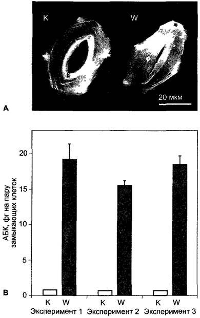
The Molecular Mechanism of ABA action on guard cells is only partially known. The processes underlying guard cell movement are discussed in more detail in Chapter 8 (see Section 8.3.2.5 and Figs. 8.32; 8.33).
The effect of ethylene on plants has been known for 100 years, but it was not until the 1960s, with the development of sensitive gas Chromatography analytical methods, that it was proven that all plants constantly produce and release small amounts of this gaseous substance with the simple structural formula H2C=CH2 into the environment. Ethylene belongs to the five "classical" groups of phytohormones. Due to its volatility, it can act not only as a hormone (signaling substance within an individual), but also as a pheromone (signaling substance between individuals of the same species) and even between individuals of different species (kairomone).
The constant synthesis of small amounts of ethylene appears to be necessary for the normal growth of higher plants. The tomato mutant diageotropica cannot form ethylene. It grows diageotropically (see 8.3.1.2) rather than orthotropically,1 but exhibits normal growth in an atmosphere containing as little as 0.005 µL of ethylene per liter of air.
1 Diageotropic growth is growth in a horizontal direction, i.e., "neutral with respect to top and bottom." It is also called plagiotropic growth. Orthotropic growth is growth in a vertical direction, i.e., "straight with respect to top and bottom." — Editor's note.
7.6.5.1. Metabolism and Transport of Ethylene
Ethylene is synthesized by bacteria, fungi, and plants from Methionine (Fig. 7.59) (some bacteria use glutamate or 2-oxoglutarate, respectively, as an initial substrate). The direct precursor of ethylene, 1-aminocyclopropane-1-carboxylic acid (ACC), is derived from S-adenosylmethionine. This reaction, catalyzed by the enzyme ACC synthase, determines The rate of ethylene formation; furthermore, the enzyme is characterized by a high metabolic turnover rate, making it a key regulatory checkpoint for ethylene biosynthesis. ACC synthase induction is triggered by various environmental factors, including mechanical wounding and stress, flooding, drought, and cold, as well as the onset of senescence in flowers and fruits. High concentrations of indole-3-acetic acid also promote ACC synthase induction. It is hypothesized that many of the physiological effects of auxin observed upon application of high concentrations of IAA are actually mediated by induced ethylene synthesis. This holds true for IAA-induced inflorescence development in bromeliads (e.g., pineapples) and growth inhibition at high auxin concentrations. Pyridoxal phosphate serves as a cofactor for the ACC synthase reaction; consequently, the formation of ACC, and thereby ethylene synthesis, can be significantly reduced using inhibitors of pyridoxal phosphate-dependent enzymes, such as aminooxyacetic acid and aminoethoxyvinylglycine.
Fig. 7.59. Biosynthesis of ethylene from L-methionine and the reaction sequence for methionine regeneration (Yang cycle)
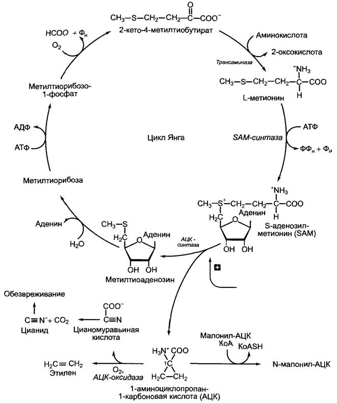
ACC is cleaved by the enzyme ACC oxidase (a dioxygenase, see Fig. 7.53) in an oxygen-dependent reaction yielding ethylene and cyanoformic acid. The latter spontaneously decomposes into CO2 and HCN. Cyanide (CN-) is detoxified by conversion into β-cyanoalanine, which is subsequently metabolized to asparagine and aspartic acid. A portion of ACC conjugates with malonic acid to form N-malonyl-ACC, which then accumulates in the vacuole. Malonyltransferase is regulated by light via the phytochrome system (see 7.7.2.4). N-malonyl-ACC undergoes no further breakdown, representing an irreversible conjugation that serves to limit ACC levels and thereby regulate ethylene formation. As a gaseous compound, ethylene readily diffuses out of the plant, meaning that degradation pathways play no role in eliminating the active phytohormone.
An essential step in ethylene biosynthesis is the regeneration of methionine from S-adenosylmethionine's second reaction product, methylthioadenosine (see Fig. 7.59). Named the Yang cycle after its discoverer, this biochemical pathway enables plants to produce ethylene over extended periods without requiring continuous de novo synthesis of methionine. This is particularly important, for instance, in fruits after they have been detached from the mother plant.
7.6.5.2. Physiological Effects of Ethylene
Like all phytohormones, ethylene influences a multitude of physiological processes across various stages of plant development. In etiolated seedlings (grown under severe light deprivation), minute amounts of ethylene (0.1–1 µl per liter of air) strongly inhibit stem elongation growth, which is accompanied by enhanced radial expansion and a disruption of the characteristic negative gravitropism. This triad of symptoms is known as the triple response (see Fig. 7.62)1. Additionally, ethylene-treated seedlings form a sharply curved apical hook. Mechanical stress (e.g., caused by soil resistance) intensifies ethylene production in etiolated seedlings; thus, the triple response can be viewed as an integrated mechanism enabling seedlings to navigate obstacles in the soil or grow along the path of least resistance. The site of maximal ethylene biosynthesis in the etiolated seedling is the region of the apical hook. Upon illumination, ethylene production decreases via engagement of the phytochrome system (see 7.7.2.4). As a result, the ethylene-induced inhibition of elongation growth in cells lateral to the hook ceases, and the hook disappears.
1 Termed the "triple response" in English literature. The effect was first described by D. N. Neljubov in 1901. — Ed. note.
However, the reduced ethylene synthesis in illuminated shoots eventually rises again in response to various stimuli, such as wounding (defense — see 9.4.1) and mechanical stress (e.g., wind action). In the latter case, the enhanced production of ethylene retards longitudinal growth while stimulating radial expansion and increased formation of mechanical tissues, collectively increasing the mechanical resistance of the plant body. Accumulating evidence suggests that oxylipins (see 7.6.6.2) are also involved in regulating these processes.
The enhanced apical growth of the shoot axis—induced by ethylene concomitant with the suppression of longitudinal growth—proceeds at THE CELLULAR LEVEL through the reorientation of cortical microtubules from a transverse to a longitudinal arrangement. According to current models, this prompts a corresponding reorientation of newly synthesized cellulose microfibrils in the cell walls, as cellulose synthase complexes embedded in the plasmalemma migrate along the peripheral Cytoplasmic microtubules (see 6.17.1.1). Mechanical resistance during elongation growth is perpendicular to the orientation of the majority of cellulose fibrils; consequently, ethylene-induced synthesis of cellulose microfibrils predominantly in a longitudinal orientation drives enhanced radial cell expansion.
In many wetland and aquatic plants that possess both submerged organs and aerial structures (such as flowers, leaves, and floating leaves), ethylene stimulates longitudinal growth of the shoot axis and the formation of air spaces (aerenchyma — see Fig. 3.8). This facilitates improved oxygen supply to deeply submerged organs.1 Certain rice varieties (deepwater rice) can elongate at rates of up to 25 cm per day, reaching lengths of up to 5 m. They are thus capable of flowering and fruiting even under prolonged submergence. It is assumed that ethylene concentrations are elevated in submerged organs because less ethylene escapes from the tissues via diffusion. Seedlings of certain mesophytes (e.g., cereals) also enhance longitudinal growth under waterlogged soil conditions.
1 Aerenchyma also improves the mechanical properties of submerged organs by acting as a "float" that prevents the current from excessively deforming the plant body. — Ed. note.
Ethylene inhibits root growth, yet is essential for the development of lateral and adventitious roots; it participates in root Hair formation and also stimulates aerenchyma development in roots (e.g., in maize).
In Cucurbitaceae, ethylene treatment significantly increases the ratio of male to female flowers, whereas in Bromeliaceae, ethylene induces flowering. This property is exploited, for example, to synchronize inflorescence emergence on pineapple plantations. Because the application of gaseous ethylene is difficult to control, 2-chloroethylphosphonic acid (ethephon) is used instead (and in many other Applications, see below); in aqueous solution, it slowly decomposes into ethylene, phosphate, and chloride (Fig. 7.60).
Fig. 7.60. Release of ethylene from ethephon (2-chloroethylphosphonic acid) in aqueous solution
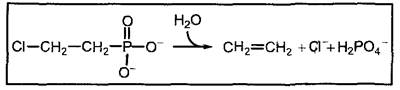
In numerous species, ethylene application breaks physiological dormancy (see 7.6.4.2), as observed in caryopses of Poaceae, peanuts, bulbs of many Liliaceae (e.g., tulip) and Amaryllidaceae (e.g., Narcissus), rhizomes and corms of Iridaceae (e.g., Iris, Gladiolus), and the axillary buds of certain species (e.g., potato).
Ethylene is a crucial regulator of senescence and the abscission of leaves, flowers, and fruits.
Many fruits undergo a phase of intense respiration during ripening—a process that can be viewed as senescence encompassing all maternal tissues (Climacteric respiration). Fruits exhibiting a pronounced respiratory climacteric include apples, pears, bananas, avocados, custard apples, peaches, and tomatoes, whereas others (such as cherries, grapes, strawberries, and citrus fruits) lack this phenomenon. In climacteric fruits, ethylene accelerates ripening, with peak endogenous ethylene synthesis occurring roughly concurrently with intense climacteric respiration. The physiological significance of ethylene in fruit ripening was elegantly demonstrated in transgenic tomatoes, where antisense suppression (see Box 7.4) drastically reduced the levels of either ACC synthase or ACC oxidase (see Fig. 7.59). This significantly lowered ethylene production and delayed fruit ripening; however, complete fruit ripening in transgenic tomatoes requires exogenous ethylene supplementation.
During the ripening of edible zoochorous fruits (see 11.2, seed and fruit dispersal), starch is hydrolyzed into sugars, organic acids are degraded via respiration, and color shifts occur due to chlorophyll breakdown and the synthesis of anthocyanins and/or carotenoids. Finally, through the partial degradation of cell walls and middle lamellas, fruit tissues soften—events that collectively enhance the appeal and palatability of the fruit. The synthesis of many enzymes involved in these processes (e.g., chlorophyllase, polygalacturonase) is induced by ethylene.
In agricultural practice, treating tomato plants with ethephon induces simultaneous fruit ripening, which facilitates harvesting. Many fruits (such as bananas) are harvested unripe and held during refrigerated ship transport in an atmosphere where ethylene levels are kept low by passing air through an activated carbon filter1. Simultaneously, CO2 is added to the atmosphere (acting as an ethylene antagonist). Raising the Temperature, removing CO2, and applying ethylene at the appropriate time prior to marketing triggers the ripening process.
1 Referring to activated charcoal. — Ed. note.
As a rule, ethylene also promotes the senescence of floral organs. For instance, in many flowers (such as orchids), perianth lobes senesce rapidly following pollination—a response triggered by ethylene release. Because floral senescence accelerates after detachment from the mother plant, cut flowers are frequently treated with ethylene inhibitors (e.g., silver thiosulfate, where Ag+ is the active ion) to extend their vase life. Inhibiting ethylene biosynthesis via antisense suppression leads to a dramatic delay in the senescence of both fruits and flowers (e.g., carnations). Conversely, leaf senescence is only weakly stimulated by ethylene: ethylene-insensitive mutants of Arabidopsis thaliana (see 7.6.5.3) display normal, albeit marginally delayed, leaf senescence.1
1 On the one hand, so-called "senescence" in Arabidopsis correlates with fruit ripening, much like in other monocarpic species, rather than representing autonomous leaf senescence (see 7.6.2.3). On the other hand, ethylene-"insensitive" mutants still produce normal amounts of ethylene, but exhibit reduced sensitivity. According to current understanding, Arabidopsis possesses at least five ethylene receptors (see 7.6.5.3), and the disruption of a single receptor through mutation has little impact on the overall phenotype (provided the other receptors remain functional). — Ed. note.
Abscission of leaves, flowers, and fruits, and occasionally branches as well (such as in poplar), represents a normal developmental phase in perennial plants. Through this mechanism, the plant can, first, discard superfluous or dysfunctional organs (e.g., unpollinated and unfertilized flowers; fruits in which seed development has ceased; spent leaves) and, second, facilitate the dispersal of mature fruits. The structural and physiological causes underlying the abscission of leaves, flowers, and fruits are very similar, with leaf abscission being the most thoroughly studied.
Deciduous woody plants shed their leaves in autumn, whereas evergreen and tropical plants do so throughout the year. Leaf abscission under specific climatic conditions (such as the onset of drought or physiological dryness resulting from lower temperatures, see 6.3.3) may be necessary to prevent excessive water loss. Furthermore, over prolonged periods of transpiration, all leaves accumulate ballast ions (e.g., Ca2+, which can no longer be transported back via the phloem, see Table 6.10), rendering them non-functional over time; therefore, leaf shedding is functionally equivalent to the Elimination of Metabolic waste.
Leaf abscission (as well as the shedding of flowers and fruits) occurs through the formation of an abscission zone at the base of the leaf petiole (Fig. 7.61). This zone consists of small parenchymal cells with narrow intercellular spaces. Although laid down during the early stages of organ development, this tissue fully differentiates only under appropriate environmental conditions. The abscission process itself is active, requiring the synthesis of specialized enzymes, primarily cellulases and polygalacturonases. Depending on the plant species, Separation proceeds either via the dissolution of middle lamellae (by polygalacturonases), middle lamellae and primary walls (by polygalacturonases and cellulases), or the entire cell.
Fig. 7.61. Longitudinal section through the basal part of a leaf petiole of a dicotyledonous plant, showing a developed yet undifferentiated abscission layer
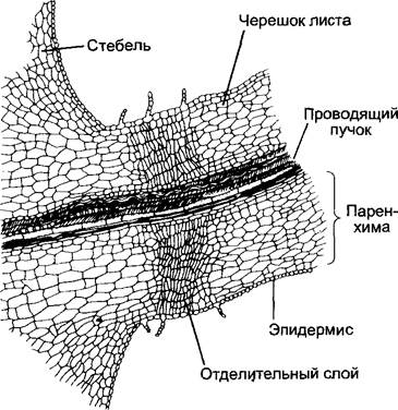
The differentiation of the abscission layer is controlled by various phytohormones, with auxin (IAA) and ethylene playing the primary roles. The process can be divided into three phases.
Phase 1. As long as there is a sufficient influx of IAA from the leaf, the cells of the abscission layer remain unresponsive and insensitive to ethylene. The differentiation of the abscission layer is thus suppressed.
Phase 2. With a declining flow of IAA from the leaf (e.g., as a result of the onset of senescence), the cells of the abscission tissue acquire sensitivity to ethylene.
Phase 3. The cells of the abscission tissue respond to endogenous ethylene by synthesizing cellulase, galacturonase, and other hydrolytic enzymes. The activity of these enzymes drives the differentiation of the abscission layer (dissolution of middle lamellae and cell walls) and subsequently leads to leaf fall. Consequently, premature fruit drop can be prevented by spraying plants with low-concentration auxin solutions (e.g., to delay the drop of mature citrus fruits)1.
1 In many citrus species, fruits do not drop simultaneously. Some may even drop In the second year following pollination. Thus, fruit abscission does not necessarily occur during its first year of development. This phenomenon is utilized for "extended tree storage of ripe fruits" ("Lagemny am Baum") to allow for the unhurried harvesting of undamaged citrus fruits. — Ed. note.
However, treating plants with high doses of auxin induces ethylene biosynthesis, leading to the opposite effect. For instance, "Agent Orange" was used as a defoliant during the Vietnam War2. One of the components of "Agent Orange" is the synthetic auxin 2,4,5-trichlorophenoxyacetic acid (2,4,5-T), a relative of 2,4-D (see Fig. 7.32).
2 "Agent Orange" is a notorious example of "environmental warfare" waged by the US military against guerrillas. Leaf defoliation facilitated the tracking of Vietnamese military movements in the rainforest. "Agent Orange" proved to be a mutagen, and its application destroyed thousands of hectares of vegetation cover, adversely affecting both wildlife and humans. — Ed. note.
Due to the multitude of agriculturally important processes regulated by ethylene, commercial preparations are widely used
that decompose to release ethylene (e.g., ethephon) or act as its antagonists (e.g., Ag+). Ethylene is used to stimulate latex flow in Hevea brasiliensis, the primary supplier of natural rubber, for which demand continues to outstrip supply. Treatment with ethephon at the cut sites can triple the yield of latex oozing from the severed laticifers compared to untreated trees.
7.6.5.3. Molecular Mechanisms of Ethylene Action
Knowledge regarding ethylene reception and signal transduction pathways has expanded significantly in recent years. This progress has been driven by The Study of tomato and Arabidopsis thaliana mutants defective in the signaling pathway. Due to their characteristic phenotypic traits, such mutants are easily identifiable (Fig. 7.62). These include, on the one hand, ethylene-resistant etr or ers mutants (from German ethylenresistent), and on the other hand, mutants exhibiting a constitutive triple response (ctr mutants).
Fig. 7.62. Phenotypes of Arabidopsis thaliana seedlings grown in the dark under specific conditions for approximately 3 days. In contrast to the wild type, the etr1 mutant shows no triple response upon the addition of exogenous ethylene; conversely, the ctr1 mutant exhibits the triple response even in the absence of exogenous ethylene. For Abbreviations, see Fig. 7.63
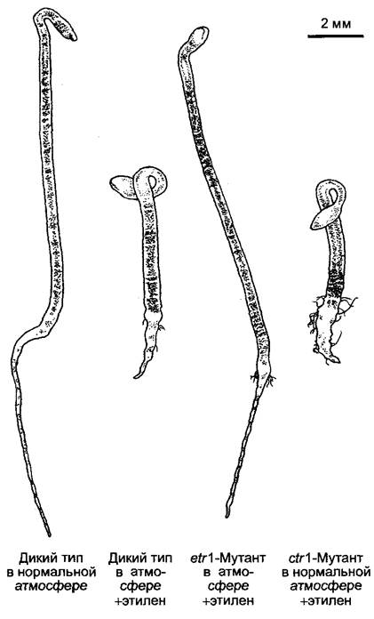
Ethylene binds to homodimeric receptors (i.e., consisting of two identical subunits) that share high sequence similarity with bacterial two-component receptor kinases and are localized in the plasma membrane. At least five ethylene receptors are currently known in Arabidopsis thaliana, which sometimes occur in different tissues or interact within the same cell via as-yet-unknown mechanisms. Ethylene binds to Cu+ ions, which are an integral part of the receptors. The binding sites are located within the transmembrane segments near the N-terminus of the receptor protein.
In the absence of ethylene, the receptors suppress the ethylene signaling pathway by activating the CTR1 gene-encoded protein kinase, which acts as a negative regulator of this pathway. When ethylene binds to the receptors, the receptors cease to interact with the CTR1 protein kinase, leading to the inactivation of the kinase. Consequently, the CTR1-mediated inhibition of the ethylene signaling pathway is lifted, thereby activating the pathway. This explains why ctr1 mutants, which carry Mutations in the CTR1 gene, exhibit a constitutive triple response: their ethylene signaling pathway is permanently active even in the absence of ethylene.1
1 In addition to the described "inhibitory" pathway, components of an "activator" pathway running from ethylene-bound receptors to other downstream effectors (possibly MAPK cascades) have recently been discovered. — Ed. note.
Several components involved in downstream signal transduction have been identified. The CTR1 kinase acts—possibly via intermediate kinases (signal Amplification?)—on a membrane protein of yet unknown function and localization, the EIN2 protein2 (ein mutants are ethylene-insensitive). Its C-terminal domain (Fig. 7.63) activates the transcription factor EIN3, which in turn induces the expression of the transcription factor ERF1 (ERF, ethylene response factor). ERF1 binds directly to the promoter element (GCC box) of ethylene-regulated genes and thus, together with general transcription factors (see 7.2.2.2), activates the transcription of these genes.
2 Its localization on the nuclear membrane is postulated. — Ed. note.
Fig. 7.63. The ethylene signaling pathway. Ethylene receptors (e.g., ETR1, ERS1) are homodimers related to bacterial two-component receptor kinases. Ethylene binds to Cu+ ions in the transmembrane region of the receptor, thereby disrupting the interaction between the receptor and the CTR1 kinase. Two domains of the receptor are involved in this process: the histidine kinase domain (H) and the aspartyl phosphate domain (D), although the latter is absent in certain receptors, such as ERS1. Upon interruption of the interaction with the ethylene receptors, the CTR1 kinase—which in its active state (in the absence of ethylene) inhibits the next step of the ethylene signaling pathway—is inactivated. In the absence of CTR1-mediated inhibition, the ethylene signaling pathway is activated: the EIN2 protein (whose mode of action remains unknown) activates the EIN3 transcription factor, which subsequently induces the transcription of the ERF1 transcription factor gene. This factor is synthesized in the cytoplasm, transported into the cell nucleus, and binds there to ethylene-specific cis-elements (ERE, ethylene response element) in the promoters of ethylene-regulated genes, thereby activating their transcription. ERF stands for ethylene response factor; other proteins derive their names from mutant phenotypes (cf. text, see Fig. 7.62): etr (ethylene-resistant); ein (ethylene-insensitive), ctr (constitutive triple response)
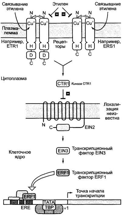
7.6.6. Other Signaling Substances with Phytohormone-Like Effects
Relatively recently1, numerous other physiologically highly active substances have been discovered in plants, and their scientific investigation is being intensified rapidly. Sometimes they are widespread in plants and are nowadays often classified as phytohormones (brassinosteroids — see 7.6.6.1, oxylipins — see 7.6.6.2), while others have a more limited distribution and regulate specific physiological functions (examples are given in Chapter 9).
1 In the 1970s–1980s, i.e., more than 20 years ago. For comparison: 1900s — ethylene, 1930s — auxin, 1950s — gibberellin and cytokinin, 1960s — abscisic acid. — Ed. note.
These triterpenes were first isolated from the pollen of Brassica species (the term brassinosteroid is derived from the Latin name of the source). Biosynthesis proceeds via squalene (see Fig. 6.122), from which the phytosterol cycloartenol is formed (Fig. 7.64). Arabidopsis thaliana mutants with impaired specific steps of brassinosteroid biosynthesis (e.g., dwf1, cbb1, cbb3; from English dwarf and cabbage, respectively) are severely stunted and morphologically resemble tiny cabbage plants. They can be restored to a normal phenotype by the application of trace amounts of brassinosteroids, growing into flowering and fruiting plants (Fig. 7.65). However, brassinosteroid-insensitive mutants (dwf1, cbb2), which exhibit a phenotype similar to that of brassinosteroid-deficient mutants, do not recover normal growth upon the addition of brassinosteroids. The sites of brassinosteroid biosynthesis remain unknown, and nothing is known about The transport of these compounds from the site of synthesis to the site of action. In the case of brassinosteroids, one might speak of locally acting growth regulators rather than phytohormones in the stricter sense. The brassinosteroid receptor is a receptor-like protein kinase localized in the plasma membrane of plant cells, belonging to the group of autophosphorylating Serine/Threonine kinases. Consequently, the binding of brassinosteroids to the receptor could affect the cellular activity state via a protein phosphorylation cascade. Details remain unclear.
Fig. 7.64. Formation of brassinolide from cycloartenol. Mutants (dwf1, cbb1, cbb3) of Arabidopsis thaliana with impaired brassinosteroid biosynthesis exhibit severe developmental defects (dwf — dwarf; cbb — cabbage)
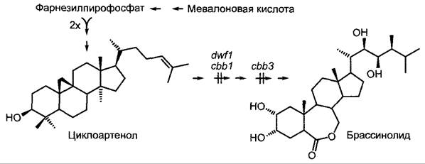
Fig. 7.65. Mutants of Arabidopsis thaliana with impaired brassinosteroid biosynthesis (cbb3). Compared to the wild type, they exhibit extreme dwarfism. They can be normalized by the application of brassinosteroids
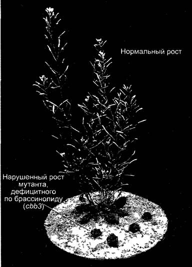
It is now well established that plants, like animals, possess signaling molecules whose biosynthesis originates from oxidized Fatty acids, and which are therefore collectively referred to as oxylipins. In animals, these are Eicosanoids derived from arachidonic acid (e.g., Prostaglandins); in plants, they are primarily octadecanoids derived from α-linolenic acid. Among these, the most important group consists of jasmonic acid and its derivatives, the jasmonates.1 Methyl jasmonate, the methyl ester of jasmonic acid, was identified in 1962 as the principal component of jasmine fragrance.
1 The Translation follows the Russian tradition of trivial names for acids (e.g., jasmonic acid), while using the standard salt/ester form (jasmonate; compare malic acid — malate, citric acid — citrate, etc.). In some publications, the term "jasmonic acid" is used for the salt/ester form, which, in our opinion, is incorrect. — Ed. note.
Fig. 7.66. Biosynthesis of jasmonic acid and its regulation in Arabidopsis thaliana (gray arrows indicate factors inducing transcription of the respective genes)
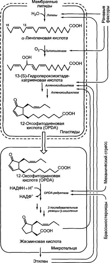
Jasmonic acid has not yet been found in prokaryotes, but it occurs in certain fungi (e.g., Lasiodiplodia theobromae), bryophytes, and ferns, as well as in all vascular plants. Biosynthesis begins in plastids with The oxidation of α-linolenic acid, which is released from Membrane Lipids, leading to the Formation of the first cyclic metabolite, 12-oxophytodienoic acid. Through the reduction of the cyclopentenone ring and side-chain shortening following three cycles of β-oxidation, 12-oxophytodienoic acid is converted into jasmonic acid in glyoxysomes or Peroxisomes, respectively (Fig. 7.66).
Jasmonic acid is synthesized in elevated amounts following wounding (e.g., herbivore feeding) and often after pathogen attack, playing a key role in inducing plant defense responses against pathogens (see 9.3) and herbivores (see 9.4). Exogenously applied jasmonic acid acts as a growth inhibitor and promotes leaf senescence. The jasmonic acid precursor 12-oxophytodienoic acid, together with ethylene and indole-3-acetic acid, is involved in controlling plant growth responses following mechanical stress (see 8.3.2.4)
Last update: 07/08/2026
Editorial and Educational Adaptation: This material has been compiled based on the primary/original source text. The project team performed an editorial review, corrected technical inaccuracies, structured sections, and adapted the content for an educational format.
What was processed:
- elimination of formatting defects (OCR errors, structural breaks, corrupted characters);
- editorial organization of content;
- standardization of terminology in accordance with academic sources;
- verification of factual statements against the original source text.
All mentions of the author, publication year, and origin of the primary text have been preserved in accordance with the source.