Vertebrate Zoology - V. M. Konstantinov 2011
Chordates
Terrestrial, or Tetrapod, Vertebrates — Amniotes
Class Mammals
Mammals represent the highest-organized Class of vertebrate animals. Their body sizes vary significantly: from 3.5 cm in the Etruscan shrew to 33 m in the blue whale, with body masses of 1.5 g and 120 t, respectively. The main Progressive features of mammals are as follows:
1) a high level of Development of the Central Nervous system, primarily the Cerebral Cortex of the Forebrain, which serves as the center of Higher Nervous Activity. Consequently, the adaptive responses of mammals to environmental conditions are highly complex and sophisticated;
2) viviparity and feeding of the young with milk, a product of the maternal Organism, which enables mammals to reproduce under extremely diverse living conditions;
3) a highly developed capacity for thermoregulation, ensuring a relatively constant body Temperature. This is achieved, on the one hand, by regulating heat production through oxidation processes (so-called chemical thermoregulation) and, on the other hand, by controlling heat retention and dissipation through changes in Skin Blood supply, Water evaporation via Respiration and sweating, as well as The Development of fur and subcutaneous fat (so-called physical thermoregulation).
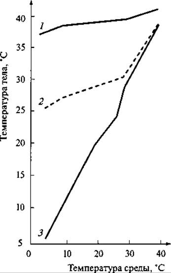
Fig. 164. Curves showing the dependence of body temperature in various animals on ambient temperature:
1 — rabbit; 2 — echidna; 3 — lizard
Thanks to these mechanisms, the body temperature of many mammals remains relatively constant, and its difference from the ambient temperature can reach approximately 100°C. For instance, in the arctic fox, which winters at temperatures down to -60°C, the body temperature is about 39°C. It should be noted, however, that homeothermy (constant body temperature) is not an absolute characteristic of all mammals.
It is fully characteristic of placental mammals of relatively large size. In lower mammals with less developed thermoregulatory mechanisms, and in small placental mammals with an unfavorable body volume-to-surface area ratio for heat conservation, body temperature fluctuates significantly depending on the ambient temperature (Fig. 164). For example, in the opossum, body temperature ranges from 37.8 to 29.3°C; in the most primitive insectivores (tenrecs), from 34 to 13°C; in one of the armadillo species, from 40 to 27°C; and in the common vole, from 37 to 32°C.
Fur, and in some species a subcutaneous fat layer, plays a crucial role in regulating heat loss.
These features, along with A number of other organizational traits, have enabled mammals to spread widely across the most diverse environments. Geographically, they inhabit almost the entire globe, with the exception of Antarctica. Even more importantly, mammals inhabit a vast array of ecological niches. In addition to numerous terrestrial species, there are flying, semi-aquatic, aquatic, and even subterranean burrowing forms. The total number of extant mammal species is approximately 4.5 thousand.
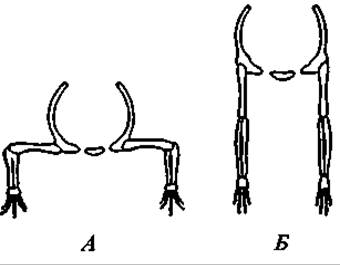
Fig. 165. Position of the trunk and limbs in a primitive terrestrial vertebrate (A) and a mammal (B)
Morphologically, mammals are characterized by the following features. The body is covered with Hair (exceptions are rare and of secondary origin). The skin is rich in glands, with Mammary Glands being particularly notable. The Skull articulates with THE Vertebral Column via two occipital condyles. The lower jaw consists solely of the dentary bone. The quadrate and articular bones are transformed into auditory ossicles located within the Middle ear cavity. Teeth are differentiated into incisors, canines, and molars, and are set in alveoli. The elbow joint points backward, and the knee joint points forward, unlike lower terrestrial vertebrates, in which both of these joints point laterally outward (Fig. 165). With the complete Separation of arterial and venous blood streams, The Heart is four-chambered, and only the left aortic arch is retained. Erythrocytes are anucleate.
Morphophysiological Overview
Integumentary System. The skin of mammals is multifunctional. It bounds and protects the body surface, participates in thermoregulation by protecting the animal from both overheating and cooling, plays a role in Sexual Dimorphism display, and facilitates respiration and excretion. Accordingly, the skin has a complex Structure (Fig. 166).
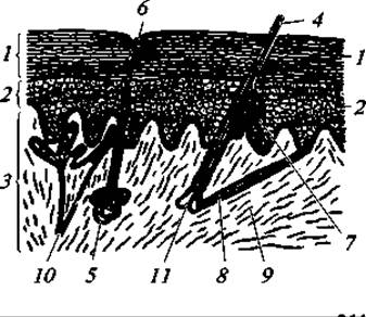
Fig. 166. Structure of mammalian skin (high magnification):
1 — outer layer of the horny epidermis, the Cells of which slough off periodically; 2 — Deep Layer of the epidermis containing living cells (Malpighian layer); 3 — dermis (cutis); 4 — hair; 5 — sweat gland; 6 — duct opening; 7 — sebaceous gland; 8 — arrector pili Muscle; 9 — Connective Tissue fibers of the dermis; 10 — blood vessel; 11 — hair papilla at the Base of the hair
Like other vertebrates, mammalian skin consists of two layers: the outer epidermis and the inner cutis, or dermis. The epidermis comprises two layers: the deep layer, represented by living cylindrical or cuboidal cells known as the Malpighian or germinative layer. Closer to the surface, the cells become flatter. The cavities of these cells gradually fill with keratohyalin, leading to keratinization and Cell death. The superficial cells gradually wear away as fine "dandruff" or in entire flakes (as seen, for example, in seals). The loss of the horny epidermal layer is replenished by continuous proliferation through Cell Division in the Malpighian layer.
The epidermis gives rise to many skin derivatives, the principal ones being hair, Nails, claws, hooves, horns (except in deer), scales, and various glands. These structures are described below.
The cutis, or true skin, is exceptionally well developed in mammals. It consists primarily of Fibrous connective tissue, whose interwoven fibers form a complex pattern. Blood Vessels branch in the upper part of the cutis, which also houses the bases of hair follicles and Sweat Glands. The lower part of the cutis is composed of very loose fibrous tissue where fat is deposited. This layer is known as the subcutaneous adipose tissue (hypodermis). It reaches its greatest development in aquatic mammals, such as whales and seals, where it serves a thermal insulation role due to the complete (in whales) or partial (in seals) reduction of the hair coat and the Physical Properties of the aquatic environment. Large subcutaneous fat deposits also occur in certain terrestrial mammals. Fat accumulation is especially pronounced in species that hibernate in winter (gophers, marmots, badgers, etc.), serving as their primary energy reserve during hibernation.
Skin thickness varies significantly among different species. As a rule, species inhabiting cold regions and possessing dense fur have thinner skin. Extremely thin and fragile skin is characteristic of hares, and it is also poorly vascularized. This has an adaptive significance: a predator that grabs a hare by the skin easily tears off a piece, letting the prey escape. The resulting wound hardly bleeds and heals quickly. A peculiar caudal autotomy of the skin is observed in certain mice, dormice, and jerboas. Their skin tail sheath easily tears off and slips off the caudal vertebrae, allowing an animal caught by the tail to escape from a predator.
The hair coat is as characteristic of mammals as feathers are of birds or scales of reptiles. Only a few species have secondarily lost it entirely or partially. For instance, dolphins lack hair altogether, while whales possess rudimentary bristles on their jaws. In pinnipeds, the hair coat is reduced—most noticeably in walruses, and least so in eared seals (such as fur seals), which are more closely tied to land than other pinniped species.
The structure of a hair is shown in Fig. 166. It consists of a shaft, which projects above the skin surface, and a ROOT embedded within the skin. The shaft is composed of the medulla, the cortex, and the cuticle. The medulla is a porous tissue containing air spaces between its cells; it is this specific layer that grants the hair its low thermal conductivity. Conversely, the cortex is extremely dense, providing the hair with structural strength. A thin outer cuticle protects the hair from mechanical and chemical damage. The upper portion of the hair root is cylindrical and forms a direct continuation of the shaft. Lower down, the root expands and terminates in a bulbous Swelling known as the hair bulb, which caps a dermal outgrowth called the hair papilla. Blood vessels entering this papilla supply the nutrients required for the metabolic activity and Maintenance of the bulb cells. Hair formation and growth occur through the division and differentiation of these bulb cells. The hair shaft itself is a dead keratinized structure incapable of further growth or morphological change.
The hair root, embedded in the skin, is housed within a hair follicle. The ducts of Sebaceous Glands open into the follicle's funnel-shaped upper part; their oily secretion lubricates the hair, increasing its durability and water repellency. Muscle fibers attached to the lower part of the follicle enable the hair to move when they contract. This movement is responsible for piloerection in animals.
Typically, hairs do not emerge perpendicular to the skin surface, but rather lie at a more or less oblique angle, closely adhering to the skin. The degree of this inclination varies among species. It is least pronounced in subterranean mammals, such as the mole.
The pelage consists of several distinct types of hair. The primary categories are down hairs (or underfur), guard hairs (or overhair), and tactile hairs (vibrissae). In most species, the coat is predominantly composed of a dense, short underfur. Interspersed among the down hairs are longer, thicker, and coarser guard hairs. In subterranean mammals like the mole and the zokor, the pelage almost entirely lacks guard hairs. Conversely, in adult deer, wild boars, and seals, the underfur is reduced, and the coat consists mainly of guard hairs (though well-developed underfur is present in the young of these species).
The coat undergoes periodic replacement. This process of hair shedding, or molting, occurs twice a year in some species—in spring and autumn—as seen in squirrels, foxes, arctic foxes, and moles. Other species molt only once a year: the old fur is shed in spring, and new fur develops during the summer, reaching full maturity only by autumn. Ground squirrels serve as an example of this pattern.
A distinct category of hair is represented by vibrissae. These are exceptionally long, stiff hairs that serve a tactile function.
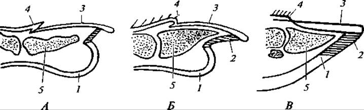
Fig. 167. Longitudinal section through the distal Phalanges of a primate (A), a carnivore (B), and an ungulate (C):
A — nail; B — claw; C — hoof: 1 — digital pad; 2 — plantar pad; 3 — nail plate; 4 — claw fold; 5 — distal phalanx
They are typically located on the HEAD (commonly referred to as whiskers), the lower neck, the chest, and, in certain arboreal climbing species (such as the squirrel), on the abdomen as well. Nerve receptors situated at the base and along the walls of the hair follicle detect contact between the vibrissa shaft and external objects.
Bristles and spines represent specialized modifications of hair.
Other epidermal keratin derivatives include epidermal scales, nails, claws, hooves, horns (hollow horns), and horny beaks. In their Cytology/cytology/67.html">Development and Structure, mammalian scales closely resemble the corresponding structures in reptiles. They are most extensively developed in pangolins, covering their entire body. Scales are also present on the feet of many murine rodents. Finally, the presence of scales on the tail is characteristic of numerous marsupials, rodents, and insectivores.
The distal phalanges of the vast majority of mammals bear keratinous appendages in the form of nails, claws, or hooves. The presence of a specific type of appendage and its structural Organization are directly related to the animal's habitat and lifestyle (Fig. 167). For instance, arboreal mammals possess sharp, curved claws; burrowing species typically have somewhat flattened and broadened claws. Large, fast-running mammals feature hooves, with forest-dwelling species (such as deer), which frequently traverse boggy terrain, possessing wider and flatter hooves. In steppe-dwelling mammals (antelopes) and especially mountain-dwelling species (wild goats, sheep), the hooves are small and narrow, offering a significantly smaller support area than those of forest ungulates that often walk on soft ground or snow. For example, the load per 1 cm2 of the sole averages 850 g in the Central Asian ibex, 500 g in the moose, and 140 g in the reindeer.
Keratinous structures also form the horns of bovids, antelopes, goats, and sheep. These develop from the epidermis and sit atop bony cores, which are independent skeletal elements fused with the frontal bones. Deer antlers have a different origin; they develop from the dermis (cutis) and consist of true Bone tissue.
Unlike those of birds and reptiles, mammalian cutaneous glands are numerous and diverse in both Structure and function. The principal types of glands are sweat, sebaceous, scent, and mammary glands.
Sweat glands are tubular, with their deep portions forming a coiled knot. They open directly onto the skin surface. Their secretory product is sweat, which consists primarily of water containing dissolved urea and salts. These substances are not synthesized by the gland cells themselves, but are extracted from the blood vessels. The function of sweat glands is twofold: cooling the body via the evaporation of moisture secreted onto the skin surface, and excreting Metabolic waste products. Thus, these glands perform simultaneous thermoregulatory and excretory Functions. While present in most mammals, sweat glands vary greatly in their degree of development. For instance, they are very sparse in dogs and cats, whereas in many rodents they are restricted to the footpads, inguinal region, and Lips. Sweat glands are entirely absent in cetaceans, pangolins, and certain other groups.
Sebaceous glands have a branched acinar (bunched) structure and almost invariably open into the funnel of the hair follicle. The fatty secretion of these glands lubricates the hair and the outer epidermal layer, protecting them against wetting and wear.
Scent glands represent modified sweat or sebaceous glands, or sometimes a combination of both (such as the anal glands of mustelids, which produce a highly pungent secretion).
Scent glands are particularly well developed in American skunks (Mephitis), which are capable of ejecting large quantities of secretion over considerable distances. Musk glands are found in musk deer, desmans, beavers, and muskrats; the exact Biological Significance of these glands is not entirely clear, but given that they reach their peak development during the mating season, their activity is evidently linked to reproduction and may serve to stimulate sexual arousal.
Mammary glands are specialized modifications of simple tubular sweat glands. In their most primitive form—found in Australian monotremes—they retain a tubular structure and open into the hair follicles of groups of hairs situated within a small area of the abdominal surface known as the mammary area. In the echidna, the mammary area is located within a specialized pouch that develops during the breeding season to incubate the egg and subsequently nurse the young. In the platypus, the mammary area is situated directly on the abdomen. Monotremes lack nipples, and the young lick the milk directly from the hairs where it flows from the follicle openings.
In marsupials and placental mammals, mammary glands have a branched acinar structure and their ducts open onto nipples. The arrangement of the glands and nipples varies considerably. Arboreal primates and bats (which hang while nursing) possess only a single pair of pectoral nipples; running ungulates have nipples restricted to the inguinal region. In insectivores and carnivores, the nipples form two rows extending along the entire ventral surface of the body. The number of nipples is directly correlated with the species' fecundity and generally corresponds to the average litter size. The minimum number of nipples (2) is characteristic of primates, sheep, goats, elephants, and certain others, whereas the maximum number (10 to 24) is typical of murine rodents, insectivores, and some marsupials.
Skin and its derivatives play a crucial role in mammals by mediating physiological thermoregulation and regulating heat loss. The cutaneous Vascular System is of major importance: the diameter of its blood vessels is neurally and reflexively regulated and can vary across a wide range. When skin vessels dilate, heat loss increases sharply, whereas vasoconstriction leads to its marked reduction.
Body cooling also occurs through the evaporation of water secreted by sweat glands onto the skin surface.
The density and length of the pelage in northern species vary substantially with the seasons. For example, on the rump (lower back) of a squirrel, the number of hairs per 1 cm2 averages 4,200 in summer and 8,100 in winter, compared to 8,000 and 14,700 in the mountain hare. Hair length (in millimeters) on the rump is as follows: in the squirrel, down hair is 9.4 and guard hair is 17.4 in summer, compared to 16.8 and 25.9 in winter; in the mountain hare, down hair is 12.3 and guard hair is 26.4 in summer, compared to 21.0 and 33.4 in winter. Tropical mammals do not exhibit such drastic variations in coat length, owing to the minimal seasonal temperature fluctuations in their environment.
Clear geographical and ecological patterns can be observed in the development of sweat glands. For instance, the average density of these glands per 1 cm2 in the zebu (farmed in the humid tropics) is 1,700, whereas in British shorthorn cattle, it is only 1,060. This same trend is evident when comparing species adapted to varying degrees of arid conditions. As an indicator, evaporation rates are expressed in milligrams per minute per 100 cm2 of skin surface. At a temperature of 37 °C, this rate reaches 17 mg/min in the donkey, compared to only 3 mg/min in the camel; at 45 °C, it is 35 mg/min for the donkey and 15 mg/min for the camel; finally, at 50 °C, it reaches 45 mg/min for the donkey and 25 mg/min for the camel (K. Schmidt-Nielsen, 1972).
The skin plays an equally vital role in mammalian chemical signaling as it does in thermoregulation.
The secretion of cutaneous glands, like other odorous excretions (for example, from the genital and digestive tracts, urine, and specialized gland secretions), serves as an essential medium for intraspecific communication—mammalian chemical signaling. The special significance of this signaling type is determined by its long-range action and the prolonged persistence of the signal. Animals with defined home ranges, individual specimens, pairs, or families mark their territory with scent marks left on conspicuous objects: hummocks, rocks, tree stumps, individual trees, or simply On the surface of the ground.
Parents leave scent marks on their offspring, within the nest, and along their travel routes beyond the nest or the offspring's Location, should no nest be built. Thanks to chemical signaling, deer, seals, and burrowing mammals such as foxes, arctic foxes, sables, martens, voles, and mice can distinguish their own young from foreign ones. Overall, olfactory signaling is of decisive importance in mammalian behavior.
Muscular System. The Muscular System of mammals is highly differentiated and features a vast number of variously positioned Muscles. A characteristic feature is the presence of a dome-shaped muscle, the Diaphragm, which separates the Abdominal cavity from the thoracic cavity. Its primary role is to alter the volume of the thoracic cavity, which is associated with the act of respiration. The cutaneous musculature, which sets various areas of the skin in motion, is significantly developed. In hedgehogs and pangolins, it enables the body to roll into a ball. The raising of spines in hedgehogs and porcupines, the bristling of hair in beasts, and the movement of tactile hairs (vibrissae) are also brought about by the action of this musculature. On the face, it is represented by facial muscles, which are particularly well developed in primates.
Skeleton. The mammalian skeleton retains the typical structural traits of the skeleton of other terrestrial vertebrates, but is more highly differentiated (Fig. 168). The Axial Skeleton comprises the Vertebral Column and the axial skull. Characteristic features in the STRUCTURE OF THE mammalian spinal column include flat articular surfaces of the vertebrae (platyspylous/amphicoelous vertebrae), separated by cartilaginous discs (menisci), a clearly defined regional segmentation of the spine (cervical, thoracic, lumbar, sacral, and caudal), and a constant number of cervical vertebrae. Deviations from these characteristics are rare and of a secondary nature.
In the cervical region of mammals, there are invariably seven vertebrae, with the atlas and axis (epistropheus) being well developed. Exceptions include the manatee, which has 6 cervical vertebrae, and certain species of sloths, which possess from six to 10 cervical vertebrae. However, the length of the neck region varies considerably. Unlike birds, the neck length in mammals is determined not by the number of cervical vertebrae, but by their body length. It is most strongly developed in ungulates, for whom head mobility is crucial when acquiring food. The neck is also well developed in carnivores. Conversely, in burrowing rodents and especially in fossorial mammals, the cervical region is short, and head mobility is limited.
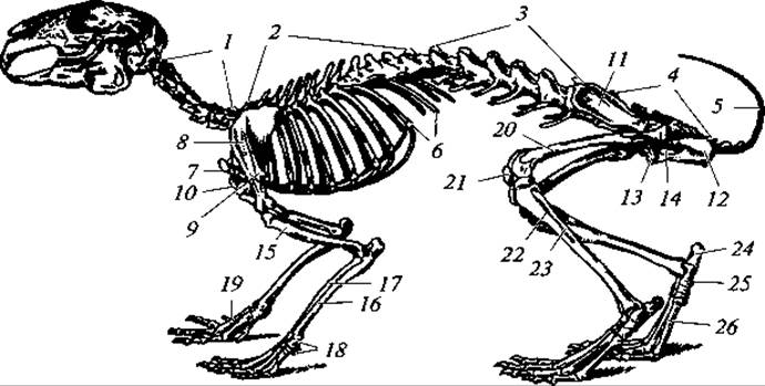
Fig. 168. Skeleton of a rabbit:
1 — cervical vertebrae; 2 — thoracic vertebrae; 3 — lumbar vertebrae; 4 — sacrum; 5 — caudal vertebrae; 6 — Ribs; 7 — manubrium sterni; 8 — scapula; 9 — acromion process of the scapula; 10 — coracoid process of the scapula; 11 — iliac region of the innominate bone; 12 — ischial region of the same bone; 13 — pubic region of the same bone; 14 — obturator foramen; 15 — humerus; 16 — ulna; 17 — radius; 18 — carpus; 19 — metacarpus; 20 — Femur; 21 — Patella; 22 — Tibia; 23 — Fibula; 24 — calcaneus; 25 — talus; 26 — metatarsus
The thoracic region typically consists of 12 to 15 vertebrae; one of the armadillo species and the bottlenose whale (Hyperoodon) have nine, whereas sloths of the genus Choloepus have 24. Ribs attach to the anterior thoracic vertebrae (usually the first seven) and connect to the Sternum (true ribs), thereby forming the rib cage. The remaining thoracic vertebrae bear ribs that do not reach the sternum (false ribs). The sternum is a segmented bony plate terminating in an elongated Cartilage known as the xiphoid process. The broadened anterior segment is termed the manubrium sterni. In bats and mammals whose limbs are adapted for digging, the sternum loses its clearly defined segmentation and bears a keel, which, as in birds, serves for the attachment of pectoral muscles.
In the lumbar region, the number of vertebrae varies from two to nine. These vertebrae bear rudimentary ribs.
The sacral region most commonly consists of four fused vertebrae. Of these, only the first two are true sacral vertebrae, while the remaining ones are caudal vertebrae fused to the sacrum.
In carnivorous mammals, there are three sacral vertebrae, whereas in the platypus, as in reptiles, There are two. The number of caudal vertebrae is the most variable. For instance, gibbons have three, whereas the long-tailed pangolin has 49.
The mobility of the spinal column varies among different animal species. Small mammals, which frequently arch their backs during locomotion, exhibit the greatest mobility. Conversely, in large ungulates, all spinal regions (except the cervical and caudal) undergo negligible movement, and only the limbs are utilized during running.
The mammalian skull (Fig. 169) is characterized by a relatively larger braincase, which is associated with the greater volume of the brain. In young animals, the braincase is generally more developed relative to the facial region than it is in adults. The number of individual bones in the mammalian skull is smaller than in preceding vertebrate groups. This is due to the fusion of several bones into complex units, a process particularly characteristic of the braincase. Thus, the basioccipital, lateral occipital, and supraoccipital bones fuse together; the fusion of the otic bones results in The formation of a single petrosal (periotic) bone. The alisphenoid fuses with the basisphenoid, and the orbitosphenoid fuses with the presphenoid. Sutures between these bony complexes obliterate relatively late, particularly in the neurocranium, which allows for the expansion of the brain volume as the animal grows.
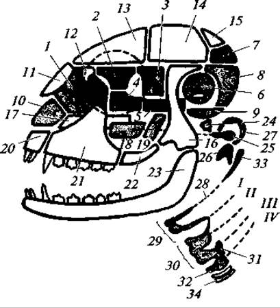
Fig. 169. Diagram of the structure of the mammalian skull:
1 — Ethmoid bone; 2 — orbitosphenoid; 3 — pterygoid; 4 — presphenoid; 5 — basisphenoid; 6 — petrosal bone; 7 — supraoccipital; 8 — exoccipital; 9 — basioccipital; 10 — cartilage of the nasal septum; 11 — Nasal bone; 12 — Lacrimal bone; 13 — Frontal bone; 14 — Parietal bone; 15 — interparietal bone; 16 — squamosal bone; 17 — Vomer; 18 — Palatine bone; 19 — pterygoid; 20 — premaxilla; 21 — Maxilla; 22 — jugal (malar) bone; 23 — dentary; 24 — stapes; 25 — incus; 26 — malleus; 27 — tympanic bone; 28 — remnants of the hyoid; 29 — hyoid apparatus; I — IV — remnants of the gill arches; 30 — thyroid cartilage; 31 — arytenoid cartilage; 32 — cricoid cartilage; 33 — styloid process; 34 — Trachea
The occipital region is formed by a single Occipital bone, as noted. It bears two condyles for articulation with the atlas. The roof of the neurocranium is formed by the paired parietal, frontal, and nasal bones, along with the unpaired interparietal bone. The sides of the braincase are formed by the squamosal bones, from which zygomatic processes extend outward and forward. The latter connect with the jugal bone, which, in turn, articulates anteriorly with the zygomatic process of the maxilla. This ultimately forms the characteristic mammalian zygomatic arch.
The floor of the braincase is formed by the basisphenoid and presphenoid bones, whereas the floor of the facial, visceral region is formed by the pterygoid, palatine, and maxillary bones. Located within the floor of the skull, in the region of the auditory capsule, is the tympanic bone, which is unique to mammals. The auditory capsules ossify from multiple centers, as already mentioned, but ultimately form only a single paired petrosal bone.
The upper jaws consist of paired premaxillary and maxillary bones. A characteristic feature is the development of a secondary bony palate, formed by the palatine processes of the premaxillae and maxillae, as well as the palatine bones. Due to the Formation of the secondary bony palate, the internal nares (choanae) open not between the maxillary bones, as in other terrestrial vertebrates (except crocodiles and turtles), but behind the palatine bones. This palatal structure prevents the interruption of breathing while a food bolus is retained in the Oral Cavity for mastication.
The lower jaw is represented by paired dentary bones that articulate with the zygomatic processes of the squamosal bones. The articular bone is transformed into an auditory ossicle—the malleus; the quadrate bone transforms into another auditory ossicle—the incus. Together with the stapes (homologous to the hyomandibula), they lie within the middle ear cavity. The outer wall of this cavity, as well as part of the external auditory meatus, is surrounded by the aforementioned tympanic bone (tympanicum), which is homologous to the angular bone of the lower jaw in other vertebrates. Thus, mammals exhibit a further evolutionary Transformation of a portion of the visceral apparatus into the auditory apparatus of the middle and External ear.
The mammalian Pectoral Girdle is relatively simple. Its foundation is the scapula, to which a rudimentary coracoid is fused. Only in monotremes does the coracoid exist as an independent bone. A clavicle is present in mammals whose forelimbs perform diverse, complex movements and where the presence of the clavicle ensures a more secure Articulation of the humerus and reinforces the entire pectoral girdle, as seen in primates. In species that move their forelimbs exclusively or predominantly in a plane parallel to the main body axis, the clavicles are rudimentary or absent. Examples include ungulates, rodents, and others.

Fig. 170. Hind limbs of plantigrade and digitigrade mammals (pedal elements are shaded in black):
A — baboon; B — dog; C — llama
The Pelvic Girdle consists of three pairs of bones typical of terrestrial vertebrates: the ilium, ischium, and pubis. In many species, these bones fuse into a single innominate bone.
The Skeleton of the paired limbs retains all the principal Structural Features of a typical pentadactyl limb. However, due to diverse living conditions and the specific ways limbs are used, their structural details vary considerably (Fig. 170). In terrestrial forms, the proximal sections—the femur and shin—are significantly elongated. In aquatic mammals, by contrast, these sections are shortened, while the distal sections—the carpus, tarsus, and especially the phalanges—are elongated. Limbs transformed into flippers in this case move independently of the body, usually acting as a single unit with the trunk, whereas the movement of limb segments relative to each other is relatively weakly developed. In bats, only the first digit of the forelimbs is normally developed, while the remaining digits are greatly elongated; a skin membrane stretches between them, forming the main surface area of the wing. In fast-running mammals, the tarsus, metatarsus, carpus, and metacarpus are positioned more or less vertically, as in the dog. In the most specialized runners—ungulates—the number of digits is reduced. The first digit atrophies, and the animals step either on equally developed third and fourth digits, with the axis of the limb passing between them (even-toed ungulates), or primarily on the single third digit, through which the limb axis passes (odd-toed ungulates).
In this regard, we may note the maximum speed of locomotion of certain mammals (in km/h): short-tailed shrew — 3, bank vole — 7, wood mouse — 10, red squirrel — 15, wild rabbit — 32–40, hare — 55–72, lion — 50, red fox — 72, cheetah — 105–112, camel — 15–65, African elephant — 24–40, Grant's gazelle — 40–50.
Digestive Organs. The digestive organs are characterized by high complexity, which is expressed in the general elongation of the digestive tract, its greater differentiation compared to other vertebrates, and the better development of digestive glands.
The digestive tract begins with the preoral cavity, or oral vestibule. It is located between the fleshy lips, Cheeks, and jaws, which are unique to mammals. In a number of species of hamsters, chipmunks, and monkeys, the oral vestibule expands to form large cheek pouches. Fleshy lips serve for suckling milk in the young and for grasping food in adults, while the oral vestibule serves as a temporary food reservoir: hamsters and chipmunks carry food supplies to their burrows in their cheek pouches. Fleshy lips are absent in monotremes and cetaceans. Behind the jaws lies the oral cavity, where food undergoes mechanical grinding and chemical Processing. Mammals have four pairs of Salivary Glands. The development of the salivary glands depends on The Nature of the diet. In cetaceans, they are practically undeveloped; in ruminants, by contrast, they are exceptionally well developed. For example, a cow produces about 56 liters of saliva per day, which is of paramount importance for moistening coarse food and filling the liquid medium of The Stomach chambers where bacterial breakdown of plant fiber takes place.
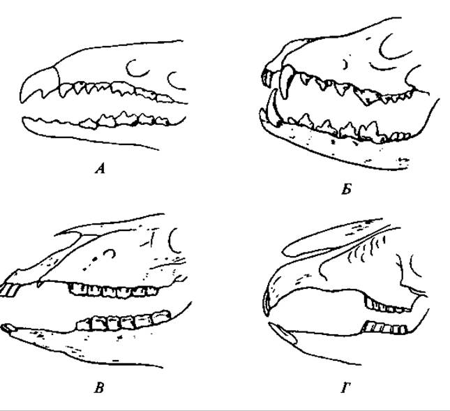
Fig. 171. Diagram of the dentition in some mammals:
A — insectivores (shrew); B — carnivores (fox); C — perissodactyls (horse); D — lagomorphs (hare)
The secretion of the cheek glands of bats, applied to the flight membranes, maintains their elasticity and prevents them from drying out. The saliva of blood-feeding vampire bats has anticoagulant properties, i.e., it prevents blood clotting. The saliva of some shrews is toxic; the secretion of their submandibular gland causes the death of a mouse in less than 1 min after injection. The toxicity of the salivary glands in primitive mammals is viewed as a reflection of their phylogenetic relationship with reptiles.
Mammals are heterodont, meaning their teeth are differentiated into incisors, canines, premolars (false molars), and molars. The number of teeth, their shape, and function vary significantly across different mammalian groups (Fig. 171). Thus, for example, less specialized insectivores possess A large number of relatively weakly differentiated teeth. Rodents and lagomorphs are characterized by strongly developed incisors, the absence of canines, and a flat chewing surface on the molar teeth. This dental structure is related to their diet: they cut or gnaw vegetation with their incisors and grind food with their molars like millstones. Carnivores are characterized by strongly developed canines used for grasping and often killing prey. The molars of carnivores have cutting peaks and flat chewing cusps. The posterior upper premolar and the first lower molar in carnivores are usually distinguished by their size and are termed "carnassial" teeth.
The total number of teeth and their distribution among groups for mammalian species is quite definite and constant, serving as an important taxonomic feature. Dental formulas, constructed as fractions, are used to denote this. The numerator shows the number of teeth in the upper jaw, and the denominator in the lower jaw. Dental groups are designated by the initial letters of their Latin names: incisors — i (incisivi), canines — c (canini), premolars — pm (praemolares), molars — m (molares). For brevity, the number of teeth in only one half of the jaw is written.
The dental formula of the wolf is as follows:

The dental formula of the rabbit:


Fig. 172. General arrangement of Internal Organs in a female rabbit:
1 — Submandibular salivary gland; 2 — Esophagus; 3 — stomach; 4 — Liver (reflected upward); 4a — Gallbladder; 5 — Pancreas; 5a — pancreatic duct; 6 — cecum; 7 — vermiform Appendix; 8 — anus; 9 — left ventricle of the heart; 10 — right ventricle of the heart; 11 — left atrium; 12 — right atrium; 13 — aortic arch; 14 — left Subclavian Artery; 15 — left subclavian vein; 16 — left jugular vein; 17 — Large Intestine; 18 — posterior vena cava; 19 — aorta; 20 — left lung; 21 — left Kidney; 22 — left Adrenal gland; 23 — right Ureter; 24 — Urinary Bladder; 25 — left Ovary; 26 — left oviduct; 27 — Spleen; 28 — Thyroid Gland; 29 — trachea; 30 — Bile duct
The teeth are set in sockets within the jawbones (thecodont dentition) and, in most mammalian species, are replaced only once in a lifetime (diphyodont dentition).
A muscular Tongue is located between the rami of the lower jaw, serving partly for grasping food (cattle, anteaters, pangolins) and lapping water, and partly for turning food over in the oral cavity during chewing.
Behind the oral region lies the Pharynx, into the upper part of which open the internal nares and Eustachian tubes. On the lower surface of the pharynx is a slit leading into the Larynx. The esophagus is well developed (Fig. 172). Its musculature is usually smooth, but in some animals, such as ruminants, striated muscle extends here from the pharyngeal region. This feature ensures voluntary contraction of the esophagus during regurgitation.
The stomach is clearly distinct from other PARTS OF THE digestive tract and is equipped with numerous glands. The volume of the stomach and its internal structure vary among different species depending on their diet (Fig. 173). The stomach is most simply structured in monotremes, where it takes the form of a simple pouch. In most mammals, the stomach is divided into a greater or lesser number of chambers.
The complex stomach is associated with dietary specialization, such as the ingestion of massive amounts of fibrous plant material (ruminants) or underdeveloped oral chewing of food (some insectivorous species). In some South American anteaters, the pyloric region of the stomach differentiates into a section with folds so hard that they function like teeth, grinding the food.
The stomach of ruminant ungulates, such as the cow, has a complex structure. It consists of four chambers: 1) the rumen, the inner surface of which bears hard papillae; 2) the reticulum, the walls of which are divided into a honeycombed network; 3) the omasum, with walls bearing longitudinal folds; and 4) the abomasum, or glandular stomach. Feed masses entering the rumen undergo Fermentation under METABOLISM/18.html">The Influence of saliva and The activity of Bacteria and Protozoa. Due to peristaltic movements, the food passes from the rumen into the reticulum, from which it is regurgitated back into the Mouth. Here the food is ground by the teeth and thoroughly moistened with saliva. The semi-liquid mass thus formed is swallowed and passes through a narrow groove connecting the esophagus to the omasum, entering it and subsequently the abomasum (see Fig. 173).
The described adaptation is of great importance, as the diet of ruminants consists of hard-to-digest plant matter, while their stomachs host a vast number of fermentative bacteria and protozoa whose activity significantly AIDS in the Digestion of food, including crude fiber.

Fig. 173. Various Forms of mammalian stomachs:
A — dog; B — rat; C — mouse; D — marten; E — ruminant (the dashed arrow indicates the direction of food passage); F — human; G — camel; H — echidna; I — three-toed sloth; 1 — esophagus; 2 — intestine; 3 — cardiac stomach; 4 — pyloric stomach; 5 — abomasum; 6 — omasum; 7 — reticulum; 8 — rumen
The intestine proper is subdivided into the small, large, and rectum sections. In species feeding on coarse plant forage (such as rodents), a long and wide cecum branches off at the junction of the small and large intestines, terminating in some animals (e.g., hares, prosimians) in a vermiform appendix. The cecum acts as a "fermentation vat" and is developed all the more extensively the more plant fiber the animal consumes. In mice, which feed on seeds and partly on the vegetative parts of plants, the cecum accounts for 7 — 10 % of the total length of all intestinal sections, whereas in voles, which feed largely on vegetative plant parts, it reaches 18 — 27 %. In carnivorous species, the cecum is poorly developed or absent. The length of the large intestine varies in the same connection. In rodents, it constitutes 29 — 53 % of the total intestinal tract length, in insectivores and bats — 26 — 30 %, and in carnivores — 13 — 22 %. The overall length of the intestine varies very considerably. As a rule, herbivorous species have a relatively longer intestine than omnivores and carnivores. Thus, in some bats, the intestine is 2.5 times longer than the body, in insectivores — 2.5 — 4.2 times, in carnivores — 2.5 times (weasel) and 6.3 times (dog), in rodents — 5.0 times (midday gerbil) and 11.5 times (guinea pig), in the horse — 12.0 times, and in the sheep — 29 times.
Table 14
Water balance of the North American desert rodent, the kangaroo rat (after K. Schmidt-Nielsen, 1982)
Water intake, % |
Water loss, % |
||
Drinking water |
0 |
Evaporation |
70 |
Food |
10 |
Via urine |
25 |
Metabolic water |
90 |
Via feces |
5 |
Having characterized the structure and functioning of the digestive tract, let us briefly Touch upon The problem of water supply in the mammalian organism.
Many species of carnivores and ungulates regularly visit watering places. Others are satisfied with the water obtained from succulent food. However, There are also those that never drink and feed on very dry forage, such as many desert rodents. In this case, the main source of water supply is the water generated through metabolic processes — the so-called metabolic water (Table 14).
Metabolic water is one of the obligatory products of organic matter transformation within the body. However, the metabolism of different substances yields varying amounts of water. Fats rank first. When consuming 1 kg of fat per day, about 1 L of water is produced, compared to 0.5 L for 1 kg of starch and 0.4 L for 1 kg of Proteins (K. Schmidt-Nielsen, 1982).
The liver is located beneath the diaphragm. The bile duct empties into the first loop of the Small Intestine. The duct of the pancreas, which lies in a fold of the Peritoneum, empties into this same intestinal section.
Respiratory organs. As in birds, essentially the sole respiratory organs of mammals are the Lungs. The Role of the skin in gas exchange is negligible: only about 1 % of oxygen enters through cutaneous blood vessels. This is easily understood when one considers, first, the keratinization of the epidermis and, second, the negligible total surface area of the skin compared to the total respiratory surface of the lungs, which in mammals is 50–100 times larger than the skin surface.
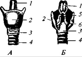
Fig. 174. Larynx of a rabbit: anterior view (A) and posterior view (B); 1 — epiglottis; 2 — thyroid cartilage; 3 — cricoid cartilage; 4 — trachea; 5 — corniculate cartilage; 6 — arytenoid cartilage

Fig. 175. Diagram of the structure of mammalian pulmonary alveoli (the left one retains only the capillary network, the right one shows an opened alveolus without vessels):
1 — bronchus; 2 — artery; 3 — vein
A characteristic feature is the increased complexity of the larynx (Fig. 174). At its base lies the ring-shaped cricoid cartilage, while the anterior and lateral walls of the larynx are formed by the thyroid cartilage, which is unique to mammals. Dorsal to the cricoid cartilage on the sides of the larynx are paired arytenoid cartilages. A thin, leaf-shaped epiglottis adjoins the anterior margin of the thyroid cartilage. Small pouch-like cavities, the laryngeal ventricles, are situated between the cricoid and thyroid cartilages. The vocal cords, appearing as paired folds of the laryngeal mucosa, lie between the thyroid and arytenoid cartilages. The Trachea and Bronchi are well developed. In the region of the lungs, the bronchi branch into a large number of smaller branches. The smallest branches, the bronchioles, terminate in sac-like structures known as alveoli, which possess a cellular structure (Fig. 175). Blood vessels branch out here. The number of alveoli is enormous: carnivores have 300 — 500 million, whereas sluggish sloths have about 6 million. The Emergence of alveoli creates a vast surface area for gas exchange. For instance, the total alveolar surface area in humans is 90 m2. Calculated per unit of respiratory surface (in cm2), the number of alveoli is 6 in the sloth, 28 in the domestic cat, 54 in the house mouse, and 100 in the bat.
Air exchange in the lungs is driven by Changes in the volume of the thoracic cavity, resulting from the movement of the ribs and a specialized, dome-shaped muscle protruding into the thoracic cavity — the diaphragm. The respiratory rate depends on the size of the animal, which is linked to differences in metabolic intensity. Thus, in the horse, it reaches 8 — 16 breaths per minute, in humans 15 — 20, in the rat 100 — 150, and in the mouse about 200. The inverse relationship between metabolic intensity and body size is clearly demonstrated in Table 15 regarding oxygen consumption by animals of various sizes.
Table 15
Oxygen consumption by mammals of various sizes
(after L. Prosser et al., 1977)
Species |
Body mass, g |
Oxygen consumption, mL/(g·h) |
Shrew |
3.5 |
7-10.6 |
Bank vole |
22 |
2.3 |
Chipmunk |
72-145 |
0.8 |
Hare |
1600 |
0.96 |
Seal |
26000 |
0.6 |
Dolphin |
170000 |
0.22-0.34 |
Camel |
17000 - 330000 |
0.03-0.04 |
Elephant |
3700000 |
0.07-0.11 |
Marsupial mouse |
8.5 |
1.26 |
Kangaroo |
33000 |
0.2 |
Echidna |
4000 |
0.22 |
The data presented in the table also indicate a lower metabolic rate in monotremes and marsupials compared to placentals, which corresponds to the primitiveness of their overall organization. For comparison, we may note that in scaled reptiles this indicator (oxygen consumption, mL/(g·h)) is only 0.12 — 0.3, and in amphibians 0.012 — 0.075 (L. Prosser et al., 1977).
Consistent with all of the above, heat production in mammals is high, although it is still clearly dependent on body size. For instance, in the shrew it is 151.2 J/(g·h), in the cat 12.6, in the dog 8.4, and in the elephant 2.1 J/(g·h) (K. Schmidt-Nielsen, 1982).
Pulmonary ventilation not only ensures gas exchange but also plays a vital role in thermoregulation. This is particularly evident in species with poorly developed sweat glands. When overheating, these animals cool their bodies largely by increasing water evaporation, with the water vapor being expelled alongside exhaled air from the lungs (a phenomenon known as panting). For example, a resting dog exhales 2 L of air per minute; with a sharp rise in ambient temperature, the volume of exhaled air reaches 50–75 L/min, and The amount of evaporated water can reach 200 cm3/h.
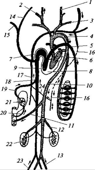
Fig. 176. Diagram of the Circulatory system in mammals: 1 — External Carotid Artery; 2 — Internal Carotid Artery; 3 — subclavian artery; 4 — aortic arch; 5 — pulmonary artery; 6 — left atrium; 7 — right atrium; 8 — left ventricle; 9 — right ventricle; 10 — dorsal aorta; 11 — visceral artery; 12 — renal artery; 13 — iliac artery; 14 — jugular vein; 15 — subclavian vein; 16 — left hemiazygos vein; 17 — right azygos vein; 18 — posterior vena cava; 19 — hepatic vein; 20 — HEPATIC PORTAL VEIN; 21 — liver; 22 — kidney; 23 — iliac vein; 24 — transverse vein
When evaluating the role of panting in enhancing heat dissipation, one must keep in mind that it is primarily respiratory tract ventilation, rather than pulmonary ventilation proper, that increases in this case. For instance, in cattle, while overall ventilation rises from 50 to 300 L/min, alveolar ventilation increases only from 25 to 75 L/min (i.e., 3-fold), whereas dead space ventilation surges from 25 to 225 L/min (i.e., 9-fold) (K. Schmidt-Nielsen, 1976).
Ecological adaptations are also apparent. For example, the desert-dwelling North American rodents known as kangaroo rats lose 50–57 mg of water per 1 mL of consumed oxygen through respiration, whereas laboratory mice and rats lose 85–94 mg (K. Schmidt-Nielsen, 1982).
Circulatory system. Just as in birds, there is only a single aortic arch (specifically the left one), originating from the thick-walled left ventricle. The main arterial vessels branch off from the aorta in various ways. Typically, a short brachiocephalic (innominate) artery departs from the aorta and divides into the right subclavian artery, as well as the right and left carotid Arteries, whereas the left subclavian artery arises independently directly from the aortic arch. In other cases, the left carotid artery originates not from the brachiocephalic artery, but independently from the aortic arch. The dorsal aorta, as in all vertebrates, lies beneath the vertebral column, giving off numerous branches to the musculature and internal organs.
The Venous system is characterized by the absence of a renal portal system. In only a few species does the left anterior vena cava enter the heart independently; more commonly, it merges with the right anterior vena cava, which channels all blood from the anterior region of the body into the right atrium. A notable feature is the presence of posterior cardinal vein remnants, known as the azygos Veins. In most species, the right azygos vein empties independently into the anterior vena cava, whereas the left azygos vein loses its connection with the vena cava and drains into the right azygos vein via a transverse vein (Fig. 176).
The relative size of the heart varies among species with different lifestyles and, ultimately, different metabolic rates. For example, the cardiac index (the relative heart mass expressed as a percentage of total body weight) is 0.3 in the sperm whale, 0.4 in the African elephant, 0.3 in the sloth, 0.6 in the tundra vole, 1.2–1.4 in the long-eared bat, and 1.4 in the common shrew.
The correlation between body size and heart size is further confirmed by comparing the cardiac indices of systematically and ecologically closely related animal species. For instance, this index is 0.47 in the large bobak marmot, 0.61 in the yellow ground squirrel, and 0.82 in the little ground squirrel.
Along with body size, the cardiac index also depends on locomotor activity. Comparing this metric in mice of similar size that move frequently and rapidly versus relatively sedentary voles yields: stripe-field mouse — 0.89, wood mouse — 0.85, common vole — 0.54, root vole — 0.47 (S. S. Shvarts, 1968).
It is also worth noting that the relative heart size in the domestic rabbit is 3 times smaller than that in the wild European hare. Approximately the same pattern emerges when comparing this indicator between pet and hunting hound dogs. Heart rate varies accordingly as well (Table 16).
Table 16
Pulse rate and metabolic rate in selected mammals
Animal species |
Body mass, g |
Heart rate, bpm |
Metabolic rate per kg of body mass per 24 h, kJ |
Mouse |
25 |
600 |
1680 |
Dog |
6500 |
120 |
277 |
Sheep |
50000 |
70-80 |
113 |
Bull |
500000 |
43 |
101 |
Table 17
Blood characteristics in various vertebrates (after P. V. Terentyev, 1961; L. Prosser and F. Brown, 1967; L. Prosser et al., 1977)
Vertebrate groups |
Blood volume, % of body mass |
Erythrocyte count per 1 mm3 of blood, millions |
Oxygen capacity of blood, % per unit volume |
Cartilaginous Fishes |
— |
0,15 |
6,0 |
Bony fishes |
1,5-3,0 |
— |
8,8 |
Caudate amphibians |
3,0 |
0,12 |
3,0-10,0 |
Anuran amphibians |
4,2-4,9 |
0,48 |
— |
Reptiles |
4,2-7,4 |
0,98 |
7,0-12,0 |
Birds |
5,7-9,0 |
2,7-3,5 |
10,0-22,0 |
Mammals |
5,5-9,5 |
8,4 |
15,0-24,0 |
Due to the high intensity of Cardiac Activity, blood pressure in mammals is just as high as in birds. In the southern elephant seal it is 120/90 mmHg, in the rat 130/90, and in the dog 112/56 mmHg. For comparison, in scaled reptiles this parameter ranges between 14/10 and 80/60, and in amphibians between 22/12 and 30/25 (L. Prosser et al., 1978).
The total blood volume in mammals is greater than in lower vertebrate groups. Mammalian blood also compares favorably in a number of biochemical properties, partly related to the absence of nuclei in erythrocytes. Mammals possess not only a relatively large volume of blood but, more importantly, a high oxygen-carrying capacity. In turn, this is associated with a high erythrocyte count and a large amount of Hemoglobin. Lower vertebrates (fish, amphibians) contain on average 5–10 g of hemoglobin, whereas mammals contain 10–15 g per 100 cm3 of blood. Some generalized data on blood characteristics across various vertebrates are summarized in Table 17.
Unique adaptations arise in aquatic lifestyles, where opportunities for atmospheric breathing are periodically interrupted. On the one hand, this is manifested by a dramatic increase in the amount of oxygen-binding globin in muscles (Myoglobin), accounting for about 50% of the organism's total globin. Furthermore, in animals that submerge for prolonged periods, peripheral Circulation is shut down, while Blood supply to the brain and heart remains constant.
On the other hand, in aquatic and semi-aquatic mammals, submergence triggers a decrease in heart rate (known as bradycardia), which leads to a slowed blood flow and a more complete utilization of blood oxygen. For instance, in an unmerged harbor seal (Phoca vitulina), the heart rate is 180 bpm. Eleven seconds after diving, it drops to 60, after 27 seconds to 35, and thereafter stabilizes at around 30 for the entire duration the animal remains underwater.
The oxygen capacity of blood is highest in diving animals. For example, it is 24 vol % in the platypus, 25 in the muskrat, 19 in Steller's sea lion, 26 in the common dolphin, and 29 vol % in the sperm whale.
The oxygen capacity of blood also varies in response to changes in atmospheric thinning relative to altitude above sea level.
Naturally, an increase in the oxygen capacity of the blood also depends on the species' level of locomotor activity. The markhor is more agile than the Caucasian tur, and even more so than domestic sheep and goats, and consequently exhibits a higher blood oxygen capacity.
Considering everything stated regarding the phenomena of gas exchange and Blood Circulation, there is every reason to conclude that the overall metabolic rate in mammals is noticeably higher than in their phylogenetic predecessors, and is close to that of birds.
The Hematopoietic organs are specialized. The Bone Marrow produces erythrocytes, granulocytes, and platelets. The spleen and Lymph Nodes produce lymphocytes.
Nervous system. The Nervous System of mammals, like that of other vertebrates, includes the central nervous system—the BRAIN AND SPINAL cord—and the Peripheral Nervous System, consisting of the nerves extending from them. The brain (Fig. 177) is relatively large, which is due to the increased volume of the forebrain hemispheres and the Cerebellum.
The development of the forebrain is manifested by the expansion of its roof—the secondary cerebral vault (neopallium)—rather than the Basal Ganglia as in birds; it consists of Nerve Cells and Unmyelinated nerve fibers. In connection with the development of the cerebral cortex, the Gray matter in mammals is located superficial to the White matter. The centers of higher nervous activity are situated in the cerebral cortex. The complex behavior of mammals and their intricate reactions to various external stimuli are directly related to the Progressive development of the cerebral hemispheres' cortex. The cortex of both hemispheres is connected by a commissure of white nerve fibers known as the corpus callosum.
The ratio of the mass of the forebrain hemispheres to the total brain mass varies among mammals of different taxonomic groups. In hedgehogs, it is 48%, in squirrels 53, in wolves 70, and in dolphins 75%.
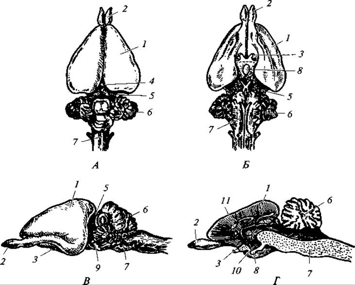
Fig. 177. Brain of a rabbit:
dorsal view (A); ventral view (B); lateral view (C); D — longitudinal section; 1 — cerebral hemispheres; 2 — olfactory lobes; 3 — Optic nerve; 4 — epiphysis; 5 — Midbrain; 6 — cerebellum; 7 — Medulla Oblongata; 8 — Pituitary Gland; 9 — Pons; 10 — infundibulum; 11 — corpus callosum
The forebrain cortex in most species is quite robust and comprises 7 layers of nerve cells. It is covered with numerous sulci that increase its surface area: the Sylvian fissure, which separates the frontal lobe of the cortex from the temporal lobe, and the central sulcus (Rolandic fissure), which separates the frontal lobe from the parietal lobe dorsally. In higher Representatives of the class, the number of sulci is large. The Diencephalon is not visible from above. The epiphysis, pituitary gland, and Hypothalamus are small but functionally extremely important.
The midbrain is characterized by its division into four colliculi (corpora quadrigemina) by two mutually perpendicular furrows. The cerebellum is large and differentiated into several sections (see Fig. 177), which is associated with the highly complex nature of movements in mammals.
The medulla oblongata is important for housing the nuclei of the centers for respiration, blood circulation, digestion, etc.
Sense Organs. Olfactory organs are very well developed in mammals—better than in other terrestrial vertebrates—and play a huge role in their lives. With the help of these organs, mammals recognize enemies, search for food, and find each other. Many species can sense odors from several hundred meters away and are capable of detecting food sources located underground. Only fully aquatic mammals (cetaceans) have a reduced SENSE OF SMELL. Seals, however, possess a very acute sense of smell.
The progressive development of the described organs is manifested in the increased volume of the olfactory capsule and its complication due to the formation of a system of turbinates (olfactory conchae). In some groups of mammals (marsupials, rodents, ungulates), There is a distinct section of the olfactory capsule that opens independently into the nasopalatine duct (Jacobson's organ), which serves for recognizing the scent of food in the mouth, as was previously described in the chapter on reptiles.
The Organ of Hearing (Fig. 178) is exceptionally well developed in the vast majority of cases. In addition to the inner and middle ear, which are also present in lower classes, it includes two new sections: the external auditory meatus and the auricle (pinna). The latter is absent only in aquatic and subterranean mammals (cetaceans, most pinnipeds, mole-rats, and certain others). The auricle directionally captures sounds and significantly enhances auditory acuity. It is particularly well developed in nocturnal mammals (bats) as well as forest ungulates, desert canids, and some others. The inner end of the auditory canal is sealed by the tympanic membrane, behind which lies the middle ear cavity. Unlike amphibians, reptiles, and birds—which possess only a single ossicle—mammals have three auditory ossicles in this cavity (Fig. 179). The malleus (homologue of the articular bone) rests against the tympanic membrane; movably attached to it is the incus (homologue of the quadrate bone), which in turn connects to the stapes (homologue of the hyomandibula), and the latter presses against the oval window of the membranous labyrinth of the Inner ear. This system provides a significantly more efficient transmission of the sound wave, captured by the auricle and passed through the auditory canal to the middle ear and then to the inner ear. In the structure of the inner ear of mammals, one's attention is drawn to the strong development of the cochlea and the presence of The Organ of Corti—extremely fine fibers, numbering several thousand, stretched within the cochlear duct. Upon sound perception, these fibers resonate, thereby ensuring the finer hearing of mammals.
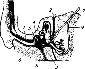
Fig. 178. Diagram of the mammalian organ of hearing:
1 — external auditory meatus; 2 — endolymphatic duct; 3 — round window; 4 — incus; 5 — malleus; 6 — tympanic membrane; 7 — auditory nerve; 8 — Eustachian tube; 9 — cochlear nerve
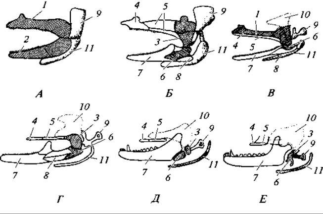
Fig. 179. Transformation of the first two visceral arches in the vertebrate Lineage:
A — shark; B — bony fish; C — amphibian; D — reptile; E — therapsid; F — mammal; 1 — palatoquadrate cartilage; 2 — Meckel's cartilage; 3 — quadrate bone — mammalian incus; 4 — palatine bone; 5 — pterygoid bones; 6 — articular bone — mammalian malleus; 7 — dentary bone; 8 — angular bone; 9 — hyomandibular cartilage — auditory ossicle (stapes) of terrestrial vertebrates; 10 — squamosal bone; 11 — hyoid
The capacity for echolocation has been discovered in a number of mammals. In addition to bats, which are well known in this regard, this ability is possessed by cetaceans (dolphins), pinnipeds (seals), and shrews. Shrews emit echolocation pulses with a frequency of 30 — 60 kHz and a duration of 5 — 33 ms. Sea lions emit localization sounds ranging from 20 to 72 kHz, and dolphins from 120 to 200 kHz. The latter are capable of locating schools of fish from a distance of up to 3 km.
Visual organs play a lesser role in the lives of mammals than they do in birds. They usually pay little attention to stationary objects, and even cautious animals such as foxes, hares, and moose can approach a standing human very closely. Visual acuity and eye development naturally vary and are linked to environmental conditions. Nocturnal mammals and animals of open landscapes (e.g., antelopes) have particularly large eyes. Forest mammals have less acute Vision, while subterranean forms have reduced eyes that are sometimes covered by a skin fold (mole-rat).
Accommodation in mammals occurs exclusively by changing the shape of the lens through the action of the ciliary muscle. Small rodents (voles, mice) practically lack accommodation abilities, which is mainly associated with their nocturnal activity and limited field of vision.
Color Vision in mammals is poorly developed compared to birds. Almost the entire spectrum can be distinguished only by higher Old World monkeys. The European bank vole distinguishes only red and yellow colors. In the opossum, European polecat, and several other species, color vision has not been detected at all.
A characteristic feature of the tactile organs in mammals is the presence of tactile hairs, or vibrissae (Fig. 180), which were described earlier in the characterization of the integument.
Excretory system. The Kidneys in mammals are pelvic and metanephric. Trunk kidneys form during embryonic development but subsequently degenerate. The kidneys are compact organs, bean-shaped or lobed. Their surface is usually smooth, occasionally bumpy (in ruminants and cats), and in some species (such as cetaceans), the kidneys are divided into lobes by constriction grooves.
A cross-section reveals that the kidney consists of layers: an outer cortex and an inner (striped) medulla (Fig. 181). The cortical layer contains convoluted tubules originating in Bowman's capsules, inside which lie capillary glomeruli (Malpighian corpuscles). Filtration takes place within the vascular glomeruli, and Blood Plasma is filtered into the renal tubules to form primary urine. Reabsorption of water, sugar, and Amino Acids from the primary urine occurs in the collecting ducts of the medulla. The number of renal tubules in mammals is exceptionally large. Both kidneys combined contain about 10,000 tubules in mice and 285,000 in rabbits. For comparison, a frog's kidneys contain 2,000 tubules, and a newt's have a mere 400.

Fig. 180. Diagram showing the arrangement of sensitive hairs (vibrissae) on a rabbit's muzzle. The dashed line outlines the "tactile zone"

Fig. 181. Longitudinal section of the kidney (A) and structural diagram of a Malpighian corpuscle (B):
1 — cortex; 2 — medulla; 3 — pyramids; 4 — renal papillae; 5 — renal pelvis; 6 — ureter; 7 — Bowman's capsule; 8 — afferent arteriole forming the glomerulus; 9 — efferent arteriole; 10 — beginning of the convoluted tubule
The relative size of the kidneys is inversely proportional to body mass. They are large in the smallest species and vice versa. This relationship makes complete sense when we consider that in phylogenetically and ecologically closely related species, metabolic rate increases as body size decreases. A clear and reliable, albeit indirect, confirmation of this is the strong correlation between the relative mass of the kidneys and the heart—two organs equally responsible for maintaining the metabolic rate.
Unlike reptiles and birds, whose primary end product of Protein metabolism is uric acid, mammals (as well as fish and amphibians) excrete urea instead. While in birds the proportion of uric acid is 63–80% and urea accounts for only 1–10%, in mammals the ratio is reversed: urea makes up 68–91%, and uric acid accounts for 0.1–8%.
This type of Protein Metabolism in mammals undoubtedly evolved in conjunction with the Placenta, through which the developing embryo can obtain an unlimited supply of water from the mother's blood. Furthermore, toxic metabolic waste products can be continuously and indefinitely eliminated from the developing embryo via the placenta (more specifically, its vascular network).
Recall that urea is significantly more toxic than uric acid, and excreting urine under this metabolic pathway requires a very high volume of water. Additionally, this serves as Evidence of the close evolutionary ties between mammals and amphibians.
The medullary layer contains straight collecting ducts that group together into pyramids and open at the tips of the papillae, which project into the renal pelvis. The ureter extends from the renal pelvis and empties into the urinary bladder. In monotremes, the ureter drains into the urogenital sinus, through which it connects to the urinary bladder. Urine is expelled from the bladder via an independent Urethra.
Sweat glands also partially contribute to the excretory function by eliminating dissolved salts and urea. However, this pathway accounts for no more than 3% of nitrogenous metabolic waste. Reproductive organs. The paired Testes of the male are typically oval-shaped (Fig. 182, A). In monotremes, certain insectivores and xenarthrans, as well as in elephants and cetaceans, the testes remain within the body cavity throughout life. In most other mammals, the testes initially develop within the body cavity, but as sexual maturity approaches, they descend into an external pouch—the Scrotum—which communicates with the body cavity via the Inguinal Canal. Adjoining each Testis along its axis is an elongated, granular structure: the Epididymis, which morphologically represents a tangled mass of highly convoluted seminiferous tubules homologous to the anterior region of the trunk kidney. Extending from the epididymis is the vas deferens (homologous to the Wolffian duct), which opens near the base of the Penis into the urogenital (urethral and ejaculatory) canal. Near their termination before entering the urogenital canal, the vasa deferentia form paired, compact, ribbed structures known as Seminal Vesicles. In mammals, these are glands whose secretions contribute to the fluid portion of semen; furthermore, their sticky consistency apparently helps prevent semen from leaking out of the female reproductive tract.
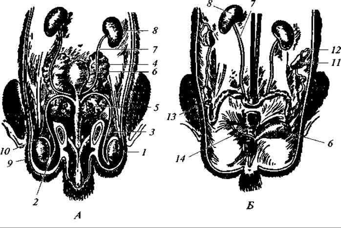
Fig. 182. Urogenital organs of a male (A) and female (B) rat:
1 — testis; 2 — epididymis; 3 — vas deferens; 4 — seminal vesicles; 5 — Prostate Gland; 6 — urinary bladder; 7 — ureter; 8 — kidney; 9 — scrotum; 10 — inguinal canal; 11 — ovary; 12 — Fallopian tube; 13 — Uterus; 14 — Vagina
At the base of the penis lies the second paired gland, the prostate, whose ducts also empty into the proximal section of the urogenital canal. Prostatic secretions form the bulk of the fluid in which the spermatozoa produced by the testes swim. Consequently, semen, or ejaculate, is a mixture of fluids secreted by the prostate, seminal vesicles (and certain other glands), and the spermatozoa themselves.
The aforementioned urogenital canal runs along the ventral side of the copulatory organ. Dorsally and laterally to this canal lie the corpora cavernosa, whose internal cavities fill with blood during sexual arousal, causing the penis to become firm and increase in size. In many mammals, the structural rigidity of the penis is further reinforced by a specialized long bone located between the corpora cavernosa. Examples include carnivores, pinnipeds, many rodents, and certain bats.
The paired Ovaries of the female always lie within the body cavity and are attached to the dorsal wall of the abdominal cavity by mesenteries (Fig. 182, B). The paired oviducts, homologous to the Müllerian ducts, open anteriorly into the body cavity in close proximity to the ovaries, forming expanded funnel-like structures. The upper, convoluted section of the oviducts constitutes the Fallopian tubes. These lead into the expanded uterine regions, which open into the vagina (a single, unpaired chamber in most mammals). The vagina transitions into a short urogenital canal, into which both the vagina and the urethra open. On the ventral side of the urogenital canal lies a small outgrowth—the Clitoris—which contains erectile tissue (corpora cavernosa) and is homologous to the male penis. Interestingly, a bone is present within the clitoris in certain species.
The Anatomy of the female reproductive tract varies significantly across mammalian groups. For instance, in monotremes, the oviducts remain paired along their entire length and are differentiated only into Fallopian tubes and uterine horns, which open independently into the urogenital sinus. In marsupials, a distinct vagina develops, though it frequently remains paired as well. In placental mammals, the vagina is always single, while the upper sections of the oviducts retain a paired configuration to varying degrees. In the simplest condition, the uterus is paired, with the left and right chambers opening independently into the vagina. Such a uterus is termed duplex and is characteristic of many rodents and certain xenarthrans. The uterine horns may be fused only in their lower portion, forming a bicornuate uterus, as seen in some rodents, bats, and carnivores. The fusion of a significant portion of the left and right uterine horns results in a bipartite uterus in carnivores, cetaceans, and ungulates. Finally, in primates, prosimians, and certain bats, the uterus is a simple, unpaired organ, with only the uppermost sections of the oviducts—the Fallopian tubes—remaining paired. Placenta. During embryonic development within the mammalian uterus, a highly specialized structure forms, known as the afterbirth or placenta (Fig. 183). Only monotremes lack a placenta, while marsupials possess rudimentary placental structures. The placenta develops through the fusion of the outer wall of the allantois with the serosa, creating a spongy structure known as the chorion. The chorion forms outgrowths, or villi, which connect or fuse with the softened areas of the uterine epithelium. At these sites, the fetal and maternal blood vessels intertwine (without fusing), thereby establishing a connection between the circulatory systems of the embryo and the mother. This ensures fetal gas exchange, Nutrition, and the removal of metabolic waste products.
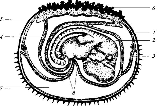
Fig. 183. Rabbit embryo at the end of the twelfth day:
1 — serous membrane; 2 — amniotic cavity; 3 — amnion; 4 — allantois; 5 — allantoic cavity; 6 — chorion; 7 — yolk sac cavity; 8 — umbilical cord
The placenta is already present in marsupial mammals, although it remains primitive there: chorionic villi do not form, and, similar to ovoviviparous lower vertebrates, there is a parietal connection between the uterine blood vessels and the yolk sac (the so-called "yolk-sac placenta"). In higher placental mammals, the chorion invariably forms villi that attach to the uterine walls. The spatial arrangement of these villi varies among mammalian groups. Based on this, Three types of placentation are recognized: diffuse, where villi are distributed evenly across the chorion (cetaceans, many ungulates, prosimians); cotyledonary, where villi are clustered into groups scattered across the chorionic surface (most ruminants); and discoidal, where villi are restricted to a localized, disc-shaped area of the chorion (insectivores, rodents, primates).
The total number of currently living mammalian species worldwide exceeds 4,000. The Class Mammalia is divided into two subclasses: Prototheria (containing a single order, Monotremata) and Theria (comprising two infraclasses and numerous orders).
Last update: 13/08/2026
Editorial and Educational Adaptation: This material has been compiled based on the primary/original source text. The project team performed an editorial review, corrected technical inaccuracies, structured sections, and adapted the content for an educational format.
What was processed:
- elimination of formatting defects (OCR errors, structural breaks, corrupted characters);
- editorial organization of content;
- standardization of terminology in accordance with academic sources;
- verification of factual statements against the original source text.
All mentions of the author, publication year, and origin of the primary text have been preserved in accordance with the source.