Medical Genetics - V. M. Zaporozhan 2005
Monogenic Disorders
Clinical Features and Genetics of Selected Monogenic Disorders
Autosomal Recessive Disorders
Autosomal recessive disorders manifest phenotypically only in the homozygous state, meaning an affected individual must possess two pathological mutant alleles. The disease Gene is designated as a, and the normal gene as A. Affected individuals have the genotype aa, whereas unaffected individuals are either AA or Aa.
Autosomal recessive disorders are characterized by the following features:
1. Typically, the parents of an affected child are unaffected, making it impossible to trace the transmission of the disorder across generations. A family may have multiple affected children, with males and females affected with equal probability. A pedigree chart is provided in section 4.2.2.
2. Children with rare disorders are more frequently born to consanguineous marriages or within relatively small, isolated populations.
3. As a rule, complete gene penetrance and relatively uniform expressivity are observed.
4. The risk of having an affected child in a family depends on the parental genotypes. Unaffected parents of an affected child are heterozygous carriers of the pathological gene (Aa). The risk of conceiving an affected child in such a family is 25 % with each Pregnancy.
If one parent suffers from an autosomal recessive disorder (aa) and the other is unaffected (AA), all children will be healthy, yet they will act as heterozygous carriers of the recessive pathological gene.
This inheritance pattern encompasses Congenital Malformations, various hereditary forms of deaf-mutism and blindness, neuromuscular disorders, inherited metabolic diseases, and others.
Autosomal Recessive Malformations
Examples of autosomal recessive malformations are presented in Table 6.3.
Autosomal Recessive Diseases of The Nervous system
Spinal Muscular Atrophies
This is a heterogeneous group of disorders, inherited predominantly in an autosomal recessive manner, characterized by the degeneration of anterior horn Cells in the Spinal Cord. The most common form of childhood spinal muscular atrophy is Werdnig — Hoffmann type I (OMIM 253300). The gene locus is 5q12.2-13.3, with a disease incidence of 1:6000.
This is the second most frequent lethal autosomal recessive disease among Caucasians, following cystic fibrosis.
The condition is associated with the degeneration of anterior horn cells resulting from a deletion of the SMN gene (Survival Motor Neuron).
Clinical Features. Pregnancy is marked by late, sluggish fetal movements. From birth, patients exhibit generalized Muscle hypotonia, delayed motor development, and Atrophy of the trunk and proximal limb Muscles. Bulbar signs include weak sucking, dysphagia, a weak cry, and Tongue fasciculations. Frequent aspiration, respiratory complications, and Pneumonia are commonly observed. Death typically occurs by 1 to 1.5 years of age.
Diagnosis is based on characteristic neurological symptoms. Electromyography indicates damage to the anterior horn Cells of the spinal cord. Blood tests show a slight elevation in creatine phosphokinase levels. Molecular genetic testing is also available.
Prenatal diagnosis is invasive and involves molecular genetic testing.
Inborn Errors of METABOLISM (IEM)
This represents the largest group of autosomal recessive disorders.
The vast majority of inherited Metabolic Disorders stem from Mutations in genes encoding Enzymes (enzymopathies). A classic example is the group of disorders caused by Amino acid metabolism defects (Fig. 6.9). Inborn errors of metabolism may also be linked to abnormalities in Cell receptors and channels, transport Proteins, immune defense mechanisms, or systems responsible for excreting metabolic end products. In enzymopathies, heterozygotes synthesize up to 50 % of the normal enzyme level, which is sufficient to maintain normal physiological function; consequently, most enzymopathies are inherited as recessive traits (either autosomal recessive or X-linked). Metabolic disorders associated with structural protein defects, however, can be either recessive or dominant.
Class="center">
Fig. 6.9. Hereditary amino acid metabolic disorders (showing Genetic Defects in a single metabolic pathway that cause phenylketonuria, albinism, alkaptonuria, and hypothyroidism)
Various classifications of IEM exist—based on inheritance patterns, The Nature of metabolic disruption, clinical manifestations, and others—though none of them are exhaustive. Examples of inborn errors of metabolism are provided in Table 6.4.
Based on the primary metabolic disturbance, the following groups of diseases are distinguished:
1. Disorders of amino acid metabolism (aminoacidurias) — phenylketonuria, homocystinuria, albinism, alkaptonuria, etc.
2. Disorders of Carbohydrate Metabolism — galactosemia, Glycogen Storage Diseases, etc.
3. Disorders of Lipid Metabolism — plasma lipidoses (familial hypercholesterolemia) and cellular lipidoses — gangliosidoses (Tay-Sachs disease), cerebrosidoses (Gaucher disease), etc.
4. Disorders of purine and pyrimidine metabolism — Lesch-Nyhan syndrome, certain forms of Gout, etc.
5. Disorders of ammonium compound metabolism — Ornithine transcarbamylase deficiency, hyperargininemia.
6. Disorders of porphyrin metabolism — acute intermittent porphyria, etc.
7. Disorders of bilirubin metabolism — Dubin-Johnson syndrome, Crigler-Najjar syndrome, etc.
8. Disorders of organic acid metabolism (organic acidemias) — propionic, methylmalonic, and isovaleric acidemias.
9. Disorders of metal metabolism — Wilson-Konovalov disease (copper metabolism), hemochromatosis (iron metabolism), etc.
10. Hormone synthesis disorders — hypothyroidism, congenital adrenal hyperplasia.
11. Hereditary Connective Tissue metabolic disorders — Marfan Syndrome, Ehlers-Danlos syndrome, etc.
12. Chloride transport disorders — cystic fibrosis.
13. Hereditary metabolic disorders (HMD) of renal transport systems (Tubulopathies) — cystinuria, vitamin-D-resistant Rickets, etc.
14. Hereditary hemoglobinopathies.
15. Lysosomal storage diseases — Mucopolysaccharidoses, sphingolipidoses.
16. Peroxisomal diseases — Zellweger syndrome.
18. Hereditary immunodeficiency states (HMD of leukocytes and lymphocytes).
19. HMD of the erythron — hemolytic anemias, glucose-6-phosphate dehydrogenase deficiency, etc.
20. HMD of the gastrointestinal tract — malabsorption syndrome due to disaccharidase deficiency, etc.
Based on their clinical manifestations, HMDs can be classified as follows:
— neuromuscular;
— endocrinopathies;
— hepatic;
— connective tissue;
— intestinal (malabsorption syndrome);
— erythrocytic (hemoglobinopathies);
— immunodeficiencies;
— DNA Repair;
— lysosomal storage diseases;
— mitochondrial;
— peroxisomal.
The Pathogenesis of enzymopathies consists of several links (Fig. 6.10). In the absence or decreased activity of the enzyme converting substance A to B, the following is observed:
1. Accumulation of substrate A. An excess of the substrate is detected in the blood, urine, or accumulates in Cells and Tissues.
2. An increase in The amount of metabolites of substrate A (metabolites E, F) and the appearance of its abnormal metabolites, which may also be detected in urine and blood.
Table 6.4. Examples of hereditary metabolic disorders (frequencies according to S. I. Kozlova et al., 1996 and N. P. Bochkov, 2001)
|
Disease name (OMIM No.) |
Population frequency |
Gene localization |
Minimum diagnostic features |
|
Amino acid metabolism disorders |
|||
|
Phenylketonuria — disorder of phenylalanine amino acid metabolism (261600) |
1:6000 1:10 000 |
12q24.1 |
Hyperexcitability, hyperreflexia, increased muscle tone, "mousy odor". Later — mental retardation, secondary microcephaly, decreased pigmentation of the Skin, Hair, and iris. With early diet prescription — normal development |
|
Oculocutaneous albinism (type I A) — disorder of Tyrosine amino acid metabolism (203100) |
1:39 000 |
11q14-q21 |
Total depigmentation of the skin, hair, and eyes; photophobia, red pupillary reflex. The iris is usually grey-blue, but may be pink |
|
Alkaptonuria — disorder of Phenylalanine and Tyrosine Metabolism (203500) |
? |
3q21-q23 |
Osteoarthritis, ochronosis (bluish pigmentation of the nasal and ear Cartilage), urine turns dark upon exposure to air |
|
Carbohydrate metabolism disorders |
|||
|
a. Defects in enzymes that cleave Monosaccharides and Disaccharides |
|||
|
Galactosemia — disorder of monosaccharide Galactose Metabolism (230400) |
1:35 000 1:150 000 |
9p13 |
Onset of the first symptoms after milk consumption. Delayed psychomotor development, cataract, hepatomegaly, jaundice. Galactosuria. With early diet prescription (special lactose-free formulas) — normal development |
|
б. Defects in enzymes that cleave Polysaccharides |
|||
|
Glycogenoses — disorders of glycogen Catabolism, storage disease. 13 types have been described |
1p21, 3p12 17q21, Xq24 etc. |
Glycogen deposition in various tissues. Growth retardation, hepatomegaly, hypoglycemia, muscle hypotonia, cardiomegaly, "doll-like face" |
|
|
Lipid Metabolism disorders |
|||
|
Familial hypercholesterolemia — disorder of LDL receptor synthesis. AD inheritance type (і4з890) |
Аа 1:200 1:500 АА 1:106 |
19p13.2 |
Early atherosclerosis, myocardial infarctions, strokes, skin and tendon xanthomas. Homozygotes (AA) have a more severe form than heterozygotes (Aa) |
|
Lysosomal storage |
diseases |
||
|
Mucopolysaccharidoses — disorders of glycosaminoglycan (GAG) catabolism, storage disease. 14 types with subtypes have been described (AR and one XR) |
4p16.3, 7q21 3p21, 12q14 Xq28 etc. |
CNS and connective tissue lesions. Deposition of glycosaminoglycans (GAGs) in the Liver, Spleen, myocardium. Joint stiffness and deformities, spinal deformities, growth retardation. Intellectual disability, cataract, Hearing loss, cardiomegaly, Heart valve lesions. Coarse facial features |
|
|
GM2-gangliosidosis (Tay-Sachs disease) — disorder of ganglioside catabolism. Storage disease (272800) |
1:3600 among Ashkenazi Jews, in other populations 1:360 000 |
15q23-q24 |
From 4-5 months of age, delayed psychomotor development, "cherry-red spot" sign on the fundus examination. Blindness, deafness, idiocy. Death at 3-4 years of age. Hexosaminidase A deficiency in blood serum and tissues |
|
Purine metabolism disorders |
|||
|
Lesch-Nyhan syndrome — XR inheritance type (300322) |
Xq26-q27.2 |
Mental retardation, choreoathetosis, self-mutilating autoaggression (biting fingers, tongue, etc.). Elevated uric acid levels in blood and urine |
|
|
Copper metabolism disorders |
|||
|
Hepatolenticular degeneration (Wilson-Konovalov disease) (277900) |
2-3:100 000 |
13q14.3-q21.1 |
Decreased plasma ceruloplasmin concentration, copper deposition in the liver, followed by the Brain and Kidneys. Hepatitis resembling chronic active hepatitis, hepato- and Splenomegaly, CNS lesions (dysphagia, dysarthria, hyperkinesia). Kayser-Fleischer ring on the iris (copper deposition). Treatment yields a good response |
|
Iron metabolism disorders |
|||
|
Primary hemochromatosis (235200) |
1:500 |
6p21.3 |
Enhanced absorption and accumulation of iron in Tissues of the liver, Pancreas, heart, and anterior Pituitary Gland. As a result — liver cirrhosis, Diabetes Mellitus, reproductive dysfunction, cardiomyopathy, skin hyperpigmentation, Arthritis. Elevated serum ferritin and serum iron levels |
|
Chloride transport disorders |
|||
|
Cystic fibrosis — disorder of chloride transport in cells, secretion of thick mucus (219700) |
1:1600 1:2500 |
7q31.2 |
Recurrent pneumonias, impaired pancreatic enzyme secretion, intestinal malabsorption. Infertility in males. Increased concentration of sodium and chloride ions in sweat |
|
Hormone synthesis disorders |
|||
|
Hypothyroidism — a heterogeneous group of diseases due to various causes; one type is caused by a defect in iodide transport (274400) |
1:3500 1:4200 (all forms) |
19p13.2-p12 |
Severe delay in psychomotor development, characteristic appearance (short neck, broad Nose, depressed nasal bridge, narrow palpebral fissures, eyelid edema, macroglossia, sparse hair). Prolonged jaundice, rough voice, bradycardia, hypothermia. Decreased thyroxine level, elevated TSH. Good response with timely treatment |
|
Adrenogenital syndrome (congenital adrenal hyperplasia); in 90% of cases, the disease is caused by 21-hydroxylase deficiency (201910) |
1:5000 |
6p21.3 |
The salt-wasting form is characterized by vomiting, tachycardia, lethargy, dehydration, hyponatremia, hyperkalemia, acidosis; the virilizing form manifests as genital masculinization in girls, premature Puberty in boys from 5-7 years of age; a mixed form also occurs |
|
Peroxisomal disorders |
|||
|
Zellweger syndrome (214100) |
7q11.23, 8q21.1, 126p21.1 |
Growth retardation, craniofacial dysmorphisms, cataract, hepatomegaly, psychomotor retardation |
|

Fig. 6.10. Pathogenesis of enzymopathies (explanation in text)
3. Deficiency of the normal metabolite B and its derivatives (C and D).
4. Disruption in adjacent metabolic pathways due to Changes in the concentrations of substances A and B (in this case, disruption of the G-H reaction).
Depending on the stage at which a given metabolic pathway is blocked, The Development of various hereditary metabolic disorders is possible.
Diagnosis of inherited metabolic disorders (IMDs) based on clinical signs is difficult because the clinical picture of hereditary metabolic diseases is polymorphic and overlapping. An accurate diagnosis of an IMD can be established using Laboratory tests (screening Biochemical Methods followed by diagnostic refinement). However, various inherited metabolic disorders share common symptoms that allow suspecting this group of diseases.
Common symptoms of hereditary metabolic disorders
A. Anamnestic data:
1. Family history of early childhood deaths.
2. Normal course of the current pregnancy. Children are generally born full-term and healthy, which is associated with the compensation of the biochemical defect by the mother's enzyme systems.
3. A characteristic feature of IMDs is the presence of an asymptomatic period (ranging from 2-3 days to several years depending on the disease). This is due to the gradual accumulation of pathological changes and their phenotypic manifestation after exceeding a certain threshold level.
B. Clinical symptoms:
1. Signs of nervous system involvement. Unexplained seizures, fainting episodes, and other neurological symptoms (lethargy, irritability, muscle hypo- and hypertonia, weak suck reflex, etc.) are observed in aminoacidurias and organic acid metabolism disorders. Athetosis, ataxia, and other neurological symptoms may also occur.
2. Psychomotor developmental delay in infants and mental retardation in older children are characteristic of aminoacidurias (phenylketonuria), storage diseases (mucopolysaccharidoses), hypothyroidism, and carbohydrate metabolism disorders (galactosemia), among others. Storage diseases are typically characterized by the loss of acquired skills.
3. Recurrent episodes of vomiting, diarrhea, and failure to thrive. Vomiting and acidosis following the Introduction of breast milk or formula may indicate amino acid or carbohydrate metabolism disorders (galactosemia). Dyspeptic symptoms may appear after the Introduction of a new food item.
4. Unusual odor of urine and sweat. This feature is typical of aminoacidurias and is associated with the urinary and sweat excretion of abnormal metabolites (Table 6.5).
5. Unusual urine color. For example, in alkaptonuria, urine darkens upon standing; in porphyria, it is red or brown; in hereditary Tryptophan transport defects, it is blue; in methemoglobinuria, it is brown; and in lipiduria, it is milky white.
Table 6.5. Unusual urine odor in inherited metabolic disorders
|
Inborn errors of metabolism |
Urine odor |
|
Phenylketonuria |
Mousy |
|
Maple syrup urine disease (leukodystrophy/leucine, isoleucine, and valine metabolism disorder) |
Maple syrup |
|
Tyrosinemia |
Rancid fish or cabbage ("boiled cabbage") |
|
Isovaleric acidemia (isovaleric acid is formed from leucine instead of 3-methylcrotonic acid) |
Sweaty feet or cheese |
|
Methionine malabsorption |
Cabbage or sour beer |
|
ß-methylcrotonylglycinuria |
Cat urine |
|
Cystinuria, homocystinuria |
Sulfurous |
6. Hair and skin changes — predisposition to diaper rash, dermatitis, and pigmentation disorders.
7. Hepatosplenomegaly may occur in storage diseases (glycogenoses) when metabolites accumulate in the cells of The Liver and spleen.
8. Growth retardation is characteristic of cystic fibrosis and other metabolic disorders, while skeletal deformities are typical of mucopolysaccharidoses, etc.
9. Cataracts are among the symptoms of galactosemia and mucopolysaccharidosis.
10. Hearing loss is characteristic of mucopolysaccharidosis.
B. Laboratory parameters:
1. High anion gap metabolic acidosis develops in aminoacidurias.
2. Hyperammonemia is usually associated with Urea Cycle and organic acid metabolism disorders.
3. Persistent abnormalities in other parameters — hypoglycemia, ketonuria, positive urine test for reducing substances, Hyperbilirubinemia (Table 6.6).
Among inherited metabolic disorders (IMDs), a specific group of conditions is characterized by an acute onset and rapid progression in the neonatal period following a brief asymptomatic interval. An apparently healthy infant refuses feeding for several days, becomes lethargic and somnolent or, conversely, extremely irritable and restless, showing depressed Reflexes, altered muscle tone (muscular hypotonia or hypertonia), and seizures. Lethargy and coma may ensue. Focal neurological signs are rare. Concurrently, the following symptoms may be observed: vomiting, anorexia, hepatomegaly, respiratory distress, hemorrhagic syndrome, vascular failure, dehydration, acidosis, Ketosis, and hyperammonemia. Sudden death may also occur.
Examples of IMDs manifesting with acute symptoms in the neonatal period include non-ketotic hyperglycinemia, Hereditary defects of urea Biosynthesis (ornithine transcarbamylase deficiency, argininosuccinic aciduria, citrullinemia), organic acidemias (tyrosinemia, maple syrup urine disease, isovaleric acidemia), and carbohydrate metabolism disorders (galactosemia), among others.
Table 6.6. Laboratory parameter changes in inherited metabolic disorders
|
Laboratory abnormalities |
Inherited metabolic disorders |
|
High anion gap metabolic acidosis |
Aminoacidurias |
|
Hypoglycemia |
Carbohydrate metabolism disorders, organic acidemias, maple syrup urine disease |
|
Hyperammonemia |
Organic acidemias, urea cycle disorders |
|
Reducing substances in urine |
Galactosemia, fructosuria |
Infants with inborn errors of metabolism are very frequently misdiagnosed with Sepsis, perinatal encephalopathy, pyloric stenosis, or Liver failure, among others. Indicators pointing toward an inborn error of metabolism in such cases include a lack of response to standard therapy and progressive symptom worsening without convincing evidence of infection or Central Nervous System damage.
Suspected IMDs necessitate targeted patient evaluation. A definitive diagnosis of an IMD can be established using laboratory methods.
MOST COMMON INHERITED METABOLIC DISORDERS
Amino acid metabolism disorders. Phenylketonuria (OMIM 261600)
Phenylketonuria (PKU) is one of the most well-known amino acid metabolism disorders. It was identified as an independent nosological entity by A. Følling in 1934. Its population incidence in European populations ranges from 1:10,000 to 1:17,000, and in Ukraine it averages 1:6,000 live births. The male-to-female sex ratio is 1:1. The heterozygote carrier frequency in European populations is 1:50 to 1:100.
The classical form of the disease (phenylketonuria I — 261600) is caused by a deficiency of the enzyme phenylalanine hydroxylase, which converts phenylalanine (PA) into tyrosine. The PKU gene is located on the long arm of chromosome 12 (12q22-24). The gene has been sequenced, enabling molecular genetic prenatal diagnosis (DNA Diagnostics) in most families. Over 250 mutations causing the disease have been described within the gene. The most frequent mutation is R408W (Arginine-to-tryptophan substitution at codon 408). The frequency of this mutation in Ukraine is 61-66 %.
The pathogenesis of phenylketonuria serves as a classic example of metabolic impairment in enzymopathies. The activity of the enzyme phenylalanine 4-hydroxylase in affected individuals may be as low as 1% of normal levels, which impairs The conversion of phenylalanine to tyrosine (see Fig. 6.9). Four distinct pathogenetic links can be identified in the pathogenesis of phenylketonuria.
1. Since patients are unable to convert phenylalanine into tyrosine, it accumulates in excess within the blood and is excreted in the urine. Normally, the blood concentration of phenylalanine is 12 mg/dL. In patients, the level of PA reaches 15 mg/dL and above. An elevated phenylalanine content is also observed in the CEREBROSPINAL FLUID.
2. Other phenylalanine metabolites (phenylpyruvic, phenyllactic, and phenylacetic acids) are formed, which are toxic to the central nervous system. Most of the phenylacetic acid binds with glutamine in the liver and is excreted in the urine as phenylacetylglutamine. Phenylalanine metabolites are excreted via urine and sweat. The presence of phenylpyruvic ketoacid in the urine gives the disease its name — phenylketonuria. The excretion of this abnormal metabolite, phenylacetic acid, causes a specific "mousy" odor of the urine, the body, and particularly the child's HEAD.
3. A decreased tyrosine content leads to impaired synthesis of melanin (resulting in poor pigmentation of the eyes and hair), adrenaline, and thyroxine.
4. High concentrations of phenylalanine disrupt the metabolism of Other Compounds:
— the Conversion of Tryptophan to serotonin is inhibited, which contributes to the progression of mental retardation;
— the conversion of Glutathione acid to (gamma)-aminobutyric acid (GABA) is suppressed; the latter acts as a neurotransmitter and anticonvulsant, and impaired GABA synthesis leads to seizures;
— the synthesis of nicotinic acid is disrupted, resulting in dermatitis and persistent diaper rash.
Clinical presentation. Newborn infants with PKU appear outwardly healthy because metabolic processes during pregnancy are compensated for by the maternal Organism. From the 6th to 7th day of life, the PA content in the blood of affected infants exceeds the normal level of 1-2 mg/dL.
The first signs of the disease begin to manifest during the second month of life. Early clinical symptoms include persistent diaper rash and a specific "mousy" odor of the urine and body (Fig. 6.11). The infant startles during Sleep and frequently regurgitates. Distinct symptoms of the disorder typically become apparent by the 4th to 6th months of life. Characteristic features include the loss of previously acquired skills and a subsequent progressive delay in psychomotor development. The child does not respond to others, fails to fix their gaze on bright toys, cannot sit unsupported, or roll over onto their Stomach. Phenylketonuria is manifested by increased excitability, hyperactivity, muscle tone disturbances, tremor, epileptiform seizure reactions (often "nodding" spasms, "absence-like" stares, psychomotor and swallowing paroxysms, etc.), and occasionally generalized seizures and dermatitis. "Fainting" of pigmentation is observed—hair and eyes become lighter (fair hair, blue or gray eyes), and the skin is poorly pigmented. The disease follows a progressive course. Without treatment, the child continues to lag behind in psychomotor and speech development. Subsequently, microcephaly and mental retardation—ranging from mild debility to severe forms of idiocy (the historical name for the condition being phenylpyruvic idiocy)—develop.

Fig. 6.11. Phenylketonuria (pale skin, eczema)
Nervous system abnormalities dominate the clinical picture, which is associated with secondary brain Damage caused by high concentrations of phenylalanine and its metabolites (phenylpyruvic, phenyllactic, and phenylacetic acids), as well as impaired metabolism of tyrosine, tryptophan, and serotonin. Myelination processes in the central nervous system are impaired, the number of Neurons in the Cerebral Cortex is reduced, and other histological changes are observed.
Early diagnosis of phenylketonuria and preventive treatment prevent the Development of the clinical manifestations of the disease.
Other variants of phenylketonuria have been described, in which insufficient activity is observed not in PA hydroxylase (type I hyperphenylalaninemia, or classical phenylketonuria), but rather in the enzymes responsible for tetrahydrobiopterin synthesis—dihydrobiopterin reductase (type II and III hyperphenylalaninemia) or dihydrobiopterin synthetase (types IV and V). Tetrahydrobiopterin deficiency (a cofactor for phenylalanine hydroxylase) leads to impaired synthesis of tyrosine and tryptophan, and subsequently of the Neurotransmitters DOPA and 5-hydroxytryptophan. The form of PKU associated with tetrahydrobiopterin deficiency has a more severe course, and dietary therapy is largely ineffective. L-DOPA, 5-hydroxytryptophan, and biopterin are used to treat these types.
Diagnosis is based on the specific clinical picture and the results of biochemical analysis of urine (for phenylpyruvic acid) and blood (hyperphenylalaninemia).
Early diagnosis and preventive treatment starting from the first month of life make it possible to prevent the development of clinical signs of the disease; therefore, Mass Newborn Screening for phenylketonuria is performed today. Elevated PA levels may not be detectable until 3 to 4 days of life, after protein feeding has commenced. For screening purposes, blood is collected from all newborns on the 4th to 5th day of life onto special chromatographic filter paper cards before discharge from the maternity hospital. The blood spots are dried and then mailed to a specialized laboratory. Screening methods may include the fluorometric ninhydrin assay, Thin-Layer Chromatography of Amino Acids, or the Guthrie microbiological test (see Section 10.6). If an analysis yields a blood phenylalanine level above the allowable threshold (2 mg/dL), an urgent recall of the infant to a genetic laboratory is required for re-examination and MEDICAL Genetic Counseling for the family. Once the diagnosis of phenylketonuria is confirmed, specific treatment is prescribed.
False-positive results may occur in premature newborns due to the delayed maturation of enzymes involved in phenylalanine metabolism.
Starting from the second month of an infant's life, the ferric chloride test (Fölling test) becomes positive: 1 drop of a 1 M HCl solution is added to 3 mL of urine, shaken, and several drops of a 10% FeCl3 solution are added. If phenylpyruvic acid is present in the urine, a persistent green coloration appears. Phenylpyruvic acid is detectable in the urine only after blood PA levels exceed 10–15 mg/dL; therefore, the test becomes informative only from the second month of life.
To verify the diagnosis, high-precision quantitative methods can be employed, such as quantitative liquid chromatography, amino acid analyzers, and molecular Genetic Methods.
Treatment for PKU consists of a low-phenylalanine diet. The intake of phenylalanine and protein is restricted to the minimum requirement for the child's age (phenylalanine is an essential Amino Acid and must be provided in the diet according to age-appropriate norms). The dietary regimen for patients includes vegetables, fruits, juices, and special low-protein products (sago, starch-based bread, vermicelli, groats). High-protein foods (meat, fish, milk, bread) are excluded. To correct the nutritional profile, special protein hydrolysates are used that are free of phenylalanine yet contain all other necessary amino acids ("Lofenalac," "Phenyl-Free," "Tetrafen," "PKU-1," "PKU-2," etc.). Treatment is carried out under the monitoring of blood phenylalanine levels.
Patients require supplemental Vitamins, minerals, and micronutrients. Symptomatic treatment with nootropic agents, anticonvulsants, neuroactive medications, and other therapies is administered as needed. Courses of massage and therapeutic exercise are also essential.
The diet is mandatory until the differentiation of Nervous Tissue is complete (until the end of adolescence); in some countries, dietary therapy is maintained throughout life.
Dietary compliance is especially crucial for women of childbearing age. Pregnancy must be planned, with continuous monitoring of phenylalanine levels. Otherwise, maternal hyperphenylalaninemia can result in the birth of children with hypoplasia, microcephaly, Congenital heart defects, and mental retardation (maternal phenylketonuria). These defects are caused by the teratogenic effect of elevated phenylalanine and its metabolites in the maternal blood.
Prenatal diagnosis is invasive, followed by molecular genetic testing.
Disorders of carbohydrate metabolism. Galactosemia (OMIM 230400)
Galactosemia is a disorder of monosaccharide galactose metabolism. The incidence of the disease ranges from 1:35,000 to 1:150,000 newborns. The condition is caused by a defect in the galactose-1-phosphate uridylyltransferase gene (9p13), which catalyzes the conversion of galactose-1-phosphate to uridine diphosphate galactose.
Galactose metabolism pathway
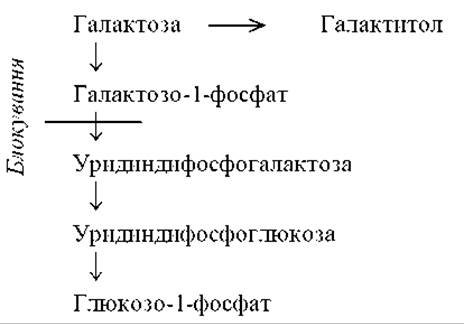
When this enzyme is blocked, its substrates (galactose, galactose-1-phosphate, and galactitol) accumulate in tissues and blood, and are excreted in the urine. These compounds are responsible for the development of most symptoms of the disease. Galactitol deposits in the lens of the eye, causing cataracts. Simultaneously, glucose metabolism is disrupted in the liver, kidneys, and brain, leading to hypoglycemia. Amino acid metabolism is also altered, resulting in the appearance of cystathionine, methionine, and other compounds in the urine.
Clinical features. The disease manifests after the newborn starts being fed milk. Symptoms include vomiting, diarrhea, jaundice, hepatomegaly, and splenomegaly. Liver failure and cataracts progressively develop. Affected children experience delayed GROWTH AND DEVELOPMENT, and older children suffer from intellectual disability. In some cases, residual enzyme activity is preserved, in which case cataracts and hepatomegaly develop gradually during the child's first year of life.
Diagnosis: elevated blood and urine galactose levels, and absent galactose-1-phosphate uridylyltransferase activity in erythrocytes. If infants have not received milk for any reason (such as hyperbilirubinemia), galactose is not excreted in the urine.
Some countries perform newborn screening for galactosemia.
Treatment: specialized lactose-free formulas. A strict diet is mandatory During the first three years of life. Later on, a secondary pathway for converting galactose-1-phosphate to uridine diphosphate galactose becomes active via the enzyme hexose-1-phosphate uridylyltransferase, which is inactive in young children.
Prenatal diagnosis is invasive, followed by molecular genetic testing.
Glycogen storage diseases
These disorders result from impaired glycogen catabolism and are classified as storage diseases. There are 13 types of glycogen storage diseases, depending on the enzymatic defect involved in the complex process of glycogen catabolism. The various types differ in the Organ Systems where glycogen accumulates, as well as in The Structure of the glycogen itself. Type II is associated with defects in lysosomal enzymes involved in glycogen catabolism. Other types are caused by low activity of cytoplasmic enzymes in the cells of the liver, kidneys, muscles, erythrocytes, and the Small Intestine wall. Clinically, glycogen storage diseases are generally divided into muscular and hepatic forms. Hepatic forms include von Gierke disease (Type 1, OMIM 232200), Cori disease (Type 3, OMIM 232400), and Andersen disease (Type 4, OMIM 232500); whereas Pompe disease (Type 2, OMIM 232300) and McArdle disease (Type 5, OMIM 232600) are characterized by predominant Skeletal Muscle involvement.
Clinical features. The disease manifests in newborns. During the neonatal period, the leading symptoms are hypoglycemic seizures and hepatomegaly. Subsequently, symptoms include growth retardation, progressive hepatosplenomegaly (Fig. 6.12), nephromegaly, cardiomegaly, muscular hypotonia, and facial dysmorphism described as a "doll-like face" or cherubic facies (chubby Cheeks, small nose, Mouth, and chin).
The central nervous system is rarely affected, meaning that intellect remains preserved in most types of the disease. In von Gierke syndrome, psychomotor retardation is observed due to hypoglycemia and hypoglycemic seizures. With most forms, patients succumb by the 12th year of life from heart or liver failure, or respiratory complications caused by respiratory muscle weakness or secondary respiratory infections. Life expectancy is significantly longer in certain forms.
Diagnosis: hypoglycemia unresponsive to Glucagon or adrenaline, hyperketonemia, persistent hyperglycemia, and elevated lactate levels following glucose administration. Increased levels of Pyruvate, triglycerides, Phospholipids, Cholesterol, and uric acid. Liver and muscle biopsy with enzyme activity assay and glycogen content determination.
Treatment: for certain forms, a high-protein diet, administration of fructose or galactose, continuous nighttime nasogastric tube feeding in infants, Antibiotic therapy for bacterial infections, and allopurinol for hyperuricemia are effective. Liver transplantation has also been proposed.
Cystic fibrosis (OMIM 219700)
This is an autosomal recessive disorder caused by defective transport of Cl- and Na+ ions across cell membranes. In Europe, the average incidence is 1:2,500, while in the Odesa region it is 1:1,600. The heterozygote carrier frequency is 1:20 in the population of Southern Ukraine. It is believed that the accumulation of heterozygotes in the population is linked to resistance against cholera or tuberculosis.

Fig. 6.12. Hepatomegaly in a patient with a glycogen storage disease (von Gierke disease)
The disease arises from mutations in the CFTR gene (7q31-32), which encodes the chloride transport membrane protein (CFTR — cystic fibrosis transmembrane conductance regulator). The gene is approximately 250 kb in size, contains 27 exons, and encodes a protein consisting of 1,480 amino acids. The function of the protein is to transport chloride ions across epithelial cell membranes. The CFTR gene also acts as a regulatory protein for other Ion Channels in The Cell membrane. The protein is located in the membranes of epithelial cells in exocrine glands (salivary, pancreas, Sweat Glands), Testes, intestines, and Lungs. Over 1,000 mutations have been identified in the gene, with about 300 of them being pathogenic. The most frequent pathological mutation (accounting for 70% of all cases) is a phenylalanine deletion at position 508 (ΔF508). These mutations lead to complete or partial cessation of Protein Synthesis, prevent its incorporation into the cell membrane (ΔF508), or impair its function.
Impaired chloride Ion transport across the apical membrane of secretory endothelial cells leads to disrupted sodium transport into the cells and altered cellular electrolyte composition. This, in turn, causes dehydration of exocrine gland secretions. Consequently, extremely viscous mucus is secreted by the exocrine Glands of the gastrointestinal tract and Bronchi. The primary manifestations of cystic fibrosis are chronic obstruction of small bronchi accompanied by chronic bacterial infection, and Digestive System impairment with exocrine pancreatic insufficiency. Because the pancreatic ducts become blocked by thick mucus, cysts form (hence the alternative name of the disease — cystic fibrosis), pancreatic enzymes fail to reach the intestinal lumen, Digestion is impaired, and malnutrition develops. In males, obstructive azoospermia and infertility occur due to congenital Aplasia of the vas deferens. Sweat contains elevated levels of Na+ and Cl-, making the patients' skin taste salty, which is used in diagnosing the condition.
Clinical features. According to the traditional Classification still found in medical literature, the following forms of cystic fibrosis are distinguished:
1. Intestinal form (5%) — manifests in infancy (often upon transition to formula feeding), caused by impaired delivery of pancreatic enzymes into the intestine. Despite a normal appetite, the child exhibits growth failure/malnutrition (Fig. 6.13). Characterized by voluminous, foul-smelling stools containing large amounts of neutral fat, and decreased fecal Elastase levels. Later on, delayed physical development, cholestatic hepatitis, cirrhosis, and fatty liver infiltration are observed. Endocrine pancreatic function may also be impaired, leading to the development of diabetes mellitus.
In nearly 5% of cases, intestinal cystic fibrosis presents as meconium ileus in newborns. The disease essentially begins in utero. Thick, putty-like meconium accumulates in the terminal ileum, leading to dilatation of the proximal intestinal segments while the distal segments remain collapsed (pencil colon). Perforation of the dilated bowel segment results in meconium Peritonitis in utero or shortly after birth. Mortality is very high. In some cases, intestinal lavage is effective, and Surgical treatment to remove meconium masses is occasionally performed.
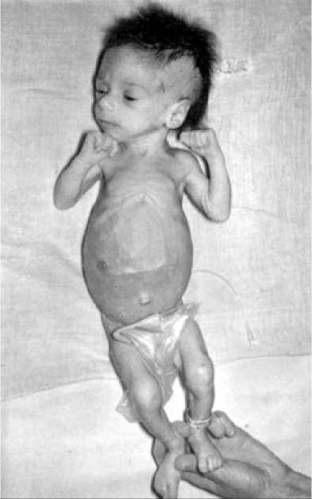
Fig. 6.13. Newborn with cystic fibrosis
2. Bronchopulmonary form (10–15%) is caused by the obstruction of small bronchi by viscous mucus. Respiratory symptoms dominate the clinical picture of cystic fibrosis in the majority of patients. In the first months of life, inflammation with thick bronchial secretion and small-bronchi obstruction develops. Subsequently, chronic infectious and inflammatory conditions join in: severe Bronchitis and pneumonia (most commonly caused by Staphylococcus aureus, Haemophilus influenzae, Pseudomonas aeruginosa). Characteristic features include a chronic cough with viscous sputum production, wheezing, intermittent Respiration, and sinusitis. Later, Bronchiectasis, emphysema, Cor Pulmonale, and pneumosclerosis develop. Children die from cardiac and respiratory failure.
3. Mixed bronchopulmonary-intestinal form (in 80–85% of all patients).
According to the modern classification adopted in Ukraine since 1999, the following forms of cystic fibrosis are distinguished:
— cystic fibrosis with pancreatic insufficiency, corresponding to the intestinal and mixed forms in the previous classification;
— cystic fibrosis without pancreatic insufficiency, corresponding to the pulmonary form in the old classification. This group also includes congenital bilateral aplasia of the vas deferens (genital form of cystic fibrosis);
— atypical forms of cystic fibrosis. The atypical phenotype in cystic fibrosis includes chronic respiratory disease of varying severity with characteristic cystic fibrosis manifestations, normal exocrine pancreatic function, and normal or borderline sweat chloride levels. Atypical forms also include cases where the patient presents with only a single clinical manifestation of the disease (sinusitis, pancreatitis, liver damage, etc.).
The clinical form of the disease correlates with the degree of protein function impairment. Less than 3% residual function causes the "classic" severe form of the disease with pancreatic insufficiency; 3–8% causes the form with predominant pulmonary involvement and preserved pancreatic function; 8–12% results in the mildest clinical manifestations of cystic fibrosis, such as Male infertility.
Diagnosis. The primary test is the determination of Na+ and Cl- levels (or chloride alone) in a sweat sample obtained via pilocarpine iontophoresis. The test is considered positive at chloride concentrations above 60 mEq/L in children and above 70 mEq/L in adults and adolescents (normal is 40; a result of 40–60 is regarded as borderline, requiring repeat testing). Diagnostic significance is attached to positive results from two or more tests performed at 2-week intervals. In infants under 3 months of age, the diagnostic threshold for sweat chloride concentration is lower at 40 mEq/L (doubtful level is 25–40 mEq/L).
A screening test can be the reduction of meconium proteolytic enzyme activity. Trypsin levels can be determined using biochemical or immunological methods, as well as by the ability of meconium dilutions to dissolve the gelatin layer of an exposed X-ray film (Schwachman test). Normally, digestion is observed at a dilution of 1/40 and higher (1/40, 1/80, 1/160, 1/320). In cystic fibrosis, the test is negative. The albumin level in meconium is elevated in patients—greater than 20 mg/g (normal is less than 3 mg/g).
Molecular genetic diagnostics—identification of mutations in the CFTR gene—has been developed. Newborn screening methods have also been proposed.
Treatment. The principles of dietary therapy involve prescribing a high-calorie diet with protein and fat content (predominantly plant-based) corresponding to age norms, an increased amount of CARBOHYDRATES (125% of age-appropriate requirements), and supplemental sodium chloride intake. Pancreatic enzyme replacement therapy (Creon) must accompany every meal. The dosage of medications is chosen individually, taking into account body weight dynamics and the cessation of steatorrhea. Protein-enriched therapeutic formulas may be used. Courses of antibacterial therapy are administered to prevent and treat pulmonary infections. Mucolytics (acetylcysteine, etc.) are used continuously via inhalation or orally; to remove sputum, vibration massage, breathing in drainage positions, postural drainage, and physical therapy (PT) are necessary. Gene Therapy methods are currently under development.
The prognosis is very grave. Previously, patients died at an early age; nowadays, life expectancy has increased up to 30 years.
Prenatal Diagnosis of cystic fibrosis is invasive, followed by molecular Genetic Testing of the biological material.
Lipid Metabolism Disorders
Plasma lipidoses are characterized by elevated serum lipid levels and are accompanied by the risk of developing CORONARY HEART DISEASE, atherosclerosis, and stroke. Among plasma Lipids, cholesterol is of primary importance in the development of the disease.
Several forms of plasma lipidoses are distinguished, which are inherited in a monogenic and multifactorial manner. One form is familial hypercholesterolemia (type II), which is inherited in an autosomal dominant manner. The disease is associated with a defect in low-density lipoprotein (LDL) receptors (19p13.2). Impaired binding and uptake of LDL lead to hyper-beta-lipoproteinemia and elevated plasma cholesterol levels. At the same time, the Circulation time of LDL prior to catabolism increases from 2.5 days (normal) to 4.5 days (in heterozygous Aa individuals) and even longer (in homozygous AA individuals).
Clinical features. Heterozygotes are more common in the population (1:200–1:500). Serum cholesterol levels in adult heterozygous patients range from 9–14 mmol/L. Characteristic features include tendon xanthomas (cholesterol deposits), primarily in the Achilles tendon and hand tendons, in the soft tissues of the eyelids, the cornea, and the skin (Fig. 6.14). Early atherosclerosis (at 30–40 years of age) leads to myocardial infarction and patient death before the age of 60.
In homozygotes, the same symptoms appear at an earlier age. Characteristic features include atheromatous plaques at the aortic ROOT and aortic Valves, which can lead to aortic stenosis and sudden death. Treatment is of low efficacy, and homozygous patients die before the age of 30 (a case of a 3-year-old girl dying from myocardial infarction has been described).
Diagnosis is based on detecting elevated serum cholesterol levels (greater than 6.7 mmol/L up to 16 years of age and 7.5 mmol/L in adults) and a family history.
Treatment. A diet restricted in cholesterol and saturated fats, along with medications that lower lipid content and prevent cholesterol Absorption in the gastrointestinal tract. Gene therapy methods are currently undergoing clinical trials.
Lysosomal Storage Diseases
This is a group of enzymopathies characterized by impaired catabolism of high-molecular-weight compounds. As a result of undegraded macromolecules accumulating in the cells and intercellular substance of various tissues and Organs, their function is impaired. When the impairment of catabolism is caused by mutations in lysosomal enzyme genes, these conditions are referred to as lysosomal storage diseases. Storage diseases are characterized by a progressive course, considerable severity, rapid disability, and early death.
Storage diseases are classified according to the accumulated macromolecules. Such diseases include mucopolysaccharidoses (disorders of glycosaminoglycan metabolism), glycogenoses (disorders of glycogen catabolism), and sphingolipidoses (disorders of Sphingolipid Metabolism), among others.

Fig. 6.14. Multiple skin xanthomas in a patient with hypercholesterolemia

Fig. 6.15. Mucopolysaccharidosis type IV, Morquio syndrome (short neck, kyphoscoliosis, knee valgus deformity, broad and flat hands and feet)

Fig. 6.16. Coarse facial features in mucopolysaccharidosis (Hunter syndrome)
Table 6.7. Types of glycosaminoglycans and their tissue localization
|
Type |
Localization |
|
Unsulfated |
|
|
1. Hyaluronic acid |
Practically in all connective tissue, including the vitreous body of the eye, loose |
|
connective tissue, synovial fluid |
|
|
2. Chondroitin |
Cartilage, skin, bones |
|
Sulfated |
|
|
3. Chondroitin-4-sulfate |
Cartilage, skin, bones, cornea |
|
Chondroitin-6-sulfate |
Cartilage, skin, aorta, dental pulp |
|
4. Dermatan sulfate |
Skin, ligaments, tendons, heart valves, Blood Vessels; stabilizes Collagen fibers |
|
5. Heparan sulfate |
Lungs, Arteries, cell membranes |
|
6. Keratan sulfate |
Skeleton, cornea, cartilage |
|
7. Heparin |
Mast cells (lungs, skin, liver, intestine); anticoagulant |
Mucopolysaccharidoses
Mucopolysaccharidosis is characterized by the accumulation of polymeric carbohydrates, known as glycosaminoglycans (GAGs), in various tissues of the body.
Glycosaminoglycans are polymers consisting of aminosugars and glucuronic acid. They are Components of the Extracellular matrix of connective tissue. Several GAG fractions (7 types) are distinguished, differing in monomer structure, glycosidic bonds, as well as the presence and localization of sulfate groups (Table 6.7).
In MPS, disorders of heparan sulfate, keratan sulfate, and dermatan sulfate catabolism are of the greatest significance. Under normal conditions, GAGs are rapidly degraded (half-life is 7-10 days). This is a multistep process involving several enzymes—lysosomal Hydrolases—whose genes are located on various Chromosomes (3, 5, 7, 22, X, etc.). Mutations in the genes encoding these enzymes lead to the development of Various Forms of mucopolysaccharidosis (a total of 9 types with subtypes; in all, 14 have been described to date). Each condition is caused by a defect in a specific lysosomal hydrolase involved in the sequential degradation of glycosaminoglycans. All mucopolysaccharidoses, except for one—Morquio syndrome (type 4)—are caused by a defect in an enzyme involved in The breakdown of dermatan sulfate and heparan (heparin) sulfate (either individually or both). In Morquio syndrome, keratan sulfate catabolism is impaired.
Non-degraded GAGs accumulate within cells and the intercellular substance of various tissues and organs, including the liver, spleen, kidneys, neurons, cartilage, Bone tissue, and cornea. Various GAG fractions are excreted in the urine.
Most types of mucopolysaccharidosis are inherited as recessive traits, whereas Hunter syndrome (type 2) is inherited as an X-linked recessive trait.
Clinical features. Since GAGs are a crucial component of the extracellular matrix of connective tissue, skeletal abnormalities are characteristic of MPS. These include growth retardation (following normal growth during infancy), a very short neck, macrocephaly, deformities of the Sternum, Ribs, and spine, shortening and deformation of the limbs, stiffness of large and small joints, and camptodactyly (Fig. 6.15). The hands and feet are broad and flat. The facial appearance is coarse, featuring a saddle nose, hypertelorism, exophthalmos, thick Lips, macroglossia, spaced Teeth, multiple caries, Hypertrophy of the alveolar processes and Gums, and low-set ears (Fig. 6.16). Hypertrichosis is present, with thick and coarse scalp hair. The eyebrows are broad, bushy, and show synophrys.
Other clinical manifestations (depending on the type of mucopolysaccharidosis) include inguinal or umbilical hernias, hepatosplenomegaly, central nervous system involvement (cognitive impairment), ocular disorders (cataracts, glaucoma), hearing loss, and cardiovascular abnormalities (cardiomegaly, heart valve disease).
Biochemical signs of the disorder can be detected immediately after birth. Clinical symptoms appear later, at varying ages depending on the disease type (ranging from the second year of life up to 4-5 years of age). The disease progresses inexorably. Patients typically succumb to respiratory complications or progressive Heart Failure before the age of 10 (in Hurler syndrome) or during the second to third decade of life, although some cases of survival up to 60 years have been documented.
Diagnosis is based on clinical symptoms and biochemical changes: urinary excretion of dermatan sulfate, heparan sulfate, keratan sulfate, and other GAGs is elevated 5-10 fold, while hydroxyproline levels are decreased. The specific type of mucopolysaccharidosis can only be determined by measuring the activity of various enzymes in blood serum, skin fibroblasts, leukocytes, and other cells (depending on the disease type).
Treatment is symptomatic. Orthopedic and audiologic care is provided as needed.
Prenatal diagnosis is feasible for certain types of mucopolysaccharidosis (mucopolysaccharidosis type I — Hurler syndrome, and mucopolysaccharidosis type IV — Morquio syndrome) by enzymatic assay in cultured Amniotic Fluid cells following amniocentesis. DNA diagnostic methods are currently under development.
Intracellular lipidoses are associated with the accumulation of lipids within cells. An example is sphingolipidoses, which are storage diseases caused by impaired metabolism of Sphingolipids (derivatives of the aminosphingosine base (C18), Fatty acids, and carbohydrates). Sphingolipids include cerebrosides, sphingomyelins, gangliosides, and others. They are essential components of the cytoplasmic membrane. A very high concentration of sphingolipids is found in the brain (particularly in myelin sheaths). Sphingolipid degradation is mediated by lysosomal enzymes. A defect in a specific enzyme leads to the intracellular accumulation of the corresponding lipid and the development of the disease. More than 15 types of sphingolipidoses have been described.
The disease manifests in the first months of life with psychomotor retardation, visceromegaly, and at least one of the following findings: vacuolation of lymphocytes, the presence of "foamy" vacuolated cells in the Cytology/practical/86.html">Red Bone Marrow (vacuoles being cell Lysosomes engorged with sphingolipids), a cherry-red spot on the retina, and slowed nerve conduction velocity. The most frequent intracellular lipidoses are Niemann-Pick, Gaucher, and Tay-Sachs diseases (Table 6.8).
Tay-Sachs disease ($GM_1$ gangliosidosis) develops as a result of a deficiency of the enzyme hexosaminidase A in cellular lysosomes. The disorder is also known as infantile familial amaurotic idiocy. Degenerative processes occur in the Gray matter of the brain, leading to blindness, deafness, spastic tetraparesis, and decerebrate rigidity.
Clinical features. The disease manifests at the age of 5-6 months. A previously healthy infant develops hypotonia, followed by hypertonia, paralysis, seizures, and loss of hearing and Vision (Fig. 6.17). Acquired skills are lost, and severe intellectual disability ensues, progressing to idiocy. Fundoscopic examination reveals a cherry-red spot in the macula lutea region, surrounded by a whitish halo—the "cherry-red spot" sign. Children typically do not survive past the second year of life. The disease is more prevalent among Ashkenazi Jews.
The incidence in this population is 1:3600, with a carrier frequency of 1:25. The high frequency of heterozygotes is attributed to isolation, Genetic Drift, and a selective advantage for heterozygotes, who may have a lower susceptibility to tuberculosis.
Diagnosis is based on determining the activity of the enzyme (hexosaminidase A) in leukocytes, fibroblast culture, and blood serum (the latter can also be used to detect heterozygous carriers). A specific clinical sign is the "cherry-red spot" on the fundus.
Prenatal diagnosis involves determining the activity of the corresponding enzyme in chorionic villus cells and amniotic fluid, as well as DNA diagnostic methods.
Peroxisomal Disorders
Peroxisomes are round or oval cellular Organelles bounded by a single membrane that contain about 40 enzymes involved in the oxidative metabolism of cells. The Functions of peroxisomes include protecting the cell from generated reactive oxygen species, breaking down hydrogen peroxide, oxidizing fatty acids, and synthesizing phospholipids. Peroxisomal disorders are associated with impaired formation and function of peroxisomes. Discovered in the early 1980s, they joined the group of inherited cellular organelle metabolic disorders, following lysosomal and mitochondrial diseases. Generalized peroxisomal disorders are associated with the complete absence of peroxisomes.
Generalized peroxisomal disorders include Zellweger syndrome (a genetically heterogeneous condition). The disease is characterized by craniofacial dysmorphisms: a high forehead, flat occiput, hypoplasia of the supraorbital ridges, broad nasal bridge, epicanthus, "gothic" palate, micrognathia, and External ear deformity. Eye anomalies include Brushfield spots, cataracts, glaucoma, corneal clouding, and pigmentary retinopathy. Marked muscular hypotonia, hyporeflexia, delayed psychomotor development, epileptic seizures, and hepatomegaly are observed. Hypotonia and a "mongoloid" facial appearance often prompt suspicion of Down syndrome. The prognosis is unfavorable, and affected children typically do not survive past a few months of age. A similar clinical picture is characteristic of other peroxisomal disorders.
Table 6.8. Characteristics of Intracellular Lipidoses
|
Disease |
Defective Enzyme |
Accumulated Compound |
Clinical Signs |
|
Niemann-Pick Disease (257220) |
Sphingomyelinase |
Sphingomyelin |
Enlargement of the liver and spleen, brown skin pigmentation, mental retardation |
|
Tay-Sachs Disease (GM2-gangliosidosis) (272800) |
Hexosaminidase A |
GM2-ganglioside |
Loss of acquired skills, blindness, seizures, muscle rigidity |
|
Gaucher Disease (231000) |
Glucocerebrosidase (ß-glucosidase) |
Glucocerebroside |
Hepatosplenomegaly, Osteoporosis, frequent fractures, hypochromic anemia, thrombocytopenia, bleeding, mental retardation. In the chronic form, intellect is preserved. Enzyme replacement therapy with ceredase has been developed |

Fig. 6.17. Tay-Sachs disease
The morphological marker of Zellweger syndrome is the absence of peroxisomes in liver biopsies and other tissues. Diagnosis of peroxisomal disorders is based on a combination of a characteristic phenotype, ocular lesions, neurological symptoms, determination of peroxisomal enzyme activities, fatty acid levels, and other biochemical parameters.
Disorders of Hormone Synthesis
Congenital Hypothyroidism
The incidence in various populations is 1:3500–1:4200, with the incidence of congenital hypothyroidism in Ukraine being 1:4200. The sex ratio is M1:F1.
Etiology. The causes of congenital hypothyroidism are diverse and may include:
1. Thyroid dysgenesis (aplasia, hypoplasia, ectopia). The disease usually occurs sporadically, though familial cases with a putative Autosomal Recessive Inheritance pattern exist. It is 4 times more frequent in females. Dysgenesis results from an embryonic developmental defect, sometimes caused by an autoimmune process. Thyroid dysgenesis is the most common cause of congenital hypothyroidism.
2. Autosomal recessive impairment of thyroid hormone synthesis (10–15% of all cases of congenital hypothyroidism). Thyroid hormone synthesis occurs in several stages, each controlled by a specific enzyme. Mutations in the genes encoding these enzymes lead to deficient or abnormal hormone synthesis. A distinctive feature of these forms of hypothyroidism is thyroid enlargement resulting from the organ's compensatory response to thyrotropin hyperstimulation.
3. Thyroid-stimulating hormone (TSH) or thyrotropin-releasing hormone (TRH) deficiency. This is rare.
4. Thyroid unresponsiveness to TSH.
5. Peripheral unresponsiveness to THYROID Hormones (symptoms of hypothyroidism develop despite elevated levels of T4 and TSH).
6. Drug exposure during pregnancy (radioactive iodine, iodides, thiouracil, methimazole, etc.).
7. Iodine deficiency (endemic cretinism).
8. Idiopathic congenital hypothyroidism.
Clinical Features. In severe cases, the disease is diagnosed in the neonatal period. Characteristic features include macrosomia, prolonged neonatal jaundice (often with elevated direct bilirubin levels), and enlarged fontanelles. The skin is pale, cold, and dry. Mucinous edema leads to macroglossia, narrowing of the nasal passages, and Swelling of the Larynx and vocal cords, causing a specific phenotype: coarse facial features, eyelid edema, and macroglossia, which typically leaves the mouth open (Fig. 6.18). Breathing is labored, and the voice is hoarse and low-pitched. Marked hyporeflexia, bradycardia, and hypotonia are present. In mild forms, the disease manifests in the 1st–2nd month of life, presenting with decreased appetite, regurgitation, lethargy, constipation, peripheral microcirculation disorders, and reduced skin Temperature. Marked muscular hypotonia, abdominal distention, and umbilical hernias are observed.
In the absence of adequate therapy, there is delayed ossification, uneven bone growth with shortened limbs and neck combined with a large head, saddle nose, and delayed physical and mental development, potentially reaching idiocy.
Diagnosis. The diagnosis is established based on elevated levels of pituitary thyrotropin (TSH) and decreased levels of thyroxine (T4) and triiodothyronine (T3). Normal values are T4 — 0.006 mg/dL and TSH — 20 IU/mL. Screening programs for neonatal hypothyroidism have been developed.
Treatment. Replacement therapy with L-thyroxine at a dose of 0.01 mg/(kg·day) is initiated immediately after diagnosis. The prognosis depends on the timing and adequacy of the replacement therapy. When treatment is started before 6 weeks of age, normal intelligence is preserved in 70% of patients. In severe cases involving intrauterine brain damage, even timely initiated therapy may not yield the desired outcome.
Prenatal diagnostics is invasive, followed by molecular genetic testing.
Congenital Adrenal Hyperplasia (Adrenogenital Syndrome)
This is a group of syndromes caused by adrenal cortex dysfunction. The term "adrenogenital syndrome" stems from the fact that adrenal hypofunction is accompanied by impaired sexual development in both character and rate. A block in the steroidogenesis metabolic pathway can occur at any of the 5 steps of cholesterol conversion to cortisol, which accounts for the Variability of clinical manifestations.
In 95% of cases, the condition is caused by a 21-hydroxylase enzyme deficiency resulting in decreased production of cortisol, or both cortisol and aldosterone, depending on the mutation type. The steroid 21-hydroxylase gene is localized on the short arm of chromosome 6 (6p) and belongs to the cytochrome P450 gene superfamily.
The disease begins developing in utero. The reduction in cortisol levels triggers pituitary hypersecretion of ACTH, which in turn increases the synthesis of Steroids prior to the metabolic block. Among these, sex steroids—specifically androgens—play the most prominent role. They cause virilization and pseudohermaphroditic genitalia in girls, alongside premature sexual and somatic maturation in boys. Mineralocorticoid synthesis is also impaired.
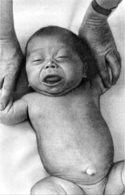
Fig. 6.18. Hypothyroidism (eyelid edema, saddle nose, macroglossia, umbilical hernia)
Clinical features. The disease presents in three main forms: virilizing, salt-wasting, and simple virilizing (or mixed). In the virilizing form, glucocorticoid production is impaired while mineralocorticoid synthesis remains intact. Affected newborn girls exhibit varying degrees of external genitalia masculinization (Fig. 6.19). In severe cases of virilization, genetically female newborns are frequently mistaken for boys. If pathogenetic treatment is not initiated, subsequent sexual development proceeds along male lines. Newborn boys have normally formed external genitalia, but premature puberty sets in by 5–6 years of age.
The salt-wasting form is characterized by a blockade in the synthesis of both glucocorticoids and mineralocorticoids. Mineralocorticoid deficiency leads to Water-electrolyte balance disorders (hyperkalemia, hyponatremia, hypochloremia). The salt-wasting syndrome typically manifests In the second week of life, though it can occasionally appear immediately after birth or later, by the end of the second month. The hallmark signs of this form include anorexia, regurgitation, nausea, diarrhea, dehydration, and arterial hypotension. Severe metabolic disturbances cause delayed physical development. Without treatment, the condition is frequently fatal.
The mixed form presents with overlapping symptoms of both types.
Diagnosis is based on detecting elevated daily urinary excretion of 17-ketosteroids.

Fig. 6.19. Virilizing form of adrenogenital syndrome in a girl (clitoromegaly, fusion of the labioscrotal folds, hyperpigmentation of the genital area)
Additional laboratory criteria include hyperkalemia, hyponatremia, and hypochloremia. In the salt-wasting form, aldosterone levels are drastically reduced. Abdominal ultrasound may reveal adrenal hyperplasia. Newborn screening methods have also been developed.
Treatment involves lifelong corticosteroid replacement therapy, with maintenance hydrocortisone therapy (1 mg/(kg·day) divided into three doses).
Prenatal diagnosis is invasive, followed by molecular genetic testing.
Last update: 11/08/2026
Editorial and Educational Adaptation: This material has been compiled based on the primary/original source text. The project team performed an editorial review, corrected technical inaccuracies, structured sections, and adapted the content for an educational format.
What was processed:
- elimination of formatting defects (OCR errors, structural breaks, corrupted characters);
- editorial organization of content;
- standardization of terminology in accordance with academic sources;
- verification of factual statements against the original source text.
All mentions of the author, publication year, and origin of the primary text have been preserved in accordance with the source.