Medical Genetics - V. M. Zaporozhan 2005
Monogenic Disorders
Clinical Features and Genetics of Certain Monogenic Disorders
Autosomal Dominant Disorders
An autosomal dominant trait is a characteristic that phenotypically manifests in the heterozygous state. In other words, an individual who carries one normal allele and one pathogenic mutant allele of a Gene will be affected by the disorder. The disease-causing gene in this case is designated as A, and the normal allele as a. The genotype of an affected individual is typically Aa, and less commonly AA; the genotype of an unaffected individual is aa.
Autosomal dominant disorders are characterized by the following features:
1. In the pedigree of an affected child, typically one of the parents is also affected; individuals of both sexes are affected with equal probability; and an affected parent transmits the disorder to both sons and daughters. A detailed pedigree analysis is provided in section 4.2.1.
2. The birth of an affected child to unaffected parents is associated with a de novo (newly arisen) mutation. This scenario is particularly typical for severe disorders associated with reduced reproductive fitness (virtually all cases in the population result from new Mutations). The probability of the same mutation recurring is extremely low, and families usually have only a single affected child. Gene Mutations occur more frequently during Spermatogenesis than oogenesis, and their likelihood increases with paternal age.
3. Lethal effect of the gene in the homozygous state. For example, in homozygotes (AA) for the Achondroplasia gene, severe skeletal deformations lead to embryonic lethality, meaning that all patients with achondroplasia are exclusively heterozygotes (Aa). Homozygous lethality has also been described in brachydactyly, Marfan Syndrome, and other conditions.
2. Reduced (incomplete) penetrance of the gene. Penetrance refers to the frequency with which a gene phenotypically expresses itself in a population of individuals who carry it. It is expressed as the percentage of carriers in whom the gene actually manifests. For instance, the penetrance of polydactyly inherited in an autosomal dominant manner is 65%. This means that out of 100 individuals carrying the dominant disease gene, 65 will be affected, while 35 will remain healthy. Incomplete penetrance is attributed to METABOLISM/18.html">The Influence of modifier genes, although its exact molecular mechanisms are not fully elucidated in every specific case.
3. Variable expressivity of the disorder. Expressivity is the individual Variability in the manifestation of a trait among different affected persons. With very low gene expressivity, mild or forme fruste clinical variants may go undiagnosed, or the observed features may not be recognized as part of the familial disorder. For example, clinical manifestations of Marfan syndrome—which may not always be readily recognized in such cases—can include an asthenic build, Scoliosis, or myopia, whereas the sole minimal clinical sign of myotonic dystrophy might be a cataract or cardiac conduction abnormalities. Adult-onset autosomal dominant Polycystic Kidney Disease may present in some patients with early renal failure, while in others of the same age, it may manifest solely as Hypertension with preserved normal renal function.
In cases of variable expressivity, the clinical manifestations of autosomal dominant syndromes in offspring generally do not depend on the severity of the disease in the parents and can exhibit varying degrees of severity.
4. Pleiotropy—the influence of a single gene on The Development of multiple traits, which clinically manifests as involvement of multiple Organ Systems.
5. The recurrence risk of having an affected child in a family is determined by the parental genotypes. With complete penetrance, the probability of inheriting the disorder is 50% if one parent is affected (Aa), and 75% if both parents are affected (Aa). If the parents of an affected child are unaffected (genotype aa) and the birth of the child is due to a new mutation, the recurrence risk is low and corresponds to the population frequency of the given disorder.
The birth of multiple children with a dominant trait to healthy parents can be explained by:
— incomplete penetrance of the gene. In this case, a clinically unaffected individual carries the pathogenic dominant gene and has a 50% probability of passing it on to their offspring;
— low expressivity of the gene, which may prevent the disease from being recognized without a targeted, detailed clinical examination;
— Gonadal Mosaicism. In this scenario, a distinct clone of Cells carrying the mutant gene exists within the Gonads, meaning the mutation is present in multiple Gametes rather than just one;
— a premutation state of the gene, which expands into a full mutation during gametogenesis (such as in triplet repeat expansion disorders).
Basic data on some common autosomal dominant disorders are presented in Table 6.1.
Achondroplasia (OMIM 100800)
An autosomal dominant disorder characterized by 100% penetrance and lethality in homozygotes. Between 80% and 95% of cases are due to new mutations, which occur more frequently during spermatogenesis. The population frequency is 1:100,000, with a male-to-female sex ratio of 1:1.
The disease results from mutations in the fibroblast growth factor receptor 3 gene (FGFR3), located at 4p16.3. Other mutations in the same gene lead to Hypochondroplasia and Thanatophoric Dysplasia (constituting an allelic series).
Characteristic Clinical Features of achondroplasia. The condition can be diagnosed at birth. Newborns present with macrocephaly, a birth length of 46–48 cm, rhizomelic shortening of the limbs (proximal segments), redundant Skin folds on the arms and thighs, broad and short hands with fingers arranged in a characteristic "trident" shape, and frequently isodactyly (Fig. 6.2). Once the child begins to walk, pronounced lumbar lordosis and genu varum (bowlegs) develop (Fig. 6.3). Motor development is delayed, but intelligence is normal. In adults, height ranges from 120–130 cm, accompanied by a specific "scooped-out" facial appearance: macrocephaly, prominent frontal bossing, a saddle Nose, and mandibular prognathism. Spinal canal stenosis can lead to Spinal Cord compression and associated neurological symptoms. Life expectancy is not reduced.
Characteristic radiographic findings: narrowing of the foramen magnum, Spinal stenosis; shortening and thickening of tubular bones; flared iliac wings with flattened acetabular roofs.
Diagnosis is based on syndromic and radiographic evaluations.
Treatment is symptomatic, and orthopedic correction has been proposed in recent years.
The genetic risk for offspring is 50% if one parent is affected.
Prenatal diagnosis: assessment of tubular bone length at 24–26 weeks of gestation (shortening of the Femur and humerus); molecular genetic testing when indicated.
Class="center">
Fig. 6.2. Trident hand sign in achondroplasia, isodactyly
Table 6.1. Monogenic Diseases and syndromes with Autosomal dominant inheritance (frequencies according to S. I. Kozlova et al., 1996, and N. P. Bochkov, 2001)
|
Name of syndromes or diseases (OMIM ID) |
Population frequency |
Gene localization |
Minimum diagnostic criteria |
|
Apert syndrome (101200) |
1:160,000 |
10q26 |
Acrocephaly, microcephaly, Syndactyly of fingers 2-5, severe intellectual disability |
|
Achondroplasia (100800) |
1:100,000 |
4p16.3 |
Dwarfism caused by shortening of the proximal limbs, characteristic facial features |
|
Marfan syndrome (154700) |
1:10,000–1:15,000 |
15q21.1 |
Asthenic build, elongation of the distal limbs, arachnodactyly, chest and spinal deformities, aortic dilation, aortic aneurysm, mitral valve prolapse, myopia |
|
Myotonic dystrophy type 1 (160900) |
1:7500–1:10,000 |
19q13.2-q13.3 (Trinucleotide Repeat Expansion disorder) |
Myotonia, Muscle weakness, muscle atrophy and paresis of skeletal Muscles (primarily in the distal limbs and face), cataract, cardiomyopathy, endocrine disorders, intellectual disability |
|
Neurofibromatosis type 1 (Recklinghausen's disease) (162200) |
1:3500–1:4000. In 50–70% of cases, it is a de novo mutation |
17q11.2 |
Benign multiple tumors of the peripheral and optic nerves. Freckling, "café-au-lait" pigment spots on the skin: at least 5 in children (diameter ≥5 mm), at least 6 in adults (diameter ≥15 mm) |
|
Polycystic kidney disease, adult type (173900, 173910) |
1:2500 (OMIM) |
16p13.3-p13.12 (type I), 4q21-q23 (type II) |
Bilateral kidney enlargement, proteinuria, Hematuria, progressive renal failure |
|
Syndactyly Type I (185900), Type II (186000), Type III (186100) |
1:2500–1:3000 |
2q34-q36 (type I), 2q31-q32 (type II), 6q21-q23.2 (type III) |
Partial or complete fusion of fingers/toes |
|
Polydactyly — postaxial, type A3 (607324), type A1 (174200) — preaxial type II (174500) type IV (174700) |
1:630–1:3300 |
19p13.2-p13.1 (type A3), 7p13 (type A1) 7q36 (type II) 7p13 (type IV) |
Supernumerary digits on the little finger side (postaxial polydactyly) or thumb side (preaxial polydactyly) |
|
Ectrodactyly |
1:90,000 |
Genetically heterogeneous disorder |
Split hand and split FOOT deformity ("lobster-claw hand") |
|
Ehlers-Danlos syndrome — autosomal dominant, autosomal recessive, and X-linked forms have been described |
1:5000 (all forms combined) |
Genetically heterogeneous disorder; 14 genes mapped including 17q21.31-q22 and 9q34.2-q34.3 |
Impaired Collagen synthesis. Joint hypermobility and recurrent dislocations, chest and spinal deformities. Multiple skin scars, skin hyperextensibility, fragility, and bruising; varicose Veins, mitral valve prolapse. Ocular and dental abnormalities |
Hypochondroplasia (OMIM 146000)
The disease is characterized by a milder clinical course compared to achondroplasia. Affected newborns have normal birth weight and length. Growth retardation becomes noticeable after 3–4 years of age. Adult height is below average, with mild shortening of the proximal limbs and short hands. The HEAD and face are of normal size and proportions.
Thanatophoric dysplasia (OMIM 187600)
It is always the result of a de novo mutation and represents a lethal syndrome. It is characterized by severe limb shortening, stubby fingers, isodactyly, a narrow Thorax, macrocephaly with a prominent forehead, and a depressed nasal bridge. Rib hypoplasia and a narrow thorax lead to neonatal death due to respiratory distress.
Acrocephalosyndactyly
A group of autosomal dominant disorders characterized by acrocephaly and varying degrees of syndactyly.
Apert syndrome (OMIM 101200)
This is type I acrocephalosyndactyly, with a population frequency of 1:160,000 and a male-to-female ratio of 1:1. The condition is caused by a mutation in the fibroblast growth factor receptor 2 gene, FGFR2 (10q26).
Clinical features. Apert syndrome is characterized by the following manifestations:
1. Specific cranial deformity (acrocephaly) and facial dysmorphism: flat forehead, exophthalmos, hypertelorism, downward-slanting palpebral fissures, depressed nasal bridge, and prognathism. A cleft palate may also be present. The cranial anomaly develops due to premature closure of the cranial sutures (Fig. 6.4).

Fig. 6.3. Patients with achondroplasia (macrocephaly, shortening of the proximal limbs, Varus deformity of the lower extremities)
2. Complete syndactyly of the hands and feet.
3. Severe intellectual disability. Social adaptation is impaired.
Malformations of the Brain, Heart, and major Blood Vessels may occur. The life prognosis is determined by the severity of visceral malformations.
Diagnosis. Based on syndromological evaluation.
Prenatal diagnosis is possible via qualified ultrasound at 24–26 weeks of gestation. Since the disease is typically the result of a de novo mutation, the recurrence risk for siblings is minimal.
Marfan syndrome (OMIM 154700)
This is a hereditary Connective Tissue disorder. The syndrome is characterized by nearly 100% penetrance, variable expressivity (Fig. 6.5), and lethality in homozygotes.
In the European population, the frequency is 1:10,000–1:15,000, with a male-to-female ratio of 1:1. The condition is usually inherited, though in 25–30% of cases it results from a de novo mutation.
The disorder is caused by a mutation in the fibrillin-1 gene (FBN1) located on the long arm of chromosome 15 (15q21.1). Fibrillin is a glycoprotein that forms an integral part of connective tissue. It is found in the Extracellular matrix, Cartilage, blood vessel walls, the crystalline lens of the eye, and elsewhere. Impaired fibrillin synthesis leads to hyperextensibility of connective tissue, which largely accounts for the characteristic clinical signs. Altered metabolism of acidic mucopolysaccharides (glycosaminoglycans) has been detected, leading to their accumulation in the body and excessive urinary excretion. Hydroxyproline metabolism is also disrupted, resulting in high urinary excretion of this essential collagen component.
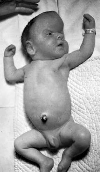
Fig. 6.4. Apert syndrome (acrocephaly, syndactyly of hands and feet)

Fig. 6.5. Variable expressivity in Marfan syndrome:
a — pedigree showing only pronounced clinical forms; b — pedigree indicating cases with a mild/subclinical presentation

Fig. 6.6. Marfan syndrome in a 9-year-old female patient (long slender limbs, narrow face, Pectus excavatum, flat feet)

Fig. 6.7. Arachnodactyly in Marfan syndrome
Clinical features. The main diagnostic criteria for Marfan syndrome are:
1. Skeletal abnormalities. Characteristic features include tall stature, asthenic habitus (Fig. 6.6), dolichostenomelia (disproportionately long arms and legs), arachnodactyly (Fig. 6.7), pectus excavatum or pectus carinatum, kyphoscoliosis, flat feet, dolichocephaly, narrow facial Skeleton ("bird-like" facies), and a high-arched ("gothic") palate. Joint hypermobility is frequently observed. Muscles often lag behind the skeleton in growth and are poorly developed.
Examples of tests used to identify characteristic musculoskeletal abnormalities are given in Table 6.2.
2. Cardiovascular involvement. Typical manifestations include mitral valve prolapse, dilation of the ascending aorta, and aortic aneurysm. Rupture of the aneurysm is the most frequent cause of early mortality in these patients.
3. Ocular pathology. Myopia due to an increased axial length of the Eyeball is the most common finding, though hyperopia may also occur. Weakness of the lens suspensory ligament leads to its subluxation or complete dislocation (ectopia lentis), which can be accompanied by iridodonesis (trembling of the iris). Secondary glaucoma, retinal detachment, and cataracts may develop. Blue sclerae are a characteristic feature.
4. Other clinical manifestations include emphysema and Spontaneous pneumothorax due to the rupture of pulmonary bullae (more common in adults), gastroptosis, gastrointestinal dysmotility, Nephroptosis, femoral, inguinal, and diaphragmatic hernias, muscle and subcutaneous fat hypoplasia, muscular hypotonia, striae cutis distensae, lumbosacral dural ectasia, etc.
5. Psychoneurological disorders may manifest as increased nervous excitability, asthenoneurotic syndrome, and emotional-volitional disturbances.
Table 6.2. Diagnostic tests for Marfan syndrome
|
Clinical sign |
Test |
|
Dolichostenomelia |
Ratio of hand length to height multiplied by 100%, greater than 11% Ratio of foot length to height multiplied by 100%, greater than 15%. Difference between arm span and height greater than 7 cm |
|
Arachnodactyly |
Thumb easily folds across the palm and extends beyond its ulnar border in this position Length of the middle finger exceeds 10 cm Patient easily wraps the wrist with the fifth digit and thumb |
|
Joint hypermobility |
Thumb touches the forearm upon wrist flexion Passive extension of the fifth finger by 90° Hyperextension of both elbows and knees by more than 10° Dorsiflexion of the foot greater than 45° |
The average life expectancy of a patient is determined by the severity of cardiovascular involvement. In recent years, it has increased significantly and is approximately 45 years. With appropriate work and rest regimens, patients can live to a ripe old age.
Diagnosis is based on family history, the patient's characteristic phenotype, and data from cardiological and ophthalmological evaluations. Biochemical Diagnostics reveal an increase in daily urinary excretion of hydroxyproline and glycosaminoglycans. Molecular genetic testing is also available.
Treatment:
— non-pharmacological therapy (Selection of an adequate daily routine, limitation of physical exertion, physical therapy, massage, physiotherapy);
— diet therapy — a diet rich in protein and collagen is recommended, supplemented with individually tailored dietary supplements containing Essential Amino Acids, Trace Elements, Unsaturated Fatty acids, and Vitamins (especially vitamins C and E);
— pharmacological therapy, including the administration of angioprotectors, venotonics, metabolic energizers, vitamins, and immunomodulators.
Patients require regular follow-up and monitoring by a multidisciplinary team of specialists, primarily a cardiologist, ophthalmologist, and orthopedist.
The genetic risk for the offspring of an affected heterozygous parent is 50%; due to variable expressivity, affected children may present with either a more severe or a milder form of the syndrome.
Prenatal diagnostics: Marfan syndrome can be diagnosed by elongated fetal long bones; molecular genetic testing is also available.
Ehlers–Danlos Syndrome
This is a genetically heterogeneous hereditary connective tissue disorder associated with impaired collagen synthesis. The collagen fibers in affected individuals are abnormally shaped and disorganized, which results from mutations in collagen genes or genes encoding Enzymes involved in collagen molecule maturation. Based on clinical and genetic criteria, the disease is classified into 11 types inherited in an autosomal dominant, autosomal recessive, or X-linked manner. The overall prevalence is 1:5,000 (all forms combined).
Ehlers–Danlos syndrome typically affects virtually all Organs and systems. The most critical diagnostic Criteria for the disease are as follows:
1. Skin abnormalities. Characterized by velvety texture, hyperextensibility, and fragility of the skin, subcutaneous nodules, multiple cigarette-paper-like scars, and increased bleeding diathesis.
2. Musculoskeletal manifestations: varying degrees of joint hypermobility and spinal deformities.
3. Cardiovascular manifestations: mitral valve prolapse, multiple aneurysms, and varicose veins.
4. Other symptoms characteristic of collagenopathies: hyperelasticity of ocular Tissues, generalized periodontitis, dental anomalies, hernias of various locations, splanchnoptosis, weakness of fetal membranes, and precipitous labor.
Diagnosis is based on Clinical and Genealogical data along with laboratory testing. A characteristic finding is increased urinary excretion of glycosaminoglycans and hydroxyproline. To precisely identify the disease type, enzyme activity assays and collagen typing are performed.
The treatment principles are the same as those for Marfan syndrome.
Ectrodactyly
This is a heterogeneous group of hand and foot malformations characterized by oligodactyly and Aplasia of the central rays of the hand and/or foot, resulting in a "lobster-claw" hand (or foot) deformity (Fig. 6.8). The population frequency is 1:90,000. Variable expressivity and penetrance are characteristic features. Treatment involves orthopedic correction.
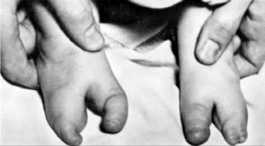
Fig. 6.8. Ectrodactyly of the feet ("lobster-claw" foot deformity)
Table 6.3. Examples of Congenital Malformations with an Autosomal Recessive Inheritance pattern
|
Syndrome Name (OMIM No.) |
Gene Localization |
Minimum Diagnostic Criteria |
|
Anophthalmia (206900) |
3q26.3-q27 |
Absence of eyes |
|
Familial alobar holoprosencephaly (236100) |
Genetically heterogeneous defect 21q22.3 2q37.1-q37.3 |
Failure of the Forebrain to divide into hemispheres, median cleft lip and palate, hypotelorism |
|
Primary microcephaly (251200) |
Genetically heterogeneous defect 8p23, 19q 13.1-q13.2 15q15-q21 |
Reduced head circumference, decreased brain mass and dimensions, intellectual disability; secondary microcephaly may occur in various single-gene, chromosomal, and teratogenic syndromes |
Malformations of the hands and feet (ectrodactyly, syndactyly, polydactyly)
These can manifest either as isolated autosomal dominant disorders or as symptoms of other monogenic and chromosomal syndromes.
Last update: 11/08/2026
Editorial and Educational Adaptation: This material has been compiled based on the primary/original source text. The project team performed an editorial review, corrected technical inaccuracies, structured sections, and adapted the content for an educational format.
What was processed:
- elimination of formatting defects (OCR errors, structural breaks, corrupted characters);
- editorial organization of content;
- standardization of terminology in accordance with academic sources;
- verification of factual statements against the original source text.
All mentions of the author, publication year, and origin of the primary text have been preserved in accordance with the source.