Fundamentals of Biochemistry - A. A. Anisimov 1986
Proteins
Amino Acids
2.2.1. Definition and Classification. Amino Acids can be regarded as carboxylic acid derivatives in which one of the hydrogen atoms in the carbon chain is replaced by an NH2 group. In most Naturally Occurring Amino Acids, the amino group is in the a-position relative to the carboxyl group:
Class="center">
Amino acids with ß- or y-positioned amino groups (such as ß-aminopropionic or y-aminobutyric acid) are significantly less common in living organisms.
Depending on The Nature of their side chains (R-groups), Amino acids are classified into acyclic (aliphatic) and cyclic (homo- and heterocyclic).
Based on the number of amino and carboxyl groups, amino acids are divided into: 1) monoaminomonocarboxylic (Glycine, Alanine, valine, leucine, isoleucine, Serine, Threonine, Cysteine, Methionine, Tryptophan, Tyrosine, phenylalanine); 2) diaminomonocarboxylic (Lysine, Arginine, citrulline); 3) monoaminodicarboxylic (aspartic and glutamic acids); 4) diaminodicarboxylic (cystine). According to the charge and polarity of their side chains, amino acids are classified as: 1) nonpolar, hydrophobic (glycine, alanine, valine, leucine, isoleucine, Proline, phenylalanine, tryptophan, methionine); 2) polar, uncharged (serine, threonine, asparagine, glutamine); 3) polar with negative (aspartic and glutamic acids, cysteine, tyrosine) or positive (lysine, arginine, Histidine) charges.
2.2.2. Isomerism, General Concepts1. Isomers are compounds that share the same molecular formula but differ in the spatial arrangement of their atoms, as well as in The Nature and sequence of the bonds between them.
1 The material in this section is presented in accordance with the nomenclature of organic chemistry proposed by the International Union of Pure and Applied Chemistry (IUPAC), Nomenclature of Organic Chemistry, Oxford–New York–Toronto–Sydney–Paris–Frankfurt, 1979.
There are two MAIN TYPES OF isomers: structural isomers and stereoisomers.
Structural isomers (which the new international nomenclature rules recommend calling constitutive isomers) have a different sequence of atomic connectivity. For example, leucine and isoleucine have the same molecular formula — С6Н13O2 — but different structural formulas;

Stereoisomers are isomers with the same sequence of atomic bonds, but with a different spatial arrangement of atoms. For example, the stereoisomers of alanine:

In the past, stereoisomerism encompassed optical isomerism and geometrical isomerism. The Use of these terms is no longer recommended today, as it is known, for instance, that optical activity can vary not only in magnitude but also in sign depending on the solvent.
If two stereoisomers are related to each other as an object and its mirror image, they are called enantiomers. Stereoisomers that are not related as an object and its mirror image are called diastereomers. Cis-trans isomers also fall under diastereomers if they differ solely in the arrangement of atoms relative to a plane passing through the molecule:

Cis-trans isomerism is possible only in structures rigidified by a double bond or a ring.
Overall, isomers can be classified as follows:

The Symmetry of any molecule can be characterized by the presence or absence of principal symmetry elements, which include: a plane of symmetry, a center of symmetry, and an axis of symmetry. If a molecule possesses neither a center nor a plane of symmetry, it is chiral and exists as a pair of enantiomers.
Chirality is therefore the property of compounds to exist as a pair of non-superimposable mirror images (from the Greek cheir, meaning hand).
Chiral compounds are also referred to as dissymmetric. Achiral molecules that lack an axis of symmetry are called asymmetric. Thus, an asymmetric molecule is always chiral, but not every chiral molecule is asymmetric. Molecules that do not have enantiomers are called achiral. Chiral molecules contain an atom surrounded by four different atoms or functional groups, known as a chiral atom or chiral center. In addition to carbon, atoms such as P, N, Si, etc., can also be chiral. If a compound contains n chiral atoms, it can exist in up to 2n stereoisomers.
The previously used term "asymmetric carbon" is no longer recommended, since chirality and Asymmetry are not synonymous.
Chiral compounds exhibit optical activity and are capable of rotating the plane of polarized light. The magnitude and direction of optical rotation are measured using a polarimeter. Enantiomers display identical chemical and physical properties, with one exception: the direction in which they rotate the plane of polarized light. An enantiomer that rotates the plane of polarization clockwise is called dextrorotatory (denoted by the "+" sign), while one that rotates it counterclockwise is called levorotatory (denoted by the "–" sign).
An equimolar mixture (i.e., a mixture containing equal amounts of molecules) of right- and left-handed enantiomers is called a racemic mixture (racemate). It lacks optical activity because the effects of the dextrorotatory and levorotatory molecules mutually cancel each other out. To characterize optically active compounds, a constant known as specific optical rotation is used, defined as the angle of rotation measured at a substance concentration of 1 g/mL and a path length of 1 dm. Specific rotation is denoted as [a]tD, where t is the Temperature at which the measurement was taken, and D is the wavelength of light (typically the sodium D-line of 546 nm).
There are two systems that describe the arrangement of atoms and groups around chiral atoms. One of these, established long ago and widely used (especially for characterizing Amino Acids and sugars), is based on comparing the configuration of the chiral compound in question with glyceraldehyde (for Monosaccharides and amino acids) or with serine (for amino acids). This relative principle for designating sugar and amino acid forms was introduced by E. Fischer in 1898. M. A. Rosanoff proposed using glyceraldehyde as the "key" in 1906. The form of glyceraldehyde in which the OH group appears on the right side of the carbon chain in a projection drawing was designated as the D-form, while the form with the OH group on the left was designated as the L-form:

To determine whether a given amino acid enantiomer belongs to the D- or L-form, its chiral center configuration is compared with glyceraldehyde enantiomers. If the amino group is located to the right of the COOH—R axis, it is a D-amino acid; if the amino group is located to the left of the COOH—R axis, it is the L-form.
To designate monosaccharide enantiomers, THE POSITION OF the OH group at the last chiral atom—i.e., the one most distant from the aldehyde or keto group—is compared with glyceraldehyde. Sometimes, the letters D and L are appended with the indices s or g to indicate whether serine (s) or glycerin (g) was used as the reference "key".
In 1951, a METHOD FOR DETERMINING absolute configuration was discovered, leading to the proposal of a new system for describing chiral centers without comparison to any "reference" compound: the RS system. We do not consider it here, since the D, L-system is customarily used when determining the configurations of amino acids and sugars.
Conformation is a specific case of stereoisomerism. The rules of international chemical nomenclature (1978) state that there is as yet no universally accepted Definition of the term conformation. The following is most frequently used: the Conformations of a molecule are defined as the various spatial arrangements of its atoms resulting from rotation about single (o) bonds.
In recent years, some authors have extended this definition to double and other bonds. In the presence of a ring, conformations are understood as the shapes of a molecule arising from bending of the ring plane and Changes in the relative positions of the ring parts. Molecules that differ in conformation are called conformational isomers or conformers. As applied to protein molecules, the term conformation characterizes the Spatial Structure of the molecule as a whole.
2.2.3. Stereochemistry of Amino Acids. All amino acids, with the exception of glycine, are optically active and can exist as a pair of enantiomers—D and L—because the carbon atom where hydrogen is replaced by the NH2 group is chiral. The direction of Rotation of the polarization plane is designated by a "+" or "—" sign, for example, D(—)-alanine and L(+)-alanine. It is necessary to specify the conditions under which the measurement was carried out: the Nature of the solvent, the reaction medium, and the presence of salts in the solution. Thus, L-histidine in aqueous solution exhibits a specific rotation of —39,3°, whereas in Hydrochloric acid solution [a]20D = +11,1°. The sign and magnitude of optical rotation also depend on the nature of The amino acid side chain (R-group).
Living organisms distinguish between the L- and D-forms of amino acids. It has been shown that Yeast and the mold fungus Penicillium glaucum utilize L-glutamic acid and L-leucine, but do not assimilate the D-forms of these amino acids. Most D-isomers possess a sweet taste, whereas L-amino acid forms are tasteless or bitter.
Only L-amino acids are found in the composition of Proteins. If Protein Hydrolysis is carried out under mild conditions, the amino acids retain their optical activity. During the Chemical synthesis of amino acids or upon boiling them in the presence of strong bases, racemization occurs, resulting in a D,L-mixture of amino acids that lacks optical activity.
D-Forms of amino acids are rarely encountered in nature. For instance, they form part of The Cell walls of certain microorganisms (see section 6.4.4) and serve as components of peptide Antibiotics (gramicidin, actinomycin D); they are very rarely found in plants.
2.2.4. Dissociation of Amino Acids. All a-amino acids exist In aqueous solutions predominantly as bipolar ions, or zwitterions, with a dissociated carboxyl group and a protonated amino group:

The dipolarity of amino acid molecular structures determines A number of their properties, notably the high solubility of Most amino acids in Water and relatively low solubility in organic Solvents, large dipole moments of their molecules, and high dielectric constant and melting point values. Depending on the pH of the medium, amino acids can exist as anions, cations, electroneutral dipolar ions, or as a mixture of these forms with one of them predominating. In strongly acidic solutions, amino acids are present as positive ions, whereas in alkaline solutions they are present as negative ions; that is, amino acids represent amphoteric electrolytes:

In accordance with their amphoteric nature, amino acids can form various salts depending on the COMPOSITION OF THE solution by reacting with both acids and bases:

The dissociation of amino acids can be clearly understood from the standpoint of the Brønsted acid-base theory, according to which an acid is any substance capable of donating a proton, and a base is any substance capable of accepting it. From this perspective, the COOH and H3N+ groups should be considered acids, while the COO- and NH2 groups are bases.
The dissociation constants of the carboxyl (K1) and amine groups (K2), or their negative Logarithms (pK1 and pK2), are numerically equal to the hydrogen ion concentration (pH values) at which The ratio of dissociated to undissociated forms is equal to unity—i.e., 50% of the amino acid exists in dipolar form, and 50% as cations and anions.
The values of pK1 and pK2 can be determined using electrometric titration. The titration curve of alanine with hydrochloric acid and sodium hydroxide is presented in Fig. 2.1. As can be seen from the figure, the titration of alanine (and other Monoaminomonocarboxylic Acids) is two-stage in nature, As a result of which the titration curve consists of two clearly distinguishable branches. The inflection points of these branches correspond to the values of pK1 and pK2 and are equal to 2,34 and 9,69, respectively. At pH values below pK1, all neutral amino acids carry a positive charge, while at pH above pK2 they carry a negative charge. At the transition point between the branches, the amino acid molecule carries no charge, since the number of negative and positive ions is equal.
The pH value at which the net charge of the amino acid is zero—i.e., the molecule is electroneutral—is called the isoelectric point pI (IEP). At this pH, no migration of the amino acid occurs in an electric field. The pH of a pure amino acid solution in water is called the isoionic point. The Isoelectric and Isoionic points of an amino acid in dilute solutions approximately coincide.
The isoelectric point for monoaminomonocarboxylic acids can be found by dividing the sum of pK1 and pK2 by 2. In our case: (2,34+9,69)/2 = 6.02. At the isoelectric point, amino acids do not exhibit buffer properties; their buffer capacity is maximal at pH values equal to the pK values of the acidic groups. If the values of pK1 and pK2 are known, the ratio of various types of ions can be calculated for any pH value.
Titration curves of amino acids with ionizable R-groups (e.g., histidine, lysine, glutamic acid) have a more complex character, because the curves describing the dissociation of R-groups overlap with the dissociation curves of a-carboxyl and a-amino groups. The dissociation of side R-groups proceeds as follows:

Fig. 2.1. Titration curve of alanine (types of ions predominating at the inflection points of the curve are indicated within the frames)

The pK values of the dissociating groups for Certain amino acids are given in Table 2.1.
Table 2.1. pK values of dissociating groups of Some amino acids (at +25 °C)
Amino acid |
pK1 |
pK2 |
R-group name and pK3 |
Alanine |
2,34 |
9,69 |
|
Leucine |
2,19 |
9,60 |
— |
Aspartic acid |
1,88 |
9,60 |
ß-Carboxyl 3,65 |
Glutamic acid |
2,19 |
9,67 |
γ-Carboxyl 3,22 |
Histidine |
1,82 |
9,17 |
Imidazole 6,00 |
Lysine |
2,18 |
8,95 |
ε-Amino group 10,53 |
Arginine |
2,17 |
9,04 |
Guanidino group 12,48 |
Tyrosine |
2,20 |
9,11 |
Phenolic hydroxyl 10,07 |
Cysteine |
1,96 |
10,28 |
Sulfhydryl 8,18 |
A comparison of the pK values of the dissociating groups across different amino acids leads to the Conclusion that for most amino acids, both the pK of the α-COOH groups (2.1–2.3) and the pK of the α-NH+3 groups (9.1–9.6) are quite similar. The carboxyl groups of monoaminomonocarboxylic acids are stronger acids than the carboxyls of the corresponding aliphatic acids (positively charged α-amino groups promote the repulsion of the carboxyl H+). For example, alanine has a pK1 of 2.34, whereas the corresponding propionic acid has a pK1 of 4.85. The amino group of lysine and the guanidino group of arginine exhibit strongly basic properties, carrying a net positive charge at pH 7. The hydroxyl group of tyrosine and the sulfhydryl group of cysteine display weakly acidic properties.
2.2.5. Absorption spectra of amino acids. None of the twenty amino acids that make up proteins absorb light in the visible spectrum; they absorb only in the far ultraviolet region (<220 nm). Tyrosine, phenylalanine, and especially tryptophan exhibit significant Absorption in the ultraviolet region (260–280 nm). Cystine shows weak absorption at 240 nm due to the presence of a disulfide group.
2.2.6. Solubility. With few exceptions, amino acids are readily soluble in water. As the hydrocarbon side chain increases in size, solubility in water decreases while solubility in alcohol increases (Fig. 2.2).
2.2.7 Chemical properties of amino acids. One of the most characteristic Reactions of the α-amino group is its interaction with ninhydrin. This reaction can be used for the Quantitative determination of amino acids present in solution at very low concentrations. At a pH above 5, the reaction proceeds in two stages. In The First stage, reduced ninhydrin is formed via the Oxidative Deamination of the amino acid, which is accompanied by its decarboxylation.

Fig. 2.2. Solubility of Amino acids

In the second stage of the reaction, the resulting ammonia reacts with equimolar amounts of oxidized and reduced ninhydrin to form a blue-purple product (Ruhemann's purple) with an absorption maximum at 580 nm, the color intensity of which is proportional to The amount of amino acid.

Proline and hydroxyproline, which contain substituted α-amino groups, react with ninhydrin to yield yellow derivatives (with an absorption maximum at 440 nm) and do not produce ammonia.
The ninhydrin reaction is not specific; it is given by many Other Compounds containing amino groups, including proteins and ammonia, with the only difference being that CO2 is not released in those cases. The formation of CO2 is specific to α-aminocarboxylic acids. This reaction is utilized in Chromatography for the detection of amino acids, whereas quantitative determinations in automated analyzers are performed by measuring the color that develops when amino acids emerging sequentially from the Column interact with ninhydrin.
Due to the presence of the amino group, amino acids also react with nitrous acid (1) and formaldehyde (2).
 (1)
(1)
 (2)
(2)
These reactions are employed for the quantitative Determination of Amino acids: the Van Slyke method measures the volume of evolved nitrogen gas, and the formol titration method determines the protons liberated after The addition of formaldehyde by titration with alkali.
Very important reactions specific to α-Amino groups are Sanger's reaction with 1-fluoro-2,4-dinitrobenzene and Edman's reaction with phenylisothiocyanate. They are of exceptional value for identifying the N-terminal amino acids of polypeptide chains (see Section 2.4.2).
Of considerable interest and importance is the reaction leading to the formation of asparagine and glutamine amides. In nature, these reactions generally proceed via the Utilization of ATP energy:

The Decarboxylation of amino Acids yields amines:

Amines typically exist as pungent-smelling liquids with an alkaline reaction. Many of them exhibit pronounced physiological activity. For instance, histamine—the decarboxylation product of histidine—displays a wide spectrum of biological effects. It possesses vasodilatory properties, which sets it apart from other biogenic amines that exert a vasoconstrictive effect. Histamine is produced in large quantities at sites of inflammation. By inducing vasodilation in the inflammatory focus, it thereby accelerates the influx of leukocytes. Histamine levels rise sharply following exposure to ionizing radiation and during Traumatic Shock. It also promotes the secretion of gastric juice with an elevated hydrochloric acid content, and plays a significant role in allergic reactions and pain syndromes. Histamine is found in bee venom and the venom of various animals, and is present in large amounts in ergot.
Of great interest are the diamines putrescine and cadaverine, which are produced as a result of the decarboxylation of Ornithine and lysine.

Cadaverine and putrescine are formed during the Breakdown of Proteins in decomposing corpses (from Latin *cadaver* — corpse, *putresko* — to rot, decay) and can cause poisoning from spoiled meat. They can also be produced in the human intestine during certain intestinal infections. Normally, small amounts of these diamines are rapidly neutralized within the Cells of the intestinal mucosa by binding amino groups—such as through Acetylation—and by degradation via oxidative deamination. The degradation products of these substances are excreted by The Kidneys in the urine. At low concentrations, putrescine stimulates RNA Synthesis and accelerates the growth of animal and bacterial cells.
Epinephrine and norepinephrine are exceptionally important amine derivatives; they are adrenal medullar Hormones synthesized from tyrosine (see Section 12.6.6).
Amino acid radicals are exceptionally diverse, which allows color reactions to be used for detecting most amino acids. Many of these reactions are highly sensitive and specific, enabling the identification of minute quantities of a particular individual amino acid within complex mixtures, biological fluids, and proteins. Certain color reactions are also utilized for the quantitative determination of Amino Acids and Proteins.
Amino acids can react with compounds containing a carbonyl group, such as reducing sugars. This results in the Formation of the corresponding aldehyde from the amino acid, and furfural or hydroxymethylfurfural from the carbohydrate. Aldehydes derived from amino acids possess pleasant aromas, and the combination of various aldehyde scents largely determines the flavor profile of food products. In turn, furfural and hydroxymethylfurfural can interact with amino acids to yield dark-colored products known as melanoidins. Melanoidin synthesis is responsible for the browning of numerous foods during preparation, drying, and storage. This browning reaction proceeds particularly intensely at elevated temperatures—for instance, during the drying of fruits, vegetables, and malt, the simmering of milk, the baking of bread (which imparts color to the crust), and the thermal Processing of wine, among other processes.
2.2.8. Characteristics of Individual Amino Acids Found in Proteins. Monoaminomonocarboxylic Acids. Glycine (Glycocoll, a-Aminoacetic Acid)

This is the only optically inactive amino acid. It has a sweet taste and is one of the most widespread amino acids, being particularly abundant in gelatin. Glycine serves as a precursor in The Biosynthesis of Purines, the porphyrin moiety of Hemoglobin, chlorophyll, and heme Enzymes. It is involved in the formation of bacterial cell walls and Functions as an inhibitory neurotransmitter in the Spinal Cord and most structures of the Brainstem, where it is present in high concentrations.
L-Alanine (a-Aminopropionic Acid)

Found in virtually all proteins, L-alanine plays a major role in Nitrogen METABOLISM and can serve as a Starting Material for the synthesis of carotenoids, rubber, fats, and CARBOHYDRATES.
L-Valine (a-Aminoisovaleric Acid)

L-Valine is capable of hydrophobic interactions, which is crucial for establishing and stabilizing Protein Structure. It is present in many proteins, typically in small quantities, and participates in the synthesis of Alkaloids, certain cyclopeptides, pantothenic acid, and penicillin.
L-Leucine (a-Aminoisocaproic Acid)

L-Isoleucine (a-Amino-β-ethyl-β-methylpropionic Acid)

Both amino acids are poorly soluble in water. They occur in proteins in insignificant amounts and are capable of hydrophobic interactions. They also serve as a source of fusel oils during Fermentation.
L-Serine (a-Amino-β-hydroxypropionic Acid)

Serine plays a major role in the metabolism of any Organism. It is a constituent of Phospholipids (phosphatidylserines) and the Polypeptides bradykinin and kallidin, participates in forming the Active Site of certain Proteolytic Enzymes, and is involved in the Synthesis of the amino alcohol sphingosine (a component of Sphingolipids). In some proteins, such as milk casein or egg yolk vitellin, it occurs as an ester—known as serine phosphoric acid—which plays an essential role in the metabolism of growing young animals. In plants, serine is produced during Photorespiration from glycine.
L-Threonine (a-Amino-β-hydroxybutyric Acid)

It participates in the synthesis of Vitamin B12 and the antibiotic actinomycin D. L-Cysteine (a-Amino-β-mercaptopropionic acid)

It plays a major role in the metabolism of plants and animals as a source of sulfur, as well as due to the presence of the sulfhydryl SH group, which acts as a reducing agent. It is a constituent of the tripeptide Glutathione, forms part of coenzyme A in amine form, and is present in the active center of numerous enzymes. In protein and peptide molecules (such as Insulin and ACTH), it participates in the formation of Disulfide Bonds between polypeptide chains or within a single chain, thereby playing a crucial role in shaping the Tertiary Structure of the protein molecule. It serves as the compound through which certain microorganisms and plants metabolize hydrogen sulfide. The oxidation of two cysteine molecules yields cystine, and the reverse transition occurs just as readily. This forms one of the most vital redox systems in living organisms:

In proteins, cystine predominates over cysteine. It is found in large quantities in the proteins of Hair, horns, and hooves.
L-Methionine (a-amino-y-methylthiobutyric acid)

It is one of the principal Donors of methyl groups in the synthesis of plant Cell wall carbohydrates, Choline, adrenaline, creatine, and sterols, as well as a source of sulfur in the formation of thiamine. It can also give rise to other Sulfur-Containing Amino Acids. Methionine is abundant in milk protein, specifically casein. Acting as a lipotropic factor, it is used as a therapeutic agent in the Treatment of atherosclerosis.
L-Lysine (a, ε-diaminocaproic acid)
It is found in almost all proteins, being particularly abundant in fish milt proteins, which belong to Protamines and Histones. It serves as a precursor in the synthesis of alkaloids (such as anabasis, nicotine, lupinine, and coniine). Through its ε-amino group, it participates in the formation of complexes between the protein moiety of an enzyme and its coenzyme, as seen in biotin-dependent enzymes.
It is present in negligible amounts in the proteins of cereal seeds, especially corn, which significantly reduces the Nutritional Value of the latter. Soviet breeders and State Prize laureates M. I. Khadzhinov and G. S. Galeev developed a number of corn varieties with an enhanced lysine content.
L-Arginine (a-amino-δ-guanidyl-n-valeric acid)

It is present in all proteins. It occurs in large quantities within basic histone and protamine proteins, which explains its Abundance in fish milt. It plays a significant role in Protein metabolism by participating in the synthesis of urea and creatine. In the form of phosphoarginine in invertebrate Muscles, it performs a function analogous to that of phosphocreatine in higher animals.
Monoaminodicarboxylic Acids. L-Aspartic acid, aspartate (a-aminosuccinic acid)

It is poorly soluble in water, and its solution exhibits an acidic reaction. It is found in large amounts in all plant proteins and plays an important role in PLANT AND ANIMAL metabolism, specifically by participating in Transamination reactions, the formation of urea, creatine, and cyclopeptides. Its decarboxylation yields a- or ß-alanine, which is necessary for the synthesis of carnosine, anserine, and coenzyme A (CoA). The amide of aspartic acid, asparagine, accumulates in very large quantities during the germination of legume seeds, particularly in the dark due to a lack of carbohydrates or in the presence of excess ammonia. Asparagine has been detected in Blood Plasma and within certain animal proteins (such as insulin, hemoglobin, and Myoglobin). In humans and animals, asparagine—much like aspartic acid—actively participates in transamination reactions, serves as a precursor in the Synthesis of purine and pyrimidine bases and nicotinic acid, and functions as a reserve and transport form of nitrogen in plants.
L-Glutamic acid (glutamate, a-aminoglutaric acid)

It gives an acidic reaction in aqueous solutions and is present in large quantities in all proteins. It is a component of the vitamin Folic acid and of glutathione. Along with aspartic acid, it plays a paramount role in Amino acid metabolism, actively participating in transamination, deamination, and direct amination reactions. The decarboxylation of glutamic acid yields γ-aminobutyric acid, which is of great importance in Brain metabolism—specifically enhancing inhibitory processes—as well as δ-aminolevulinic acid, which is involved in porphyrin synthesis. The amide of glutamic acid, glutamine, serves as a nitrogen transport form in animals and plants, as well as a starting compound for the synthesis of purine and pyrimidine bases and nicotinic acid. Monosodium glutamate is used as a food seasoning, possessing the taste and aroma of chicken broth.
Cyclic amino acids L-Phenylalanine
(a-amino-β-phenylpropionic acid)
It is found in all proteins and participates in the biosynthesis of Flavonoids and alkaloids. It exhibits a capacity for hydrophobic interactions and is responsible for the xanthoproteic reaction for proteins. The molecules of gramicidin and the antibiotic tyrocidine contain D-phenylalanine, which has not been found in a free state in nature.

L-Tyrosine (a-amino-β-hydroxyphenylpropionic acid)

One of the most widespread amino acids in nature; found in all proteins (with the exception of certain protamines). It serves as a primary precursor for the synthesis of hormones (thyroxine, noradrenaline, adrenaline) and alkaloids (morphine, codeine, papaverine). The oxidation of tyrosine mediated by tyrosinase leads to the formation of Melanins (pigments of the Skin, hair, and feathers).
L-Tryptophan (a-amino-β-indolylpropionic acid)
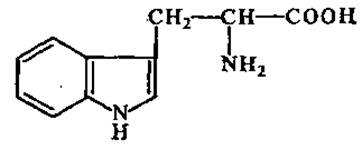
Found in nearly all proteins. Cereal grains are deficient in tryptophan. It decomposes upon acid Hydrolysis of Proteins. Tryptophan serves as a starting material for the synthesis of nicotinic acid (Vitamin PP) and heteroauxin.
L-Histidine (a-amino-β-imidazolepropionic acid)

Belongs to the group of basic amino acids, yielding an alkaline reaction in aqueous solutions. Significant amounts of histidine are found in globin, the protein component of blood hemoglobin. It is a constituent of the active sites of certain proteolytic enzymes.
L-Proline (pyrrolidine-a-carboxylic acid)

An imino acid that is highly soluble in alcohol. The proline content is particularly high in cereal grain proteins (prolamins), Collagen, Elastin, and tooth enamel protein. It is a component of several cyclic peptide antibiotics, such as gramicidin, licheniformin, and actinomycin D.
L-Hydroxyproline

A derivative of L-proline. Significant quantities of it have been detected in cell wall proteins, gelatin, and collagen.
2.2.9. Rare amino acids in proteins and non-protein amino acids. In addition to the usual 20 amino acids, small amounts of other, less common Amino acids have been isolated from proteins:

Over 150 amino acids discovered in Living organisms are not constituents of proteins. Some of them play a vital role in metabolism.
ß-Alanine (β-aminopropionic acid)
Н2N—CH2—CH2—COOH
A constituent part of coenzyme A (see Section 3.4.1), vitamin B3 (pantothenic acid), carnosine, and anserine. It also occurs in a free state and exerts an inhibitory effect on the Central Nervous system.
L-Ornithine (a, δ-diaminovaleric acid)

Easily formed from arginine through the action of the enzyme arginase. It participates in urea formation as well as in the synthesis of alkaloids and the antibiotic gramicidin.
L-Citrulline (a-amino-δ-carbamidovaleric acid)

It serves as an intermediate in urea synthesis. It occurs in a free state in the juice of watermelon fruits (Citrullus), from which it derives its name.
γ-Aminobutyric Acid (GABA)
H2N—СHЗ—CH2—CH2—COOH
It is found in many plants, as well as in the brain Tissues of mammals, certain amphibians, and birds. It is synthesized in the brain through the action of glutamate decarboxylase:

In animals, it functions as a chemical agent in Nerve Impulse transmission, acting as a neurotransmitter. Quantitatively, GABA is believed to be the principal inhibitory neurotransmitter in The Nervous System. The presence of GABA in the central nervous system is also essential for normal metabolic processes: it enhances Energy Metabolism, increases the respiratory activity of brain tissue, improves glucose utilization by the brain, activates numerous respiratory enzymes, enhances cerebral blood supply, and stimulates the removal of toxic metabolic waste from the brain. GABA is used as a pharmaceutical preparation under the trade names Aminalon or Gamalon.
Last update: 06/08/2026
Editorial and Educational Adaptation: This material has been compiled based on the primary/original source text. The project team performed an editorial review, corrected technical inaccuracies, structured sections, and adapted the content for an educational format.
What was processed:
- elimination of formatting defects (OCR errors, structural breaks, corrupted characters);
- editorial organization of content;
- standardization of terminology in accordance with academic sources;
- verification of factual statements against the original source text.
All mentions of the author, publication year, and origin of the primary text have been preserved in accordance with the source.