Molecular Biology of the Cell - Volume 2 - Alberts B., Bray D., Lewis J., Raff M., Roberts K., Watson J. 1993
Intracellular macromolecular sorting and maintenance of cellular compartments
RNA synthesis and processing
Up to this point, we have discussed the chromosome as a very large complex of DNA and protein that replicates prior to Cell Division. However, the primary purpose of a chromosome is to serve as a template for RNA Synthesis, since this is the only form in which the Genetic information contained within Chromosomes can be directly utilized by The Cell. RNA synthesis is extremely intensive: the overall rate of nucleotide incorporation into RNA during interphase is approximately 20 times higher than The rate of nucleotide incorporation into DNA during the S phase.
RNA synthesis (Introduction/24.html">DNA METABOLISM/31.html">Transcription) exhibits a high degree of Specificity. For example, in most mammalian Cells, functional RNA sequences are copied from roughly 1% of the DNA sequences (mature Messenger RNA or structural RNA). Selection occurs at two levels, which will be discussed in turn in this section: 1) only a portion of the DNA sequence is transcribed to generate nuclear RNAs, and 2) only a minor fraction of the nuclear RNA nucleotide sequences is retained following the Processing that precedes the export of RNA molecules into the Cytoplasm. Let us begin with a description of RNA polymerase, the enzyme that catalyzes all DNA transcription.
9.4.1. RNA Polymerase Subunit Exchange at the Initiation of Each New RNA Chain [42]
A general description of DNA transcription is provided in Chapter 5. Transcription begins when an RNA polymerase molecule binds to a promoter sequence in DNA. During initiation, the two DNA strands locally unwind to form an open complex in which the template strand becomes exposed. Following The formation of this complex, the polymerase begins to move along the DNA, elongating the growing RNA chain in the 5'-to-3' direction by the sequential addition of ribonucleoside triphosphates. This process continues until a stop (termination) signal is reached, at which point the newly synthesized RNA chain and the polymerase dissociate from the DNA. Thus, each RNA molecule is a single-stranded copy of a DNA nucleotide sequence representing a relatively short region of The Genome (see Fig. 5-1).
RNA polymerases typically consist of multiple polypeptide chains and have a Molecular Weight of 500,000 daltons or more. Bacterial and eukaryotic Enzymes are evolutionarily related (Fig. 9-64). Because the bacterial enzyme is much easier to study, its properties have served as a foundation for understanding how the analogous eukaryotic enzyme Functions. E. coli RNA polymerase contains five subunits: α, β, β', σ, and ω (with two copies of α and one of each other subunit). The complete Amino Acid Sequence of each subunit has been deduced from The nucleotide sequence of its Gene, and DNA footprinting data indicate that upon binding to DNA, the enzyme covers 60 nucleotide pairs.
The sigma (σ) subunit of E. coli RNA polymerase has a specific function and acts as a Transcription initiation factor. This subunit enables the enzyme to locate consensus promoter sequences. RNA polymerases that recognize different promoters contain distinct forms of the σ subunit. Upon binding to the promoter, the enzyme undergoes a series of reactions to initiate RNA synthesis (Fig. 9-65). After approximately eight NUCLEOTIDES of the RNA molecule have been synthesized (step 4, Fig. 9-65), the σ subunit dissociates and is replaced by several elongation factors required for chain elongation and termination. Elongation factors include several well-characterized Proteins, although the exact mechanism of their action remains to be fully elucidated.
Class="center">
Fig. 9-64. Amino acid sequence similarity between bacterial and eukaryotic RNA polymerases suggests that these enzymes share a common evolutionary origin. The largest RNA polymerase subunits from E. coli, Yeast, and Drosophila (compared in the figure) are thought to bind to DNA. Conserved sequence blocks are more than 70% homologous between yeast and Drosophila, and more than 40% homologous between Drosophila and E. coli. The carboxyl terminus of the eukaryotic subunits contains a unique sequence, Ser-Pro-Ser-Tyr-Ser-Pro-Thr, whose function is unknown; it is repeated 26 times in yeast and more than 40 times in Drosophila (indicated by circles). (From A. Z. Greenleaf et al., In: RNA Polymerase and the Regulation of transcription [W. S. Reznikoff et al., eds.], pp. 459–464. New York: Elsevier, 1987.)

Fig. 9-65. Schematic diagram of RNA synthesis initiation catalyzed by RNA polymerase. The indicated steps were elucidated through studies of the E. coli enzyme. A DNA molecule carrying a promoter sequence for E. coli polymerase is shown (see Fig. 5-6). Initially, the enzyme forms a closed complex in which the two DNA strands remain paired throughout their length. In the next step, the enzyme catalyzes the unwinding of slightly more than one turn of the DNA helix, generating an open complex in which the template is accessible for the initiation of RNA chain synthesis. However, the polymerase with its associated sigma subunit behaves as though it were anchored to the promoter region: it is incapable of RNA chain elongation and frequently reverts to the closed complex. As shown in the figure, Conversion of the enzyme into an actively elongating polymerase requires the release of initiation factors (such as the sigma factor in E. coli) and the association of other proteins acting as elongation factors (e.g., the nusA protein in E. coli). (Adapted from D. C. Straney and D. M. Crothers, J. Mol. Biol. 193: 267–278, 1987.)
In all organisms, different Genes are transcribed at widely varying rates. At some promoters, a new RNA chain is initiated every 1–2 seconds, whereas at others, the onset of synthesis may take an hour. Typically, the transcription rate of each gene is governed by gene-regulatory proteins that influence transcription initiation (Chapter 10). In general, the action of these proteins involves accelerating or decelerating one or more of the steps outlined in Fig. 9-65.
9-25
9.4.2. Eukaryotic RNA Synthesis Is Carried Out by Three Distinct RNA Polymerases [43]
Although transcription in eukaryotes shares certain similarities with that in a prokaryote such as E. coli, the mechanism is considerably more complex in Eukaryotic cells. Both yeast and humans possess Three types of RNA polymerases, each responsible for transcribing different sets of genes. The structures of these enzymes—designated RNA polymerases I, II, and III—are closely related; some of their subunits are identical, while others differ (Fig. 9-66). Each is more complex than E. coli RNA polymerase and is believed to comprise 10 or more polypeptide chains. The most important distinction between bacterial and eukaryotic RNA polymerases is that while the bacterial enzyme binds directly to the promoter, eukaryotic RNA polymerases attach to promoters only in the presence of auxiliary protein factors. Partly for this reason, systems capable of studying The Mechanism of Eukaryotic Transcription initiation in vitro were not successfully developed until 1979. The three eukaryotic RNA polymerases were originally identified based on their differential sensitivity to α-amanitin, a highly toxic substance isolated from the death cap mushroom. α-Amanitin was found to have no effect on RNA polymerase I, whereas RNA polymerase II is extremely sensitive to it, and RNA polymerase III displays intermediate sensitivity. The α-amanitin sensitivity of RNA synthesis is still used to determine which polymerase transcribes a given gene. Thus, it was established that only RNA polymerase II transcribes genes whose transcripts are subsequently translated into proteins. The other two polymerases synthesize exclusively RNAs with structural or catalytic functions: RNA polymerase I synthesizes large Ribosomal RNAs, while RNA polymerase III produces a variety of small stable RNAs, including 5S ribosomal RNA and Transfer RNAs. However, the majority of Small nuclear RNAs (snRNAs) are synthesized by polymerase II.

Fig. 9-66. Selected protein subunits composing the three yeast eukaryotic RNA polymerases. Only the best-characterized subunits are shown. Related subunits are shaded identically, and their molecular weights are given in kilodaltons. All three enzymes share three subunits of identical mass (highlighted in black).
Mammalian cells typically contain about 40,000 molecules of RNA polymerase II, roughly the same number of RNA polymerase I, and approximately 20,000 molecules of RNA polymerase III. Studies of cultured cells have demonstrated that the concentrations of RNA polymerases are regulated independently in response to the Cell Growth Rate.
9-26
9-27
9.4.3. Transcription Factors Form Stable Complexes on Eukaryotic Promoters [44]
As noted above, eukaryotic RNA polymerases do not recognize their promoters on purified DNA molecules. For this to occur, one or more site-specific proteins must bind to the DNA. These proteins are termed transcription factors (TFs). They differ from prokaryotic sigma factors (σ factors) in that they can bind to DNA independently of RNA polymerase. Polymerases I, II, and III require distinct sets of transcription factors, designated TFI, TFII, and TFIII, respectively. The letter generally following the Roman numeral in a transcription factor's name indicates the chronological order in which that factor was isolated. For example, TFIIIA is the first characterized transcription factor that acts on a gene transcribed by polymerase III (the 5S rRNA gene).
In vitro, transcription factors appear to form relatively stable transcription complexes that selectively recruit RNA polymerase molecules to their promoters. Different factors bind to DNA at varying positions relative to the transcription start site. Polymerase I and polymerase II form complexes with transcription factors that bind immediately upstream of the transcription start site. In contrast, the major transcription factor for genes read by polymerase III binds to DNA immediately downstream of the transcription start site; consequently, RNA polymerase III must carry out its function without displacing this protein from the DNA (Fig. 9-67). It is believed that this factor (TFIIIC) wraps the DNA around itself, forming a large nucleoprotein particle.
The transcriptional activity of DNA is assessed by examining its interaction with RNA polymerase, comparing DNA molecules that have been pre-incubated with transcription factors against those that have not undergone such Treatment. It has been found that RNA polymerase readily utilizes DNA molecules complexed with specific factors for RNA synthesis, whereas in the latter case—despite the DNA molecules being otherwise identical to the first group—no RNA polymerase binding occurs (Fig. 9-68). A similar situation is characteristic of DNA molecules injected into Xenopus oocytes, indicating that an analogous mechanism operates in vivo.

Fig. 9-67. Selected stable complexes formed by transcription factors with eukaryotic genes. Specific DNA sequences thought to be essential for promoter function are marked in black. For each gene, the regions "protected" by bound factors are highlighted in color (based on DNA footprinting data, see Fig. 4-69). Transcription factors are protein molecules likely composed of multiple subunits, although the exact Structure of these factors remains unknown. Numbers indicate positions on the DNA molecule (in nucleotide pairs) relative to the transcription start site, designated as +1.

Fig. 9-68. In vitro assays demonstrating The Importance of stable transcription complex formation on a eukaryotic promoter. The experiments were performed using transcription factors and promoters specific to each of the three eukaryotic RNA polymerases. Examples of the cloned genes used in these experiments are shown in Fig. 9-67.
An important transcription factor for many RNA polymerase II promoters is TFIID. It is a large protein complex commonly referred to as the TATA factor because it can bind to a conserved AT-rich sequence known as the TATA box, which is located approximately 25 nucleotides upstream of the transcription start site. The MECHANISM OF ACTION of the TATA factor in stimulating polymerase II transcription is illustrated in Fig. 9-69.
Because RNA polymerase II is responsible for generating all mRNA precursors—and thus determines which specific proteins will be produced in the cell—we will henceforth focus our Discussion on the synthesis and fate of RNA transcripts produced exclusively by this enzyme.

Fig. 9-69. Minimum requirements for promoter recognition by eukaryotic RNA polymerase II. For the promoter to be recognized by the polymerase, the TATA-binding factor (TFIID) must form a stable transcription complex. The consensus sequence of the TATA box is T82A97T93A85 (A or T)83; the numbers indicate the probability (in percent) of the specified nucleotide being present. See also Fig. 10-27. (From J.J. Workman and R.G. Roeder, Cell 51: 613-622, 1987.)

Fig. 9-70. Electron micrograph of a typical Cell Nucleus preparation. An intricately complex network of intertwined Chromatin fibers released from the lysed nucleus is visible. Only at the very periphery of this network is the chromatin of sufficiently low density to be examined at high magnification. (Courtesy of Victoria Foe.)
9-28
9.4.4. RNA Polymerase II Transcribes Certain DNA Sequences Much More Frequently Than Others [45]
While experiments performed with purified polymerase preparations and transcription factors in vitro are undoubtedly crucial for studying the conditions underoperative in transcription, much of what we know about DNA transcription can only be uncovered using Electron Microscopy, which allows active genes to be visualized alongside the RNA polymerase transcribing them.
Conventional thin-section electron microscopy reveals only granular clumps of chromatin, pointing to the existence of a highly developed intranuclear structure (Fig. 9-100), but provides very little insight into how genes are actually transcribed. Much more informative in this regard are micrographs of isolated nuclear contents deposited directly onto an Electron microscope grid after nuclear disruption (Figs. 9-70 and 9-71). At a sufficient distance from the center of the disrupted nucleus, the chromatin concentration is relatively low, making it possible to resolve individual chromatin fibrils with their characteristic "beads-on-a-string" appearance.
Transcriptionally active RNA polymerase molecules appear as large globular particles, each trailing a long molecule of newly synthesized RNA. Typically, RNA polymerase II molecules appear as single particles. This indicates that most genes are transcribed into mRNA precursors relatively infrequently, allowing one RNA polymerase to complete transcription before the next one begins. In some cases, however, numerous RNA polymerase particles (and RNA transcripts) can be seen clustered together. Such clusters are typical of only a relatively small number of genes transcribed at high frequencies (Fig. 9-72). The length of the RNA molecules attached to such a cluster increases in the direction of polymerase movement, giving the entire structure a characteristic appearance. Each such region carries specific start and stop signals for RNA polymerase II transcription and constitutes a transcription unit (Fig. 9-73).
Biochemical studies have confirmed the results obtained by electron microscopy and have led to three main Conclusions.
1. Eukaryotic RNA polymerases, like their prokaryotic counterparts, initiate and terminate transcription only at specific sites on the chromosome.
2. The average length of a newly formed RNA molecule synthesized by RNA polymerase on a transcription unit is about 8,000 nucleotides, though molecules ranging from 10,000 to 20,000 nucleotides are by no means rare. This is vastly greater than the 1,200 nucleotides required to encode an average protein molecule containing 400 amino acid residues; this large size of RNA molecules reflects the Structural Features of eukaryotic genes, which we will discuss in detail later.
3. Although a high rate of chain elongation (about 30 nucleotides per second) is characteristic of all RNAs, different transcription start sites for RNA polymerase II vary dramatically in efficiency, meaning that some genes are transcribed much faster than others. The electron microscopy data are in excellent agreement with biochemical analyses, which indicate

Fig. 9-71. Method for electron microscopic examination of chromatin based on gentle lysis followed by purification of the lysed nucleus from cell debris.
that while many Different types of mRNA molecules accumulate within the cell, the majority of them are present in relatively low copy numbers (Table 9-2).
9.4.5. Messenger RNA Precursors Are Covalently Modified at Both Ends [46]
Transcripts synthesized in The Nucleus by RNA polymerase II are referred to as heterogeneous nuclear RNA (hnRNA), because the defining feature distinguishing these molecules from other nuclear RNAs is the heterogeneity of their size. Many of these heterogeneous nuclear transcripts will eventually leave the nucleus to become messenger RNA (mRNA) molecules. However, before exiting the nucleus, mRNA molecules undergo a series of covalent modifications that endow them with properties Setting them apart from transcripts synthesized by all other RNA polymerases. These modifications prove essential later when they function as mRNAs in the cytoplasm.
The 5' end of the RNA molecule (the end synthesized first during transcription) is first capped—that is, elaborated with a specialized structure responsible for the subsequent binding of the mRNA molecule to the ribosome. Capping (The addition of a methylated G nucleotide) occurs almost immediately after the Synthesis of the first 30 nucleotides of RNA and is accomplished by joining the triphosphate group of a GTP molecule to the diphosphate at the 5' end of the primary transcript (Fig. 9-74). In addition to the crucial role that the 5' cap plays in initiating Protein Synthesis, its function is to protect the RNA transcript from degradation.
The 3' end of most transcripts synthesized by RNA polymerase II is generated not by transcription termination (the corresponding site is located further downstream), but rather by a secondary modification in which the growing transcript is cleaved at a specific site and a special polymerase adds a poly(A) sequence to the 3' end at the Cleavage point. The signal for cleavage is the appearance of the AAUAAA sequence in the RNA chain (10–30 nucleotides upstream of the cleavage site) along with another, less well-characterized sequence. Having made the cut, the enzyme poly(A) polymerase adds 100 to 200 adenylic acid residues (the so-called poly(A) tail) to the 3' end of the RNA chain, thus completing the Formation of the primary RNA transcript. Meanwhile, RNA polymerase continues transcribing until it encounters a termination signal. However, these extra RNA transcript fragments lack a cap and presumably disintegrate rapidly As a result (Fig. 9-75).
The function of the poly(A) tail remains somewhat unclear. According to one hypothesis, this sequence is involved in The transport of mature mRNA out of the nucleus. There is also evidence suggesting that the poly(A) end slows down the cytoplasmic degradation of certain mRNA molecules, thereby contributing to their stabilization.
Although transcripts synthesized by RNA polymerase II account for more than half of the cellular RNA, we have already seen that the bulk of these transcripts are unstable and therefore short-lived. As a result, nuclear mRNA and the cytoplasmic mRNA derived from it make up only a small fraction of the total cellular RNA (Table 9-3). Despite being relatively scarce, mRNA molecules are quite easy to isolate thanks to the long polyA sequence at their 3' end. By passing total cellular RNA through a Column packed with electron microscopic data can determine the direction of RNA chain synthesis and identify the start and end sites of this unit. polyT-linked matrix, molecules with a polyA "tail" are selectively retained on the column due to complementary T-A base pairing. This method is widely used to separate hnRNA and mRNA from ribosomal and Transfer RNA molecules, which constitute the major mass of cellular RNA.

Fig. 9-72. An unusual chromatin region in which one of the genes is transcribed at a high frequency; A large number of RNA polymerase II molecules can be clearly seen along with growing RNA transcript chains. Transcription proceeds from left to right (see Fig. 9-73). From V. E. Foe, Z. E. Wilkinson and C. D. Laird, Cell 9: 131-146. © 1976 Cell Press.)

Fig. 9-73. Schematic diagram of an ideal transcriptional unit showing how, based on
9-30
9.4.6. Capping and Polyadenylation Require RNA Polymerase II [47]
5'-caps and 3'-polyA sequences are found exclusively in molecules transcribed by RNA polymerase II. Genetic Engineering techniques now make it possible to separate a gene normally transcribed by RNA polymerase II from its promoter and place it under the control of a promoter recognized by RNA polymerase I or III. It turns out that when such a modified gene is reintroduced into a cell, it is transcribed by one of these polymerases, but the newly synthesized RNA molecules are neither capped nor polyadenylated. Consequently, both capping and polyadenylation are carried out by enzymes that selectively associate with RNA polymerase II and are active only when complexed with it. Such Enzymes can be regarded as RNA polymerase II elongation factors. This necessity to cap and polyadenylate mRNA precursors likely explains why these molecules are synthesized by a dedicated RNA polymerase in eukaryotes.
Table 9-2. The mRNA Population in a Typical Mammalian Cell
|
Class |
Total copies of each individual mRNA sequence per cell |
Number of individual mRNA sequences in each class |
Total number of mRNA molecules in each class |
|
Abundant |
12000 |
X 4 |
= 48 000 |
|
Moderate |
300 |
X 500 |
= 150000 |
|
Scarce |
15 |
X 1 1 000 |
= 165000 |
This division of cellular mRNA into three discrete classes is somewhat artificial: in many cases, a more continuous distribution of various mRNA molecules is observed according to their Abundance in the cell. In total, each normal cell contains between 10,000 and 20,000 different species of mRNA, the majority of which are represented by a small number of copies (5 to 15 molecules per cell). Typically, total cytoplasmic RNA consists predominantly of rRNA, with mRNA accounting for a mere 3% to 5%, which translates to approximately 10 Ribosomes per mRNA molecule. A cell of this type contains a total of about 360,000 cytoplasmic mRNA molecules.

Fig. 9-74. Capping of the 5' ends of RNA molecules synthesized by RNA polymerase II. The attached cap contains a novel 5' → 5' linkage between the positively charged 7-methyl-G residue and the initial 5' end of the RNA transcript (see Fig. 5-24). It is believed that at least some of the enzymes involved in this process are bound to polymerase II, since transcripts produced by polymerases I and III are not capped. The letter N denotes any of the four ribonucleotides, although the RNA chain typically begins with a purine (A or G). (From A. J. Shatkin, Bioessays 7: 275-277, 1987.)
Evidence that cleavage and polyadenylation reactions presumably require a polymerase-associated elongation factor came from several findings. Multiple studies have shown that each transcribing RNA polymerase II molecule is capable of generating only a single polyadenylated 3' end—that is, all AAUAAA signals encountered by the polymerase after the first such site are ignored. This gives rise to the hypothesis that the polymerase carries a factor that is lost upon completion of the cleavage and polyadenylation reactions (Fig. 9-75).
9.4.7. The Bulk of RNA Synthesized by RNA Polymerase II Rapidly Degrades in the Nucleus [48]
The first evidence concerning the instability of transcripts synthesized by RNA polymerase II in the nucleus came from cell culture experiments. [3H]-uridine was introduced into the culture medium for a short pulse. This incorporates a radioactive label into hnRNA molecules, whose subsequent fate can then be tracked over a longer period. These experiments led to two important conclusions.

Fig. 9-75. Synthesis of an hnRNA molecule (an mRNA precursor) by RNA polymerase II. The polymerase is shown just as it has begun synthesizing the RNA chain (stage 5, Fig. 9-65). Recognition of an additional polyadenylation signal on the growing RNA transcript leads to cleavage and polyadenylation of the chain. In yeast, the polymerase terminates RNA synthesis immediately thereafter, but in higher eukaryotes, transcription often continues. It appears likely that The properties of the polymerase change as soon as a single RNA chain is cleaved; thus, the polymerase can no longer participate in polyadenylation of subsequently transcribed segments. It is quite possible that in this modified state, the polymerase more readily recognizes sequences that dictate termination and polymerase release. The simplest explanation, illustrated in this figure, is that an elongation factor (or factors) dissociates from the polymerase following transcript cleavage.
Table 9-3. Abundance of Various RNA Species in a Typical Mammalian Cell.

These data were obtained from the analysis of cultured mouse fibroblasts (L cells). Each such cell contains 26 pg of RNA (5 x 10¹⁰ ribonucleotides), with 14% of this amount located in the nucleus. (Thus, the cell nucleus contains twice as much DNA as RNA.) This is approximately 20 times the average rate of DNA Synthesis in the S phase. It should be emphasized that although hnRNA accounts for the vast majority of all cellular RNA synthesized, only a very small fraction of it escapes degradation during nuclear RNA Processing. Therefore, the mRNA derived from hnRNA represents a minor fraction of cellular RNA (after Brandhorst B. P., McConkey E. H. J. Mol. Biol. 85: 451-463, 1974)
1. The length of newly synthesized hnRNA molecules rapidly decreases, reaching the size of cytoplasmic RNA in about 30 minutes. Primary RNA transcripts contain an average of about 6000 nucleotides, whereas mRNA molecules are approximately 1500 nucleotides long.
2. After roughly 30 minutes, radiolabeled RNA molecules begin to leave The Nucleus as mRNA. However, only about 5% of the total labeled hnRNA reaches the Cell Cytoplasm. The remaining molecules degrade into small fragments within the nucleus over the course of about an hour. When it was discovered in the early 1970s that both mRNA and hnRNA contain polyA at their 3' ends, it was natural to assume that mRNA is formed by extensive degradation of hnRNA at its 5' end—in other words, that hnRNA molecules consist of a very long "5' leader sequence" preceding the coding region. However, this hypothesis had to be abandoned when caps were discovered at the 5' end, which turned out to be largely preserved during The conversion of hnRNA into mRNA. In hindsight, one might have guessed that the middle of the RNA molecule is removed while its 3' and 5' ends remain intact. But at the time, such a suggestion would have seemed absurd. Furthermore, researchers were at a loss to understand why a cell should discard most of its newly synthesized RNA. This puzzle was solved only when it became possible to compare the nucleotide sequence of an individual mRNA molecule with The sequence of its genomic DNA.

Fig. 9-76. The first Evidence for the existence of introns in eukaryotic genes was obtained using the R-loop method. Complexes containing base-paired mRNA and DNA molecules were visualized using an electron microscope. Highly abundant mRNA molecules, such as those for β-globin or Ovalbumin, are readily isolated from the specialized cells that synthesize them. When such single-stranded mRNA preparations are hybridized with cloned double-stranded DNA containing the corresponding gene, the RNA can displace the DNA in regions of Homology to form an RNA-DNA hybrid helix. Regions where base pairing has failed to occur are clearly distinguishable: they appear as large loops. Each of these loops, numbered 1 through 6, represents
an intron contained within a gene sequence.
9.4.8. During RNA processing, long nucleotide sequences are excised from the middle of the molecule [48]
The discovery of split genes in 1977 came as a complete surprise. Years of prior research on Bacteria had led investigators to the Conclusion that genes consist of continuous sequences required for coding The amino acid residues of a protein. There was no reason to suspect that eukaryotic genes might be organized any differently. The first indication that eukaryotic genes, unlike bacterial ones, are discontinuous came from studies of RNAs synthesized by human adenovirus (a large DNA-containing virus). It turned out that the region of viral DNA encoding these mRNAs contains sequences never found in mature RNA. The assumption that this phenomenon was unique to Viruses was quickly dismissed after analogous interruptions were discovered in vertebrate ovalbumin and ß-globin genes. As mentioned above, the sequences present in DNA but absent from mRNA were named introns, and the sequences present in mature mRNA were named exons (Figs. 9-76 and 9-77).
It is now known that the primary RNA transcript is an exact copy of the gene containing both exons and introns. Intron sequences are spliced out from the middle of the RNA transcript, yielding an mRNA molecule that directly encodes the protein (Fig. 3-13). Because the coding sequences on either side of the intron are joined together following its removal, this reaction was termed RNA splicing. RNA splicing takes place in the cell nucleus, far from the ribosomes, and RNA is transported to the cytoplasm only after this process is complete.
It is currently established that mammalian genes contain far more intronic sequences than exons (see Table 9-1). This is why very long hnRNA molecules (exceeding 50,000 nucleotides) are converted via splicing into much shorter cytoplasmic mRNA molecules (ranging from 500 to 3,000 nucleotides).
Before discussing the distribution patterns of introns within eukaryotic genes and their significance for eukaryotic cells, it is necessary to explain how intron sequences are recognized and removed during splicing.
9-31
9.4.9. Proteins and snRNPs associate immediately with hnRNA transcripts [49]
In eukaryotes, unlike bacteria, newly synthesized RNA apparently condenses immediately to form numerous closely spaced protein-containing particles. These particles contain RNA approximately 5,000 nucleotides long, wound around a protein core. Heterogeneous nuclear ribonucleoprotein particles (hnRNP particles) can be isolated by treating nuclei with Ribonuclease. The Enzyme Concentration must be carefully adjusted to degrade the RNA that links these particles together. The sedimentation coefficient of hnRNP particles is about 30S, and their diameter is roughly twice that of nucleosomes (20 nm). Consequently, the protein core of these particles is larger and more complex in composition, comprising at least 8 distinct proteins with molecular weights ranging from 34,000 to 120,000 daltons. All of them belong to the most abundant classes of Nuclear Proteins. The particle proteins characterized to date have been found to contain one or more copies of a short amino acid sequence frequently encountered in many RNA-binding proteins (Fig. 9-78).

Fig. 9-77. The transcribed region of the human ß-globin gene. The sequence shown corresponds to the DNA strand matching the mRNA sequence. The primary RNA transcript is outlined with a colored line, and the nucleotides belonging to the three coding regions (exons) are highlighted in color. Note that exon 1 contains the 5' leader sequence, whereas exon 3 includes the 3' trailer (downstream) sequence. Although these sequences are part of the mRNA, they do not encode Amino Acids. The highly conserved GT and AG nucleotides at the ends of each intron are circled (see Fig. 9-79); cleavage and polyadenylation signals near the 3' end of the gene (AATAAA, see Fig. 9-75) are also circled.

Fig. 9-78. An amino acid sequence present in many eukaryotic RNA-binding proteins. This sequence is found in organisms as diverse as yeast, Drosophila, and humans. It is a component of hnRNP particle proteins, proteins binding to the polyA tail of hnRNA, and snRNP proteins, and is also present in nucleolin, an abundant nuclear protein. The identification of this sequence in a protein of unknown function suggests that the protein binds to RNA.
Standard spreading techniques that allow transcribing genes to be viewed under the electron microscope unfortunately often result in the disruption of hnRNP particles (see Fig. 9-70). Nevertheless, unusual-looking particles can be detected in these preparations. Their arrangement provides compelling evidence for their involvement in RNA splicing. Stable particles form very rapidly at the junctions of intron and exon sequences, and as the RNA transcript elongates, they coalesce in pairs to form larger aggregates. These aggregates are thought to represent spliceosomes that catalyze RNA splicing (Fig. 9-79).
Biochemical analysis has revealed another class of particles in the cell nucleus consisting of protein and small RNA molecules (250 nucleotides or less), which were arbitrarily designated as U1, U2, ..., U12 RNAs. These complexes, termed small nuclear ribonucleoproteins (snRNPs), resemble ribosomes in that each particle contains a set of proteins associated with a stable RNA molecule. In size, however, these particles are much smaller than ribosomes (about 250,000 daltons compared to 4.5 million daltons for a ribosome), and the protein-to-RNA ratio is higher in these particles than in ribosomes. Some proteins are common to various types of snRNPs, whereas others are unique to a single type of particle. This was first demonstrated in experiments using serum Antibodies obtained from patients with systemic lupus erythematosus. The Blood of such patients contains antibodies against one or more proteins found in snRNPs. For instance, the same antibody was found to bind U1, U2, U5, and U4/U6 snRNPs. This is presumably because they share a common protein component. Individual snRNPs are believed to recognize specific nucleic acid sequences via base-pairing complementarity. Some particles likely participate in RNA splicing; certain snRNPs are involved in cleavage reactions that generate the 3' ends of newly synthesized RNAs; the functions of other snRNPs remain unknown. Evidence for the involvement of snRNPs in splicing has been obtained from in vitro RNA processing studies.
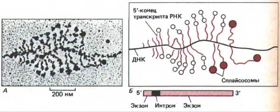
Fig. 9-79. Electron micrograph of stretched chromatin. Large ribonucleoprotein particles can be seen assembling at 5' and 3' splice sites to form spliceosomes. Panel (A) shows the gene encoding Drosophila chorion protein, with the positions of splice sites on its primary transcripts known. (B) Most RNA transcripts contain one or two large RNP particles near their 5' ends. When two particles are present on a transcript (open circles in diagram B), they typically have a diameter of 25 nm and are located near or at the 5' and 3' splice sites of individual short intron sequences (228 nucleotides long) in the 5'-terminal region of the transcripts. Longer, more mature transcripts of two genes frequently bear a single larger particle (colored circles in diagram B) in the intron region. This likely results from the stable association of two smaller particles, representing an assembled spliceosome. Because splicing sometimes occurs while the 3' end of the RNA is still being transcribed, it can be concluded that a polyA sequence at the 3' end of the hnRNA molecule is not required for splicing. (Adapted from I. N. Osheim, O. Z. Miller, and A. L. Beyer, Cell 43: 143-151, 1985.)

Fig. 9-80. Consensus sequences of 5' and 3' RNA splice sites. The sequence shown is for the RNA strand. The invariant GU and AG dinucleotides at each intron end are highlighted in color (see also Fig. 9-77).
9.4.10. Intron sequences are excised as lariat-like RNA structures [50]
Introns vary in length from 80 to 1,000 or more nucleotides. Introns differ fundamentally from exons in that a strictly defined nucleotide sequence appears to be much less critical for them. Over the course of evolution, Mutations have likely accumulated precisely within these regions; it is often possible to alter a large portion of an intron sequence without affecting gene activity. Based on these findings, it has been suggested that intron sequences are functionally inactive and serve primarily as genetic "spacers." The only highly conserved regions within introns are the sequences required for their removal. At each end

Fig. 9-81. Method for obtaining RNA suitable for in vitro RNA splicing analysis. First, it is necessary to obtain a large quantity of the appropriate DNA sequence (constructed via Genetic Engineering and cloning); second, relatively simple RNA polymerases from Bacteriophages T7 or SP6 are required, which transcribe RNA efficiently in vitro. By joining a eukaryotic DNA fragment to a bacteriophage promoter, RNA polymerase can synthesize large amounts of RNA encoded by eukaryotic DNA fragments in vitro. The 5' cap, which is part of hnRNA and essential for transcription initiation, can be attached to such RNAs using an artificially synthesized capped nucleotide (not shown).

Fig. 9-82. Catalysis of RNA splicing by a spliceosome formed through the assembly of U1, U2, U5, and U4/U6 snRNPs (indicated by circles) and other components (not shown). Following spliceosome assembly, the reaction proceeds in two stages: in stage 1, a specific A nucleotide located within the intron sequence near the 3' splice site attacks the 5' splice site, resulting in its cleavage.
The cut 5'-end of the intron sequence is covalently linked to this A-nucleotide, forming the branched structure shown in Fig. 9-83. At stage 2, the 3'-OH end of the first exon, generated in The First stage, joins the beginning of the second exon, thereby cleaving the RNA molecule at the 3' splice site. Thus, the two exon sequences are joined together, while the intron sequence is released as a lariat-like structure. The complete spliceosome complex sediments at 60S, making it comparable in size to a ribosome. RNA processing takes place in the nucleus, where these events convert primary RNA transcripts (mRNA precursors) into mature mRNA molecules.
There are consensus regions within the intron that are nearly identical across all introns. Alterations in these sequences affect the splicing process, by which intron sequences are removed from the primary RNA transcript. Figure 9-80 illustrates these conserved sequences for the 5' splice site (the donor site) and the 3' splice site (the acceptor site). The reactions of RNA cleavage and reunion must occur with absolute precision, as an error of even a single nucleotide will shift the reading frame of the resulting RNA molecule and render the information encoded within it meaningless.
The mechanism by which introns are removed from primary transcripts has been analyzed using in vitro experiments. To this end, specifically designed DNA was incubated with highly purified RNA polymerase to generate individual RNAs containing a single intron (Fig. 9-81). Splicing occurred when these RNA molecules were added to a cell extract. The reaction proceeded in a single step and required prolonged incubation with ATP, specific proteins within the extract, and the U1, U2, U5, and U4/6 snRNPs. These components assemble to form a multi-component ribonucleoprotein complex, or spliceosome. Analysis of the RNA intermediates in this reaction, as well as the snRNPs required for their formation, led to the conclusion that intron excision is accompanied by the formation of a lariat-like structure (Figs. 9-82 and 9-83).
Recently, the functions of several snRNPs have been clarified. For instance, the U1 snRNP binds to the 5' splice site owing to a nucleotide sequence in U1 RNA that is complementary to the nine-nucleotide consensus sequence of the splicing signal (see Fig. 9-80). Because RNA molecules are known to possess catalytic activity (see Section 3.2.11), either the RNA itself or a protein component of the spliceosome may catalyze the cleavage and formation of the covalent bonds required for RNA splicing.

Fig. 9-83. STRUCTURE OF THE branched RNA chain formed during RNA splicing. The highlighted A-nucleotide is the exact same nucleotide featured in Fig. 9-82; here, it shows the branch point formed During the first stage of the splicing reaction. At this stage, the 5'-end of the intron sequence is cleaved, and its phosphate group is covalently linked to the 2'-OH ribose group of the A-nucleotide located 30 nucleotides away from the 3'-end of the intron sequence. The branched chain remains within the excised intron sequence, giving rise to its characteristic lariat shape (see Fig. 9-82).
9-32
9.4.11. Multiple intron sequences are typically removed from each RNA transcript [51]
Because the spliceosome primarily recognizes consensus sequences at the intron boundaries, There is a potential risk that the 5' donor site at the end of any given intron might join with the 3' splice site of a different intron rather than its own. When the 5' and 3' halves of two different introns are experimentally joined together, the resulting hybrid intron sequences are recognized and removed by the RNA-splicing machinery.
Given these findings, it may seem surprising that vertebrate genes sometimes contain as many as 50 introns (see Table 9-1). After all, if any two 5' and 3' splice sites were to pair up randomly by mistake, it would result in the loss of functionally active mRNA sequences, potentially with catastrophic consequences. Somehow, such errors are averted: the normal processing mechanism ensures that each 5' splice site pairs specifically with the nearest 3' splice site located downstream in the 5'-to-3' direction along the linear RNA sequence (Fig. 9-84). It remains unclear exactly how this sequential joining is achieved. It is likely that spliceosome assembly during the elongation of the RNA transcript plays a crucial role (see Fig. 9-79). There is also evidence indicating that the precise three-dimensional conformation of the intron and exon sequences is vital for the correct pairing of splice sites.

Fig. 9-84. Primary transcript of the chicken ovalbumin gene. Seven Introns must be removed from this transcript to yield an active mRNA molecule. The 5' splice sites (donor sites) are designated by D, and the 3' splice sites (acceptor sites) are designated by A.
9-33
10-23
9.4.12. The same RNA transcript can be spliced in different ways to yield mRNAs encoding distinct proteins [52]
Although most intron sequences themselves appear to lack specific functions, alternative pathways during splicing can generate several different mRNAs. Consequently, a single transcript can give rise to multiple distinct proteins, providing the cell with an extra layer of genetic plasticity. As will be discussed in Chapter 10, the pattern of splicing for many RNA transcripts changes during Cell Differentiation; thus, as the cell develops, the same DNA coding sequences can be deployed for different purposes (see Section 10.4.2).
The plasticity afforded by RNA splicing was first discovered in Adenoviruses, which were, in fact, where the process was originally uncovered. The adenovirus genome directs the synthesis of very long RNA transcripts that encode a variety of proteins. This does not happen in normal eukaryotic cells, where each individual mRNA molecule encodes only a single protein, with Translation initiating exclusively near the 5' cap and terminating at the very first stop codon. Adenoviruses, however, employ a specialized RNA-splicing mechanism that can treat coding sequences as introns and excise them, allowing the same 5' cap to join with any downstream coding sequence and thereby produce various mRNAs in the ratios required for viral survival. This alternative RNA splicing enables a single 5' cap to serve as the initiation signal for the synthesis of multiple different proteins (Fig. 9-85). This strategy is widely utilized by viruses, allowing a small number of distinct RNA transcripts to encode a substantial repertoire of proteins.

Fig. 9-85. In certain viruses, the same primary transcript is spliced in alternative ways to yield three (or more) mRNA molecules, each encoding a different protein. In all cases, only the coding sequence adjacent to the 5' cap is translated.
9-34
9.4.13. mRNA alterations in thalassemia illustrate how novel proteins can arise via splicing [53]
Advances in genetic engineering have empowered researchers to use human mutations as a tool for probing cellular mechanisms. For instance, a group of inherited blood disorders collectively known as thalassemias is characterized by a drastic drop in Hemoglobin levels. DNA Sequencing of 50 thalassemia patients revealed that, in most cases, this reduction in hemoglobin was caused by defects in RNA splicing. Single nucleotide substitutions found in the DNA either inactivated a splice site or created a de novo one. Strikingly, analysis of the mRNA from these same patients demonstrated that the loss of a splice site does not halt the process entirely; the remaining normal splice site partners with a nearby suitable region and joins to it. Consequently, multiple splicing pathways can operate, meaning that a mutant gene is capable of specifying several altered proteins (Fig. 9-86). Splicing thus proves to be a remarkably flexible process. Ultimately, a mutation in a Eukaryotic Cell can result in the synthesis of several distinct novel proteins from a single gene, endowing the cell with a powerful capacity to test out its underlying genetic variations. Because of this, RNA splicing may play a pivotal role in the evolution of higher eukaryotes. In contrast, splicing in lower eukaryotes, such as yeast, is regulated much more strictly, which limits the likelihood that aberrant processing will generate novel mRNAs. As a result, the rate of divergence in form and function must be slower in lower eukaryotes than in higher eukaryotes.

Fig. 9-86. Examples of aberrant processing of primary $\beta$-globin RNA transcripts in thalassemia patients. Mutant sites are indicated by black arrows. Colored boxes correspond to the three normal exons depicted in Fig. 9-77. Colored lines connect the 5' and 3' splice sites involved in processing the primary RNA transcript. Unfilled regions represent novel nucleotide sequences incorporated into the final mRNA molecule as a consequence of the mutation. Note that if a normal splice site is left without a partner due to a mutation, one of the nearby aberrant "cryptic" splice sites is utilized as a partner instead. (From S. H. Orkin, In: The Molecular Basis of Blood Diseases [G. Stamatoyannopoulos et al., eds.] pp. 106-126, Philadelphia: Saunders, 1987.)
9.4.14. Spliceosome-catalyzed RNA splicing may have evolved from self-splicing [54]
The discovery of lariat-shaped intermediate structures during nuclear RNA splicing left molecular biologists puzzled. Why would a cell use such an elaborate splicing mechanism instead of simply joining the corresponding 5' and 3' sites? The answer likely lies in the evolutionary origin of spliceosomes.
According to the hypothesis discussed in Chapter 1, early cells used RNA molecules rather than proteins as their primary catalysts and stored genetic information in the form of RNA rather than DNA. RNA-catalyzed splicing probably played a vital role in these primitive cells, and some self-splicing introns have survived to the present day—for example, in the nuclear rRNA genes of Tetrahymena, in bacteriophage T4, and in certain Mitochondrial and Chloroplast genes. In these cases, large fragments of intron sequences have remained exceptionally well-conserved, presumably because they had to fold into specific Conformations to form a catalytic surface on the RNA molecule. Two distinct classes of self-splicing introns are readily apparent: Group I introns initiate the splicing reaction by binding to a guanine nucleotide within the intron sequence, which activates G to form a reactive attacking group that cleaves the first of the phosphodiester bonds broken during splicing (the bond at the 5' splice site). In Group II introns, an adenine nucleotide performs this role, resulting in the formation of a lariat-like structure. In other words, the reaction pathway is similar in both cases, and both likely represent remnants of ancient mechanisms (Fig. 9-87).

Fig. 9-87. Two known classes of self-splicing introns. For group I introns, a free G nucleotide binds to a specific site to initiate splicing (see Fig. 3-19), whereas group II introns utilize a specially activated A nucleotide. These two groups of introns are depicted to highlight their shared properties. In both cases, the reaction is accelerated by proteins, but the RNA residing within the intron also participates directly in catalysis. The mechanism used by group II introns leads to the formation of lariat-like structures and resembles the reaction catalyzed by the spliceosome (cf. Fig. 9-82). (From T.C. Cech, Cell 44: 207-210, 1986.)
During the evolution of nuclear RNA splicing, the reactions utilized by group II self-splicing introns were conserved, but the catalytic function of the intron sequences was transferred to separate Components of the spliceosomes. Consequently, the small U1 and U2 RNAs may well be evolutionary relics of catalytic sequences. This shift of catalytic function from the intron to the spliceosome likely relieved most constraints on intron evolution, thereby enabling The Emergence of new intron sequences.
9.4.15. mRNA Export to the Cytoplasm Occurs Only After the Completion of Splicing [55]
According to the widely accepted hypothesis (Chapter 8), mature mRNA molecules are recognized by receptor proteins (components of the nuclear pores) that facilitate mRNA transport into the cytoplasm via active translocation. However, the major proteins composing heterogeneous nuclear ribonucleoproteins (hnRNPs) and various processing molecules associated with RNA in the nucleus apparently never leave the nucleus; instead, they likely slide off the RNA as it threads through the nuclear pores (Fig. 9-88). Studies of yeast mutants have demonstrated that for RNAs containing splice sites, the transport process can begin only after splicing is complete. In Temperature-sensitive yeast mutants that fail to splice their RNA at high temperatures, all unspliced mRNA precursors are retained in the nucleus, whereas mRNAs that do not require splicing (which constitute the majority in this unicellular eukaryote) are successfully transported to the cytoplasm. This observation is entirely consistent with the hypothesis that RNAs are temporarily detained by their associated spliceosomes, which form numerous aggregates within the higher eukaryotic nucleus. Such aggregates may function as "splicing islands," although their exact formation and function remain to be elucidated (Fig. 9-89). They might represent functional analogs of the nucleolus—a much larger and more prominent nuclear structure whose Organization and function are well understood.
The nucleolus is known to be the nuclear site where precursor RNA molecules are processed into ribosomal RNA (rRNA) molecules, which then bind to specific proteins and assemble into ribosomal subunits. However, before discussing The structure of the nucleolus, we must examine the synthesis of precursor rRNA molecules on rRNA genes.
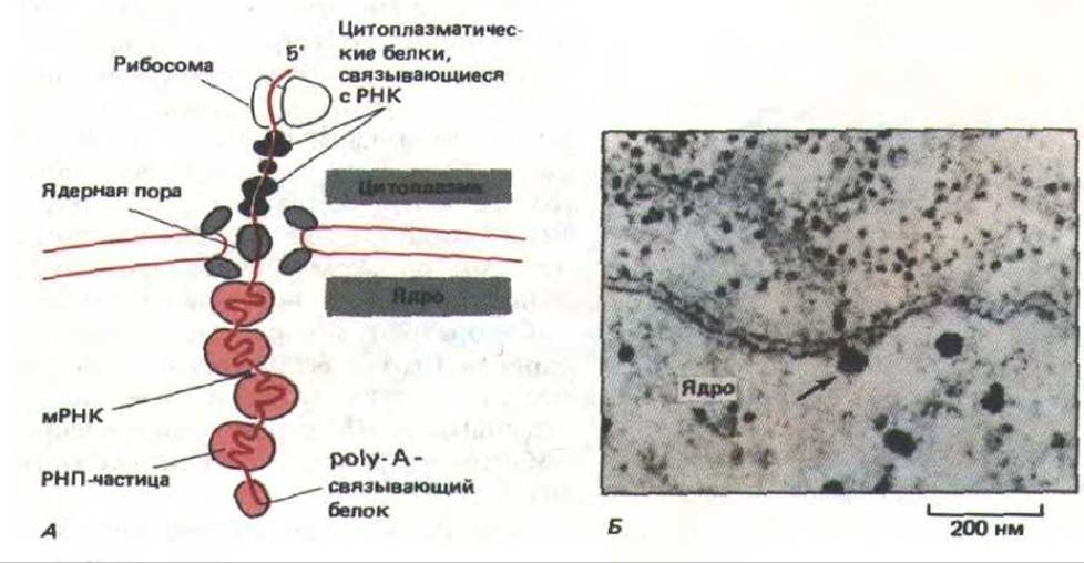
Fig. 9-88. Movement of mRNA molecules through nuclear pores. (A) Changes occurring in the proteins bound to the RNA molecule as it exits the nucleus. (B) Electron micrograph of a large mRNA molecule transcribed in an insect salivary gland cell. This molecule is apparently "caught" in the act of emerging into the cytoplasm (indicated by the arrow). (From B.J. Stevens and H. Swift, J. Cell Biol. 31: 55-77, 1966.)

Fig. 9-89. Immunofluorescence labeling of a human fibroblast nucleus with Monoclonal Antibodies against snRNP particles involved in pre-mRNA splicing. The snRNP particles form large aggregates that may function as "splicing islands." The antibodies reveal specific proteins present in a subset of snRNAs that participate in spliceosome function. (Courtesy of N. Ringertz.)
10-23
9.4.16. Ribosomal RNAs Are Synthesized from Tandemly Arranged Copies of Identical Genes [56]
Many proteins present in large amounts in differentiating cells (such as hemoglobin in red Blood Cells and Myoglobin in Muscle cells) are synthesized from genes represented by only a single copy in the haploid genome. The abundance of these proteins is achieved because each mRNA transcribed from their coding genes can be translated into up to 10 proteins per minute. Normally, in each cell generation, this yields over 10,000 protein molecules per mRNA molecule. However, such Amplification is impossible for ribosomal RNA, because these molecules themselves represent the final gene product. Nonetheless, to produce the required 10 million ribosomes per generation in a growing higher eukaryotic cell, 10 million copies of each type of ribosomal RNA molecule must be synthesized. Such quantities can only be generated if the cell contains multiple copies of the Genes encoding ribosomal RNA (rRNA genes).
Even E. coli requires seven copies of the rRNA gene to supply the cell with an adequate number of ribosomes. Human cells contain about 200 copies of the rRNA gene per haploid genome, distributed in small clusters across five different chromosomes. In Cells of the African clawed frog Xenopus, approximately 600 copies of the rRNA gene are found on a single chromosome. In any chromosome, multiple copies of these highly conserved rRNA genes are arranged as tandem repeats separated from one another by non-transcribed stretches of DNA called spacers. In Chapter 10, we will discuss current models for how such tandemly arrayed genes arise.
Because clusters of tandemly repeated rRNA genes are grouped together and transcribed at extremely high rates, they are readily visible in Cytology/cytology/93.html">ELECTRON MICROGRAPHS OF chromatin. RNA polymerase molecules and their associated nascent transcripts (numbering 100 or more per gene) are packed in such a way that the structure resembles a "Christmas tree" (Fig. 9-90). As noted earlier (see Fig. 9-73), the apex of each such tree marks the transcription initiation site, whereas the end of the rRNA gene is clearly delineated by the abrupt disappearance of RNA polymerase molecules and their corresponding transcripts.
rRNA genes are transcribed by RNA polymerase I to yield identical transcripts. In human cells, these primary rRNA transcripts, known as 45S RNA, are approximately 13,000 nucleotides long. Before leaving the nucleus as part of an assembled ribosomal subunit, the 45S RNA molecule undergoes specific cleavage to yield one copy each of 28S RNA (about 5,000 nucleotides), 18S RNA (about 2,000 nucleotides), and 5.8S RNA (about 160 nucleotides), which constitute the functional components of ribosomes. The common origin of all three rRNAs from the same primary transcript ensures that they are produced in equimolar amounts. The remainder of the transcript (about 6,000 nucleotides) is degraded within the nucleus (Fig. 9-91). It is possible that these "excess" sequences of the rRNA precursor molecule play a role in the Early stages of ribosome assembly, which take place immediately upon completion of 45S RNA synthesis.
Other tandemly repeated genes, likewise separated by non-translated spacers, encode the 5S rRNA of the large ribosomal subunit (this is the only rRNA transcribed independently of the others). The 5S rRNA genes are only about 120 nucleotide pairs long and, like most other genes encoding small stable RNAs (the best-known examples being tRNA genes), are transcribed by RNA polymerase III. Humans possess approximately 2,000 5S rRNA genes, tandemly clustered within a single region located far from the other rRNA genes. It remains unknown why this particular type of rRNA is transcribed separately.
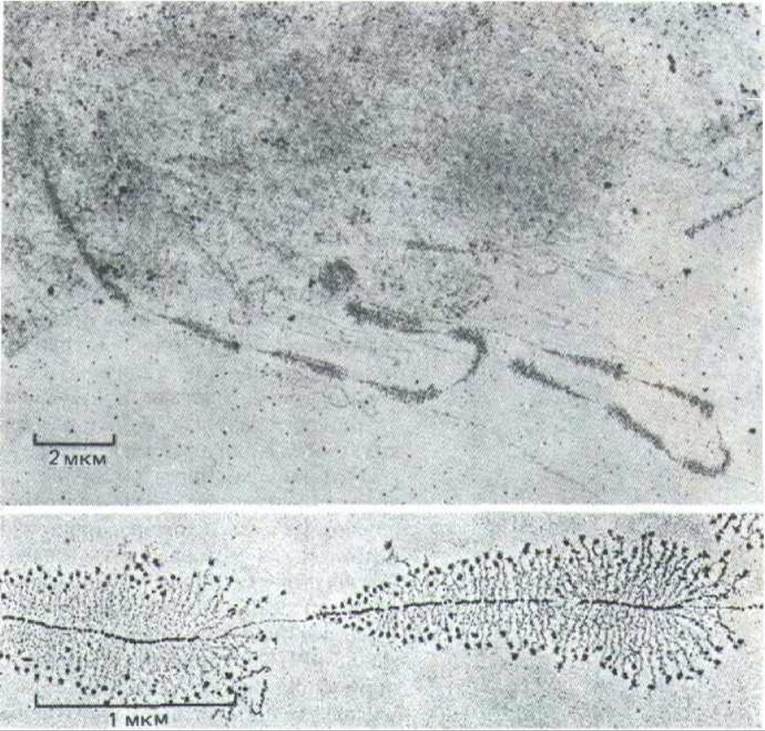
Fig. 9-90. Transcription of tandemly arrayed rRNA genes visualized by electron microscopy. The upper micrograph at lower magnification clearly shows alternating transcribed genes and non-transcribed spacers. The large particles at the 5' end of each rRNA transcript (lower micrograph) are thought to be associated with the onset of ribosome assembly. RNA polymerase molecules are also clearly visible. (Top micrograph from V.E. Foe, Cold Spring Harbor Symp. Quant. Biol. 42: 723-740, 1978; bottom micrograph courtesy of Ulrich Scheer.)

Fig. 9-91. Processing of the 45S ribosomal RNA precursor to generate three distinct RNA molecules. Note that nearly half of the pre-rRNA nucleotide sequence is degraded in the nucleus.
9.4.17. The Nucleolus Is the Center of Ribosome Biogenesis [57]
Continuous transcription of duplicated genes supplies the cell with sufficient amounts of rRNA. Newly synthesized transcripts rapidly bind to ribosomal proteins to form ribosomes. Ribosome assembly takes place in the nucleus—specifically, within a specialized, diffuse region known as the nucleolus. The nucleolus contains large DNA loops encompassing the rRNA genes, which are transcribed by RNA polymerase I at an extraordinarily high rate. These loops are termed "nucleolar organizers." The very beginning of ribosome assembly can be observed in the electron microscope: the 5' ends of the rRNA transcripts forming the tops of the "Christmas trees" become compacted into protein-containing granules (Fig. 9-90). Such granules do not form on Other types of RNA transcripts; presumably, they reflect the Initial Stages of RNA-Protein Interactions occurring within the nucleolus.
The biosynthetic Functions of the nucleolus can be studied using short-term radioactive labeling of newly synthesized RNA with [3H]-uridine. At various time intervals following the incorporation of the label, the cellular contents are fractionated, and the labeled nucleoli are isolated (Fig. 9-92). These experiments have demonstrated that intact 45S transcripts initially form large complexes by associating with a large number of diverse proteins imported from the cytoplasm, where all cellular proteins are synthesized. Most of the 70 distinct polypeptide chains that constitute the ribosome, as well as 5S rRNA, are incorporated precisely at this stage. Additional molecules are required for the correct progression of the assembly process. For instance, the nucleolus contains small nuclear ribonucleoproteins (such as U3 snRNPs), which are believed to catalyze ribosome assembly. These components remain in the nucleolus, whereas the completed ribosomal subunits are transported into the cytoplasm. A particularly prominent nucleolar component is nucleolin, a well-characterized and abundant protein that appears to bind exclusively to ribosomal RNA transcripts. Nucleolin is specifically stained by silver, a staining characteristic that is also shared by the nucleolus as a whole.
During 45S RNA processing, this giant ribonucleoprotein complex gradually sheds some of its Protein and RNA sequences and subsequently undergoes specific cleavage to generate independent precursors for the large and small ribosomal subunits (Fig. 9-93). Within 30 minutes of introducing the radioactive label, the first mature small ribosomal subunits, containing labeled 18S rRNA, emerge from the nucleolus and appear in the cytoplasm. The assembly of the large ribosomal subunits, which contain 28S, 5.8S, and 5S RNAs, requires somewhat more time (approximately 1 hour); consequently, a much larger pool of incomplete large subunits accumulates in the nucleolus compared to small ones.

Fig. 9-92. Simplified diagram of a human cell illustrating the participation of chromatin loops belonging to 10 distinct chromosomes and containing rRNA genes in the formation of a single large nucleolus. Preparations of purified nucleoli are extremely useful in biochemical studies of nucleolar function; such preparations are obtained by mechanically detaching the nucleolus-forming chromatin loops from the chromosomes, as depicted in the figure.

Fig. 9-93. Diagram illustrating The Role of the nucleolus in ribosome Biosynthesis. The 45S rRNA transcript is packaged within a large ribonucleoprotein particle containing numerous ribosomal proteins imported from the cytoplasm. During the processing of this RNP particle within the nucleolus, precursors to the large and small ribosomal subunits are formed, while a portion of the giant RNP complex is degraded. It is hypothesized that the final maturation of the ribosomal subunits occurs only after they are individually transported through the nuclear pores into the cytoplasm.
The final stages of ribosome maturation take place only after the ribosomal subunits have exited the nucleus into the cytoplasm. This ensures the isolation of functioning ribosomes from immature nuclear transcripts.
9.4.18. The nucleolus is a highly organized structure within the nucleus [57]
As observed by light microscopy, the large spheroidal nucleolus is the most readily identifiable structure within the nucleus of a non-mitotic cell. It is hardly surprising, therefore, that it was meticulously investigated by early cytologists: a review on the subject dating back to 1898 contained approximately 700 References. By 1940, it had become clear that the nucleolus contains large amounts of RNA and proteins; however, its biological role in ribosomal RNA synthesis and ribosome assembly remained unknown until the 1960s.
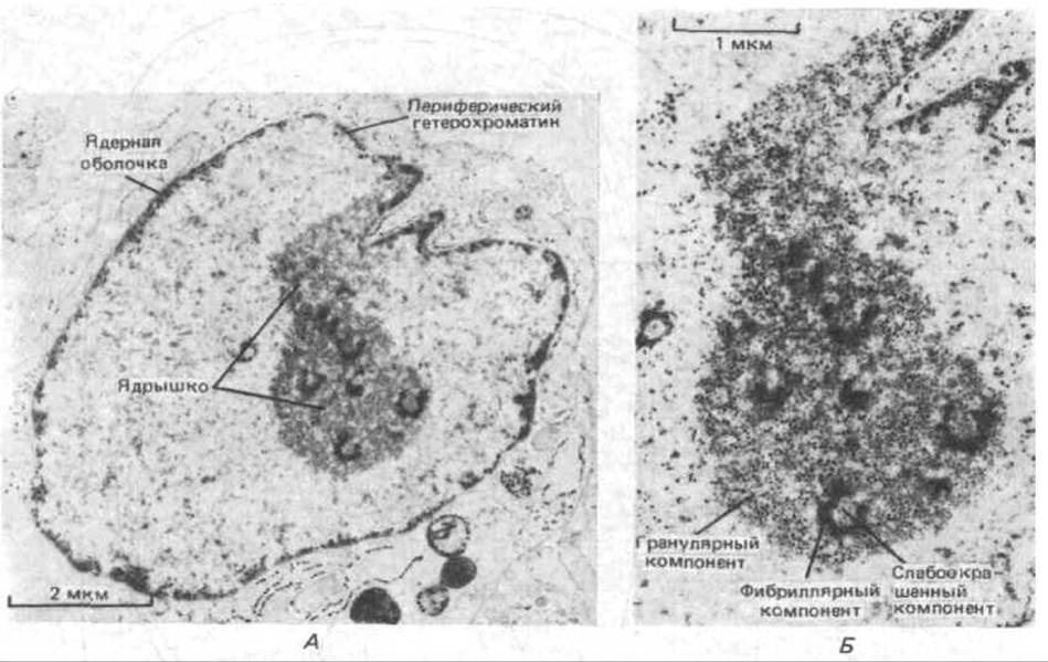
Fig. 9-94. Electron micrograph of a thin section of a nucleolus in a human fibroblast. Three distinct zones are clearly visible. A. General view of the nucleolus. B. Detailed view. (Kindly provided by E. G. Jordan and J. McGovern.)
Certain ultrastructural details of the nucleolus can be revealed using electron microscopy. Unlike cytoplasmic Organelles, the nucleolus lacks a surrounding membrane enclosing its contents. Instead, it appears to be formed by immature ribosomal precursors specifically associated with one another in a manner that remains incompletely understood. In a typical electron micrograph of a nucleolus, three distinct zones can be distinguished (Fig. 9-94): (1) a lightly stained component containing DNA from the chromosomal nucleolar organizer region; (2) a granular component consisting of 15 nm particles, which represent the most mature precursors of ribosomal particles; and (3) a dense fibrillar component composed of numerous thin (5 nm) ribonucleoprotein fibrils corresponding to RNA transcripts.
The size of the nucleolus reflects its level of functional activity, which varies widely among different cell types and can fluctuate within an individual cell. For example, in some quiescent plant cells, the nucleolus is very small, whereas in cells producing massive amounts of protein, it can occupy up to 25% of the total nuclear volume. Variations in nucleolar size are primarily associated with the expansion or reduction of the granular component, which in turn is likely regulated at the level of ribosomal gene transcription: according to electron microscopic data, the proportion of active ribosomal genes, as well as the transcription efficiency of each gene, varies depending on cellular circumstances.
9.4.19. Following each mitosis, the nucleolus re-forms from specific chromosomal regions [58]
The appearance of the nucleolus changes markedly depending on the phase of the Cell Cycle. With the onset of mitosis, the nucleolus decreases in size and eventually disappears altogether as chromosomes condense and the synthesis of all RNA types ceases; as a rule, no nucleolus is detectable in a metaphase cell. At the end of mitosis (in telophase), when ribosomal RNA synthesis resumes, miniature nucleoli reappear at the chromosomal sites containing the ribosomal RNA genes (Fig. 9-95).
In human cells, rRNA genes are located at the ends of five pairs of chromosomes, i.e., on 10 out of the 46 chromosomes (see Fig. 9-40). Consequently, immediately after mitosis, 10 small nucleoli form in a human cell; these are rarely observed because they rapidly grow and fuse with one another into a single large nucleolus typical of most interphase cells (Fig. 9-96).
What happens to the RNA and Protein components of the nucleolus after its disassembly during mitosis? Apparently, a fraction of these components is distributed among all metaphase chromosomes and carried into the daughter cell nuclei. During telophase, as the chromosomes decondense, these "old" nucleolar components may participate in the construction of new nucleoli.
9.4.20. During interphase, individual chromosomes occupy specific positions within the nucleus [59]
As noted above, during nucleolar formation, the required genes from interphase chromosomes gather together in a specific region of the nucleus. This raises the question of whether chromosomes are arranged within the nucleus in a non-random manner. This crucial question was first posed by biologists as early as the late 19th century, yet a definitive answer remains elusive to this day.
A degree of order in chromosome positioning is related to the configuration they adopt at the end of mitosis. It is known that immediately prior to cell division, condensed chromosomes are pulled toward the poles by microtubules attached to the centromeres. Thus, the centromeres lead the way, while the distal chromosome arms terminating in telomeres trail behind. In many nuclei, chromosomes tend to retain this so-called Rabl orientation throughout interphase, with their centromeres facing one pole of the nucleus and their telomeres directed toward the opposite pole (Fig. 9-97, A). In some instances, the nuclear poles exhibit a fixed orientation within the cell: for example, in early Drosophila embryos, all centromeres are directed toward the apical end (Fig. 9-97, A). Such fixed nuclear polarization can significantly influence cell polarity, although it remains unclear what experiment might be devised to test this hypothesis.

Fig. 9-95. The appearance of the nucleolus in human cells changes across different Stages of the cell cycle. The diagram depicts only the cell nucleus.

Fig. 9-96. Micrographs of cultured human fibroblasts demonstrating various stages of nucleolar association. (Kindly provided by E. G. Jordan and J. McGovern.)

Fig. 9-97. Polarized chromosome orientation in early Drosophila embryo interphase cells. A. Rabl orientation: all centromeres are directed toward one pole of the nucleus, and all telomeres toward the other. Note that all nuclei in the embryo are elongated in shape. B. A light micrograph of a Drosophila embryo at the blastoderm cell stage, obtained at low magnification; chromosomes are stained with a fluorescent dye. It should be noted that the most brightly stained regions (chromocenters) are oriented toward the outer surface of the embryo and, consequently, face the apical Plasma Membrane of each cell. (Courtesy of John Sedat.)
In most interphase cells, individual chromosomes cannot be distinguished from one another, making it difficult to determine their arrangement more precisely than described above. An exception is found in the giant polytene chromosomes of interphase cells in Drosophila larvae. In these chromosomes, individual bands are sufficiently distinct to allow precise mapping of specific gene locations within the intact nucleus. Studies have shown that the set of interphase chromosomes as a whole is not characterized by high ordering, although the Rabl orientation is preserved. Frequently, different chromosomes are located next to each other in two seemingly identical cells.
Analysis of polytene chromosomes indicates that in the interphase nucleus, each chromosome occupies a separate territory—meaning that different chromosomes are not extensively intertwined with one another (Fig. 9-98). Furthermore, it has been established that in the interphase nucleus, other types of chromosomes besides polytene ones also tend to occupy discrete domains. For example, using in situ Hybridization with appropriate DNA probes, an individual chromosome can be visualized in cultured mammalian hybrid cells (Fig. 9-99). It has been observed that the bulk of such a chromosome's DNA occupies only a very small fraction of the interphase nucleus. This suggests that each individual chromosome remains compact and structured, allowing its specific regions to actively participate in RNA synthesis.

Fig. 9-98. Stereo view of polytene chromosomes in an isolated nucleus from a Drosophila salivary gland cell. The large sphere is the nucleolus. Telomeres are typically located On the surface of the nuclear envelope opposite the region beneath which the nucleolus resides; all centromeres are clustered near the nucleolus. Chromosomes within the nucleus never become entangled; however, the details of their folding and their spatial neighbors vary between identical nuclei. (Courtesy of Mark Hochstrasser and John W. Sedat.)
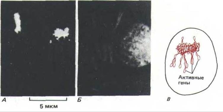
Fig. 9-99. Selective labeling of an individual chromosome in the interphase nucleus of cultured mammalian cells. A. Results of in situ hybridization (using a fluorescent probe) identifying individual Human chromosomes in a human-hamster hybrid cell line. B. The same preparation with all DNA labeled with a fluorescent marker. C. Diagram of the human chromosome in the interphase nucleus shown in part A. (A and B courtesy of Joyce A. Kobori and David R. Cox.)
9.4.21. How highly structured is the nucleus? [60]
The interior of the nucleus is far from being a random mixture of its constituent RNA, DNA, and protein molecules. As discussed above, the nucleolus functions as an efficient ribosome assembly plant, and spliceosome clusters appear to be organized into discrete islands where RNA splicing takes place (see Fig. 9-89). This structural organization is also clearly visible in electron micrographs of nuclear pores: chromatin is distributed along the inner nuclear membrane, but is absent around and directly beneath each nuclear pore (i.e., the pathway between the cytoplasm and nucleoplasm remains unobstructed; Fig. 9-100). Moreover, it turns out that in some cases, THE POSITION OF nuclear pores on the nuclear envelope is not random, but strictly ordered (Fig. 9-101). Such patterning points to a corresponding ORGANIZATION OF THE nuclear lamina, to which the pores are anchored.
Does there exist any intranuclear structure analogous to a Cytoskeleton upon which nuclear components are arranged? Many cell biologists are convinced that such a structure exists. The insoluble material remaining in the nucleus following a series of biochemical extractions is referred to as the nuclear matrix or nuclear Skeleton. It can be demonstrated that the proteins comprising this matrix bind to specific DNA sequences known as SARs or MARs (scaffold-associated regions or matrix-associated regions). It is hypothesized that these sequences form the base of DNA loops (see Fig. 9-34). Through these chromosome attachment sites, the matrix may participate in chromosome organization, determine gene localization, and regulate transcription and DNA Replication within the nucleus. However, because the Structural components of the matrix have not yet been fully identified, it remains unknown whether the matrix obtained experimentally is identical in structure to that present in intact cells.

Fig. 9-100. Electron micrograph of a mammalian cell nucleus. It is clearly visible that the condensed chromatin lining the nuclear envelope is absent around the nuclear pores. (Courtesy of Larry Gerace.)

Fig. 9-101. Freeze-fracture electron micrograph of a fern spore nuclear envelope. The ordered arrangement of nuclear pore complexes lying in parallel rows is clearly visible. On the nuclear envelopes of other cells, either clusters of nuclear pores concentrated in a single region or unusual pore-free zones (oriented in a specific manner relative to other cellular structures) have been observed. (Courtesy of Don H. Northcote, after K. Roberts and D. H. Northcote, Microsc. Acta 71: 102-120, 1971.)
Conclusion
RNA polymerase, the enzyme that catalyzes DNA transcription, is a complex molecule composed of many polypeptide chains. Eukaryotic cells contain three RNA polymerases: I, II, and III. These enzymes are evolutionarily related to each other and to bacterial RNA polymerase, sharing identical subunits. Apparently, following the initiation of transcription, one or more subunits known as initiation factors dissociate from each enzyme. They are replaced by subunits called elongation factors, which are required for RNA chain elongation, termination, and modification. Elongation factors likely differ among various types of polymerases, which may explain why the transcripts synthesized by each enzyme undergo different modifications.
The bulk of cellular mRNA is generated through a complex pathway beginning with the synthesis of heterogeneous nuclear RNA (hnRNA). Primary hnRNA transcripts are synthesized by RNA polymerase II. These transcripts are subsequently capped by the addition of a special nucleotide to the 5' end and then polyadenylated at the 3' end. The modified RNA molecules typically undergo processing, during which intron sequences are excised from the middle of the hnRNA. This reaction is catalyzed by a large ribonucleoprotein complex known as the spliceosome. In the process, a significant portion of the primary RNA transcript is removed and degraded within the nucleus. Although hnRNA typically accounts for about half of all RNA synthesized by the cell, the resulting processed mRNA represents a mere 3% of cellular RNA.
Unlike protein-coding genes transcribed by polymerase II, the genes encoding most structural RNAs are transcribed by polymerases I and III. These genes are generally amplified and organized into tandem clusters. RNA polymerase III synthesizes small stable RNAs, including tRNA and 5S rRNA. RNA polymerase I is responsible for producing the large rRNA precursor molecule (45S rRNA). All cellular ribosomes, with the exception of mitochondrial and chloroplast ones, are assembled in the nucleolus—a distinct nuclear organelle that forms around tandemly arranged rRNA genes brought together from multiple chromosomes.
References
General
Lewin B. Gene Expression, Vol. 2. Eucaryotic Chromosomes, 2nd ed. New York, Wiley, 1980.
Lewin B. Genes, 3rd ed. New York, Wiley, 1987.
Newport J. W., Forbes D. J. The nucleus: structure, function, and dynamics. Annu. Rev. Biochem, 56, 535--566, 1987.
Watson J. D., Hopkins N. H., Roberts J. W., Steitz J. A., Weiner A. M. Molecular Biology of the Gene, 4th ed. Menlo Park, CA, Benjamin-Cummings, 1987.
Cited
1. Adolph К. W., ed. Chromosomes and Chromatin, Vols. 1-3. Boca Raton FL, CRC Press, 1988.
Felsenfeld G. DNA. Sci. Am., 253 (4), 58-67, 1985.
Hsu T.C. Human and Mammalian Cytogenetics: A Historical Perspective. New York, Springer-Verlag, 1979.
2. Kacenoff R., Klotz L. C., Zimm В. Н. On The Nature of chromosome-sized DNA molecules. Cold Spring Harbor Symp. Quant. Biol., 38, 1-8, 1974.
3. Burke D. Т., Carle G. F., Olson M. V. Cloning of large segments of exogenous DNA into yeast by means of artificial chromosome vectors. Science, 236, 806-812, 1987.
Murray A. W. Chromosome structure and behavior. Trends Biochem. Sci., 10, 112-115, 1985.
4. Gall J. G. Chromosome structure and the C-value paradox. J. Cell Biol., 91, 3s-14s, 1981.
Ohta Т., Kimura M. Functional organization of genetic material as a product of molecular evolution. Nature, 233, 118-119, 1971.
5. Mapping and Sequencing The Human Genome. Washington DC, National Academy Press, 1988.
Wilson A. C., Ochman H., Prager Е. М., Molecular time scale for evolution. Trends Genet., 3, 241-247, 1987.
6. Fried, M., Crothers D. M. Equilibria and kinetics of lac-repressor-operator interaction by Polyacrylamide gel Electrophoresis. Nucleic Acids Res., 9, 6505-6525, 1981.
7. Kadonaga J. Т., Tjian R. Affinity purification of sequence-specific DNA binding proteins. Proc. Natl. Acad. Sci. USA, 83, 5889-5893, 1986. Rosenfeld P.J., Kelly T.J. Purification of nuclear factor I by DNA recognition site Affinity Chromatography. J. Biol. Chem., 261, 1398-1408, 1986.
Staudt L. M. et al. Cloning of a lymphoid-specific cDNA encoding a protein binding the regulatory octamer DNA motif. Science, 241, 577-580, 1988.
8. Pabo С. Т., Sauer R. T. Protein-DNA interactions. Annu. Rev. Biochem., 53, 293-321, 1984. Schief R. DNA binding by proteins. Science, 241, 1182-1187, 1988.
9. Ptashne M. A. Genetic Switch: Gene Control and Phage Lambda. Palo Alto CA, Blackwell, 1986.
Takeda Y., Ohlendorf D. H., Anderson W. F., Matthews R. W. DNA-binding proteins. Science, 221, 1020-1026, 1983.
10. Berg О. G., von Hippel P. H. Selection of DNA binding sites by regulatory proteins. Trends Biochem. Sci., 13, 207-211, 1988.
von Hippel P. H., Bear D. G., Morgan W. D., McSwiggen J. A. Protein-nucleic acid interaction in transcription. Annu. Rev. Biochem., 53, 389-446, 1984.
11. Dickerson R. E. The DNA helix and how it is read. Sci. Am., 249(6), 94-111, 1983.
Drew H. R., McCall M. J., Calladine C. R. Recent studies of DNA in the crystal. Annu. Rev. Cell Biol., 4, 1-20, 1988.
12. Koo H. S., Crothers D. M. Calibration of DNA curvature and a unified description of sequence-directed bending. Proc. Natl. Acad. Sci. USA, 85, 1763-1767, 1988.
Lilley D. Bent molecules-how and why? Nature, 320, 487, 1986.
Marini J. C., Levene S. D., Crothers D. M., Englund P. T. Bent helical structure in kinetoplast DNA. Proc. Natl. Acad. Sci. USA, 80, 7678-7682, 1982.
13. Echols H. Multiple DNA-protein interactions governing high-precision DNA transactions. Science, 233, 1050-1056, 1986.
Thompson J. F., de Vargas L. M., Koch C., Kahmann R., Landy A. Cellular factors couple recombination with growth phase: characterization of a new component in the lambda Site-Specific Recombination pathway. Cell, 50, 901-908, 1987.
14. Isenberg I. Histones. Annu. Rev. Biochem., 48, 159-191, 1979.
van Holt C. Histones in perspective. Bioessays, 3, 120-124, 1985.
Wells D. E. Compilation analysis of histones and histone genes. Nucleic Acids Res., 14, rll9-rl49, 1986.
Wu R. S., Panusz H. Т., Hatch C. L., Banner W. M. Histones and their modifications. CRC Crit. Rev. Biochem., 20, 201-263, 1986.
15. Chromatin. Cold Spring Harbor Symp. Quant. Biol., Vol. 42, 1978.
Kornberg R.D., Klug A. The nucleosome. Sci. Am., 244 (2), 52-64, 1981.
McGhee, J.D., Felsenfeld G. Nucleosome structure. Annu. Rev. Biochem., 49, 1115-1156, 1980.
Richmond T.J., Finch J.T., Rushton В., Rhodes D., Klug A. Structure of the nucleosome core particle at 7 A resolution. Nature, 311, 532-537, 1984.
16. Simpson R. T. Nucleosome positioning in vivo and in vitro. Bioessays, 4, 172-176, 1986. Trovers A. A. DNA bending and nucleosome positioning. Trends Biochem. Sci., 12, 108-112, 1987.
17. Eissenberg J.C., Cartwright I.L., Thomas G.H., Elgin S.C. Selected topics in Chromatin Structure. Annu. Rev. Genet., 19, 485-536, 1985.
Emerson B. M. Lewis C. D., Felsenfeld G. Interaction of specific nuclear factors with the nuclease-hypersensitive region of the chicken adult p-globin gene: Nature of the binding domain. Cell 41, 21-30, 1985.
Gross D. S., Garrad W. T. Nuclease hypersensitive sites in chromatin. Annu. Rev. Biochem., 57, 159-198, 1988.
18. Belmont A.S., Sedat J.W., Agard D.A. A three-dimensional approach to mitotic chromosome structure: evidence for a complex hierarchial organization. J. Cell Biol., 105, 77-92, 1987.
Pederson D.S., Thoma F., Simpson R. Core particle, fiber, and transcriptionally active chromatine structure. Annu. Rev. Cell Biol., 2, 117-147, 1986.
19. Allan J., Hartman P. G., Crane-Robinson C., Aviles F. X. The structure ofhistone HI and its Location in chromatin. Nature, 288, 675-679, 1980.
Clark D.J., Thomas J.O. Salt-dependent cooperative interaction ofhistone HI with linear DNA. J. Мої. Biol., 187, 569-580, 1986.
Coles L.S., Robins A.J., Madley L.K. Wells J.R. Characterization of the chiken histone HI gene Complement: generation of a complete set of vertebrate HI protein sequences. J. Biol. Chem, 262, 9656 9663, 1987.
Thoma F., Koller Т., Klug A. Involvement of histone HI in the organization of the nucleosome and of the salt-dependent superstructures of chromatin. J. Cell Biol., 83, 403-427, 1979.
20. De Bernardin W., Koller Т., Sogo J. M. Structure of in vivo transcribing chromatin as studied in simian virus 40 minichromosomes. J. Мої. Biol. 191, 469-482, 1986.
Losa R., Brown D.D. A bacteriphage RNA polymerase in vitro through a nucleosome core without displacing it. Cell, 50, 801-808, 1987.
21. Benyajati C., Worcel A. Isolation, characterization, and structure of the folded interphase genome of Drosophila melanogaster. Cell, 9, 393-408, 1976.
Gasser S. M., Laemmli U. K. A glimpse at chromosomal order. Trends Genet., 3, 16-22, 1987.
SchmidM. B. Structure and funcfion ofthe bacterial chromosome. Trends Biochem. Sci., 13, 131-135, 1988.
22. Marsden M., Laemmli U. K. Metaphase chromosome structure: evidence for a radial loop model. Cell, 17, 849-858, 1979.
Georgiev G.P., Nedospasov S.A., Bakaev V. V. Supranucleosomal levels of chromatin organization. In: The Cell Nucleus (H. Busch, ed.), Vol. 6, pp. 3-34. New York, Academic Press, 1978.
23. Holmquist G. DNA sequences in G-bands and R-bands. In: Chromosomes and Chromatin Structure (K.W. Adolph, ed.), Vol. 2, pp. 75-122. Boca Raton, FL, CRC Press, 1988.
Lewin B. Gene Expression, Vol. 2. Eucaryotic Chromosomes, 2nd ed., pp. 428-440, New York, Wiley, 1980.
24. Bostock C. J., Sumner A. T. The Eucaryotic Chromosome, pp. 347-374. Amsterdam, North-Holland, 1978. Callan H.G. Lampbrush chromosomes. Proc. R. Soc. Lond. (Biol.), 214, 417-448, 1982.
Roth M.B., Gall J.G. Monoclonal antibodies that recognize transcription unit proteins on newt lampbrush chromosomes. J. Cell Biol., 105, 1047-1054, 1987.
25. Agard D. A., Sedat J. W. Three dimensional architecture of a polytene nucleus. Nature, 302, 676-681, 1983.
Beermann W. Chromosomes and genes. In: Developmental Studies on Giant Chromosomes (W. Beermann, ed.), pp. 1-33. New York, Springer-Verlag, 1972.
26. Ashburner M., Chihara C., Meltzer P., Richards G. Temporal control of puffing activity in polytene chromosomes. Cold Spring Harbor Symp.
Quant. Biol, 38, 655-662, 1974.
Lamb M. M., Daneholt В., Characterization of active transcription units in Balbiani rings of Chironomus tentans. Cell, 17, 835-848, 1979.
27. Bossy В., Hall L.M., Spierer P. Genetic activity along 315 kb of the Drosophila chromosome. EMBO J., 3, 2537-2541, 1984.
Hill R.J., Rudkin G. Polytene chromosomes: the status of the band-interband question. Bioessays, 7, 35-40, 1987.
Judd B. H., Young M. W. An examination of the one Cistron: one chromomere concept. Cold Spring Harbor Symp. Quant. Biol. 38, 573-579, 1974.
28. Garel A., Zolan M., Axel R. Genes transcribed at divers rates have a similar conformation in chromatin. Proc. Natl. Acad. Sci. USA, 74, 4867 4871, 1977.
Weintraub H., Groudine M. Chromosomal subunits in active genes have an altered conformation. Science, 193, 848-856, 1976.
Yaniv M., Cereghini S. Structure of transcriptionally active chromatin. CRC Crit. Rev. Biochem. 21, 1-26, 1986.
29. Allis C.D. et al. hvl is an evolutionary conserved H2A variant that is preferentially associated with active genes. J. Biol. Chem., 261, 1941-1948, 1986.
Dorbic Т., Wittig В. Chromatin from transcribed genes contains HMG17 only downstream from the starting point of transcription. EMBO J., 6, 2393-2399, 1987.
Hebbes T.R., Thorne A. W., Crane-Robinson C. A direct link between core histone Acetylation and transcriptionally active chromatin. EMBO J., 7, 1395-1402, 1988.
Rose S. M., Garrard W. T. Differentiation-dependent chromatin alterations precede and accompany transcription of immunoglobulin light chain genes. J. Biol. Chem., 259, 8534-8544, 1984.
30. Brown S. W. Heterochromatin. Science, 151, 417-425, 1966.
James T. C., Elgin S. C. R. Identification of a nonhistone chromosomal protein associated with heterochromatin in Drosophila melanogaster and its gene. Mol. Cell Biol., 6, 3862-3872, 1986.
Pimpinelli S., Bonaccorsi S., Gatti M., Sandier L. The peculiar Genetic Organization of Drosophila heterochromatin. Trends Genet., 2, 17-20, 1986.
31. Hand R. Eucaryotic DNA: organization of the genome for replication. Cell, 15, 317-325, 1978.
Huberman J. A., Riggs A. D. On the mechanism of DNA replication in mammalian chromosomes. J. Моl. Biol., 32, 327-341, 1968.
32. Campbell J. Eucaryotic DNA replication: yeast bares its ARSs. Trends Biochem. Sci., 13, 212-217, 1988.
Palzkill T. G., Newlon C. S. A yeast replication origin consists of multiple copies of a small conserved sequence. Cell, 53, 441-450, 1988.
Struhl K., Stinchcomb D. T., Sherer S., Davis R. W. High-frequency transformation of yeast: autonomous replication of hybrid DNA molecules. Proc. Natl. Acad. Sci. USA, 71, 1035-1039, 1979.
33. Dodson M., Dean F. B., Bullock P., Echols H, Hurwitz J. Unwinding of duplex DNA from the SV40 origin of replication by T antigen. Science, 238, 964-967, 1987.
Li J. J., Kelly T. J. Simian virus 40 DNA replication in vitro. Proc. Natl. Acad. Sci. USA, 81, 6973-6977, 1984.
So A. G., Downey K. M. Mammalian DNA polymerases alpha and delta: current status in DNA replication. Biochemistry, 27, 4591-4595, 1988. Stahl H, Droge P., Knippers R. DNA helicase activity of SV40 large T antigen. EMBO J. 5, 1939-1944, 1986.
34. Russev G., Hancock R. Assembly of new histones into nucleosomes and their distribution in replicating chromatin. Proc. Natl. Acad Sci. USA, 79, 3143-3147, 1982.
Sariban E. R., Wu R. S., Erickson L. C., Banner W. M. Interrelationships of protein and DNA synthesis during replication in mammalian cells. Mol. Cell. Biol. 5, 1279-1286, 1985.
Weintraub H., Worcel A., Alberts B. M. A model for chromatin based upon two symmetrically paired half-nucleosomes. Cell, 9, 409-417, 1976. Worcel A., Han S., Wong M. L. Assembly of newly replicated chromatin. Cell, 15, 969-977, 1978.
35. Blackburn E. H., Szostak J. W. The molecular structure of centromeres and telomeres. Annu. Rev. Biochem., 53, 163-194, 1984.
Greider C. W., Blackburn E. H. The telomere terminal transferase of Tetrahymena is a ribonucleoprotein enzyme with two kinds of primer specificity. Cell, 51, 887-898, 1987.
36. Stubblefield E. Analysis of the replication pattern of Chinese hamster chromosomes using 5-bromodeoxyuridine suppression of 33285 Hoechst fluorescence. Chromosoma, 53, 209-221, 1975.
37. Lima-de-Faria A., Jaworska H. Late DNA synthesis in heterochromatin. Nature, 217, 138-142, 1968.
38. Brown E. H., et al. Rate of replication of the murine immunoglobulin heavy-chain locus: evidence that the region is part of a single replicon.
Mol. Cell. Biol., 7, 450-457, 1987.
39. Callan H. G. DNA replication in the chromosomes of eukaryotes. Cold Spring Harbor Symp. Quant. Biol., 38, 195-203, 1974.
Kriegstein H. J., Hogness D. S. Mechanism of DNA replication in Drosophila chromosomes: structure of replication forks and evidence for bidirectionality. Proc. Natl. Acad. Sci. USA, 71, 135-139, 1974.
Mechali M., Kearsey S. Lack of specific sequence requirement for DNA replication in Xenopus eggs compared with high sequence specificity in yeast. Cell, 38, 55-64, 1984.
40. Blow J. J., Laskey R. A. A role for the nuclear envelope in controlling DNA replication within the cell cycle. Nature, 332, 546-548, 1988.
Harland R. Initiation of DNA Replication in eukaryotic chromosomes. Trends Biochem. Sci. 6, 71-74, 1981.
Rao P. N., Johnson R. T. Mammalian Cell Fusion: studies on The regulation of DNA SYNTHESIS AND mitosis. Nature, 225, 159-164, 1970.
41. Watson J. D., Hopkins N. H., Roberts J. W., Steitz J. A., Weiner A. M. Molecular Biology of the Gene, 4th ed. Menlo Park, CA, Benjamin-Cummings, 1987. (Chapters 13, 20 and 21).
42. Chamberlin M. Bacterial DNA-dependent RNA polymerases. In: The Enzymes, 3rd ed. (P. Boyer, ed.), Vol. 15B, pp. 61-108, New York, Academic Press, 1982.
Greenblatt J., Li J. Interaction of the sigma factor and the nusA gene protein of E. coli with RNA polymerase in the initiation-termination cycle of transcription. Cell, 24, 421-428, 1981.
McClure W. Mechanism and Control of transcription initiation in prokaryotes. Annu. Rev. Biochem., 54, 171-204, 1985.
Yager T. D., von Hippel P. H. Transcript elongation and termination in E. coli. In: Escherichia coli and Salmonella typhimurium: CELLULAR AND MOLECULAR Biology (F. C. Neidhardt, ed.), pp. 1241-1275. Washington DC, American Society for Microbiology, 1987.
43. Chambon P. Eucaryotic nuclear RNA polymerases. Annu. Rev. Biochem., 44, 613-638, 1975.
Geiduschek E. P., Tocchim Valentini G.P. Transcription by RNA polymerase III. Annu. Rev. Biochem., 57, 873-914, 1988.
Sentenac A. Eucaryotic RNA polymerases. CRC Crit. Rev. Biochem., 18, 31-91, 1985.
Sollner-Webb В., Tower J. Transcription of cloned eucaryotic ribosomal RNA genes. Annu. Rev. Biochem, 55, 801-830, 1986.
44. Brown D. D. The role of stable complexes that repress and activate eucaryotic genes. Cell, 37, 359-365, 1984.
Workman J. L., Roeder R. G., Binding of transcription factor TFIID to the major late promotor during in vitro nucleosome assembly potentiates subsequent initiation by RNA polymerase II. Cell, 51, 613-622, 1987.
45. Foe V. E., Wilkinson L. E., Laird C. D. Comparative organization of active transcription units in Oncopeltus fasciatus. Cell, 9, 131 146, 1976.
Hastie N.D., Bishop J.O. The expression of three abundance classes of mRNA in mouse Tissues. Cell, 9, 761-774, 1976.
Lewin B. Gene Expression, Vol. 2. Eucaryotic Chromosomes, 2nd ed., pp. 708-719. New York, Wiley, 1980.
Miller O. L. The nucleolus, chromosomes, and visualization of genetic activity. J. Cell Biol. 91, 15s-27s, 1981. (A review.)
46. Birnstiel M. L., Busslinger M., Struh K. Transcription termination and 3' processing: the end is in site! Cell, 41, 349-359, 1985.
Friedman D.I., Imperiale M.J., Adhya S.L. RNA 3' end formation in the Control of Gene Expression. Annu. Rev. Genet., 21, 453-488, 1987. Nevins J. R. The pathway of eukaryotic mRNA formation. Annu. Rev. Biochem. 52, 441-466, 1983.
Takagaki Y., Ryner L. C., Manley J. L. Separation and characterization of a poly(A) polymerase and a cleavage/specificity factor required for pre-mRNA polyadenylation. Cell, 52, 731-742, 1988.
47. Sisodia S.S., Sollner-Webb В., Cleveland D.W. Specificity of RNA maturation pathways: RNAs transcribed by RNA polymerase III are not substrates for splicing or polyadenylation. Mol. Cell.. Biol., 7, 3602-3612, 1987.
Smale S. Т., Tjian R. Transcription of Herpes simplex virus tk sequences under the control of wild-type and mutant human RNA polymerase I promoters. Mol. Cell. Biol. 5, 352-362, 1985.
48. Chambon P. Split genes. Sci. Am. 244(5), 60-71, 1981.
Crick F. Split genes and RNA splicing. Science, 204, 264-271, 1979.
Darnell J. E., Jr. Variety in the level of gene control in eucaryotic cells. Nature, 297, 365-371, 1982. Perry R. P. RNA processing comes of age. J. Cell Biol., 91, 28s-38s, 1981. (Includes a historical review.)
49. Guthrie C., Patterson B. Splice-ossomal snRNAs. Annu. Rev. Genet., 22, 387-419, 1988.
Dreyfuss G., Swanson M.S., Pinol-Roma S. Heterogeneous nuclear ribonucleoprotein particles and the pathway of mRNA formation. Trends Biochem. Sci. 13, 86-91, 1988.
Osheim Y. N.. Miller O. L., Beyer A. L. RNP particles at splice junction sequences on Drosophila chorion transcripts. Cell, 43, 143-151, 1985. Samarina O.P., Krichevskaya A.A., Georgiev G.P. Nuclear ribonucleoprotein particles containing messenger ribonucleic acid. Nature, 210, 1319-1322, 1966.
Steitz J.A., "Snurps". Sci. Am. 258 (6). 56-63, 1988.
50. Edmonds M. Branched RNA. Bioessays, 6, 22-216, 1987.
Maniatis Т., Reed R. The role of small nuclear ribonucleoprotein particles in pre-mRNA splicing. Nature, 325, 673-678, 1987.
Padgett R. A., Grabowski P. J., Konarska M. M., Seller S., Sharp P. A. Splicing of mRNA precursors. Annu. Rev. Biochem. 55, 1119-1150, 1986.
51. Aebi M., Weissman C. Precision and orderliness in splicing. Trends Genet., 3, 102-107, 1987.
52. Andreadis A., College M.E., Nadal-Ginard B. Generation of protein isoform diversity by Alternative Splicing: mechanistic and biological implications. Annu. Rev. Cell Biol., 3, 207-242, 1987.
53. Orkin S. H., Kazazian H. H. The mutation and polymorphism of the human p-globin gene and its surrounding DNA. Annu. Rev. Genet., 18, 131-171, 1984.
54. Cech T. R. The generality of self-splicing RNA: relationship to nuclear RNA splicing. Cell, 44, 207-210, 1986.
Ringertz N. et al. Computer analysis of the distribution of nuclear antigenes: studies on the spatial and functional organization of the interphase nucleus. J. Cell Sci. Suppl. 4, 11-28, 1986.
Warner J. Applying genetics to the splicing problem. Genes Dev., 1, 1-3, 1987.
56. Long E. О., Dawid I. B. Repeated genes in eucaryotes. Annu. Rev. Biochem., 49, 727-764, 1980.
Miller O. L. The nucleolus, chromosomes and visualization of genetic activity. J. Cell Biol., 91, 15s-27s, 1981.
57. Hadjiolov A. A. The Nucleolus and Ribosome Biogenesis. New York, Springer-Verlag, 1985.
Jordan E. G., Cullis C. A., eds. The Nucleolus. Cambridge, U. K., Cambridge University Press, 1982.
Sommerville J. Nucleolar structure and ribosome biogenesis. Trends Biochem. Sci., 11, 438-442, 1986.
58. Anastassova-Kristeva M. The nucleolar cycle in man. J. Cell Sci., 25, 103, 1977.
McClintock B. The relation of a particular chromosomal element to the develope-ment of the nucleoli in Zea Mays. Z. Zellforsch. Mikrosk. Anat., 21, 294-323, 1934.
59. ComingsD.E. Arrangement of chromatin in the nucleus. Hum. Genet., 53, 131-143, 1980.
Cremer T. et al. Rabl's model of the interphase chromosome arrangement tested in Chinese hamster cells by premature chromosome Condensation and laser-UV-microbeam experiments. Hum. Genet., 60, 46-56, 1980.
Hochstrasser M., Sedat J. W. Three-dimensional organization of Drosophila melano-gaster interphase nuclei. II. Chromosome spatial organization and gene regulation. J. Cell Biol., 104, 1471-1483, 1987.
Manuelidis L. Individual interphase chromosome domains revealed by in situ hubridization. Hum. Genet., 71, 288-293, 1985.
60. Gasser S. M., Laemmli U. K. A glimpse at chromosome order. Trends Genet., 3, 16-22, 1987.
Gerace L., Burke B. Functional organization of the nuclear envelope. Annu. Rev. Cell Biol., 4, 335-374, 1988.
Newport J. W., Forbes D. J. The nucleus: structure, function, and dynamics. Annu. Rev. Biochem., 56, 535-566, 1987.
Last update: 12/08/2026
Editorial and Educational Adaptation: This material has been compiled based on the primary/original source text. The project team performed an editorial review, corrected technical inaccuracies, structured sections, and adapted the content for an educational format.
What was processed:
- elimination of formatting defects (OCR errors, structural breaks, corrupted characters);
- editorial organization of content;
- standardization of terminology in accordance with academic sources;
- verification of factual statements against the original source text.
All mentions of the author, publication year, and origin of the primary text have been preserved in accordance with the source.