Human Histology - O.D. Lutsyk 2003
Systemic Histology
Nervous System
The Nervous system (Fig. 4.114) integrates a range of Organs and structures that collectively link the Organism to its external environment, regulate all vital processes, and coordinate and integrate the activities of various Organ Systems. Thanks to the nervous system, the body Functions as a unified whole. The structural foundation of the nervous system is Nervous Tissue, which is capable of receiving stimuli from the external environment, transforming them into sensations, and forming response reactions.
The nerve Cell—the neurocyte—generates and conducts electrical impulses and serves as a key link in the vast array of processes collectively known as nervous activity. The Doctrine of the neuron as the fundamental morphological unit of the nervous system was established through the groundbreaking research of the eminent Spanish neurohistologist Santiago Ramón y Cajal. Slightly prior to Cajal's work, the Italian morphologist Camillo Golgi formulated the reticular theory, according to which the elements of the nervous system form an unbroken network encompassing every single organ system without exception. Both scientists were awarded the Nobel Prize in 1906 for their pioneering work on the nervous system.
Class="center">
Fig. 4.114. General structural layout of the nervous system
There are two classifications of the Organs of the nervous system: Anatomical and physiological. According to the anatomical Classification, the nervous system is divided into the central and peripheral divisions (Fig. 4.115, A). The Central nervous system includes the BRAIN AND SPINAL cord, while the peripheral division comprises ganglia, nerve trunks, and nerve endings. According to the physiological classification, the nervous system is divided into somatic and autonomic (vegetative) systems. The former innervates the entire body except for Internal Organs, Blood Vessels, and glands, whereas the latter innervates these specific organs. It should be noted that both classifications are somewhat conventional, as the functioning of the nervous system is fundamentally based on reflex arcs that span its various divisions and organs (Fig. 4.115, B).
Central Nervous System
The brain (encephalon, cerebrum) comprises the right and left cerebral hemispheres and the Brainstem, which is subdivided into the Diencephalon, Midbrain, and Hindbrain (Pons, Cerebellum, and Medulla Oblongata). All of these structures are housed within the cranial cavity and are composed of multipolar Nerve Cells, numbering up to 100 billion. The average mass of the human brain is 1100–1800 g, exhibiting significant individual variation (for instance, the brain mass of Ivan Turgenev was 2016 g, and that of Anatole France was 1017 g). No correlation has been found between brain mass and a person's creative output. The absolute brain mass in women is 190–200 g less than that in men.
The processes of neurocytes form the White matter OF the brain. Aggregations of nerve cell bodies (perikarya) constitute nerve centers, or Gray matter (Fig. 4.116). The interwoven network of neuronal processes and neuroglia within the gray matter resembles felt and is termed the neuropil. Two Types of Nerve centers are distinguished: nuclear and cortical (layered). Nuclear-type centers have a diverse shape and are surrounded by white matter. Cortical-type centers are superficial aggregations of neurocytes that collectively form the cortex of the cerebrum and cerebellum.
The cerebrum, or Telencephalon. This specific part of the central nervous system is primarily responsible for uniquely human traits. The surface of the cerebrum forms sulcus-separated gyri, which significantly increases the surface area of the cortex. The gray matter On the surface of the cerebrum is about 3 mm thick, reaching its maximum thickness of nearly 5 mm in the precentral gyrus. The human Cerebral Cortex contains approximately 50 billion nerve cells, where the higher analysis and synthesis of nerve impulses—Higher Nervous Activity—take place. Specific areas of the cortex responsible for distinct manifestations of higher nervous activity (speech, Vision, Hearing, Olfaction, etc.) are called fields or centers. The Topography of the human cerebral cortex was mapped in detail by the German neurologist K. Brodmann, who divided the entire cortical surface into 52 fields differing in cellular composition, Structure, and function.

Fig. 4.115. Functional Morphology of the nervous system: A – division of the nervous system into central and peripheral parts; B – schematic diagram of a simple reflex arc: reflex arcs underlying the functioning of the nervous system integrate its central and peripheral components, as well as the body's organs and systems, into a single whole. Arrows indicate the direction of Nerve Impulse propagation

Fig. 4.116. Gray and white matter of the central nervous system organs: A – sagittal section of the telencephalon and cerebellum: gray matter forms the superficial cortex and basal nuclei, white matter is localized subcortically, x0.25; B – transverse section of the cervical Spinal Cord: gray matter forms the anterior, lateral, and posterior horns, white matter forms the anterior, lateral, and posterior funiculi, x12; C – structural diagram of gray and white matter
Based on morphological features, Neurons of the cerebral cortex are divided into pyramidal and non-pyramidal cells. Pyramidal cells have a characteristic pyramidal shape, with heights ranging from 10 to 120 µm. The non-pyramidal neuron group includes various types: basket cells, spinous stellate cells, neurogliomorphic (spider) cells, axon-brush cells, chandelier cells, axo-axonic cells, double-bouquet cells, and fusiform cells with long horizontal axons. Both the distinctive names of these cells and their classificatory characteristics are determined primarily by the number, morphology, and branching pattern of their processes. Neurocytes and their processes within the cerebral cortex are arranged in indistinctly separated layers, or laminae. This layered arrangement of neurocytes is known as cytoarchitectonics. The cerebral cortex comprises six layers: molecular, external granular, pyramidal, internal granular, ganglionic (ganglion), and multiform (polymorphic) cell layer (Fig. 4.117).
The molecular layer lies directly beneath the pia mater, separated from it by a glial membrane. It is composed primarily of fusiform cells with long horizontal dendrites and descending axons that form horizontal collaterals. The molecular layer is the sparsest in cellular elements, giving it the appearance of a pale band on the cortical surface in histological preparations. The external granular layer is formed by small cells of round, polygonal, stellate, and pyramidal shapes, not exceeding 10 µm in size.
The pyramidal layer is the thickest. It is formed by pyramidal-shaped cells whose dimensions gradually increase from 10 to 40 µm from the cortical surface toward the deep gray matter. The apices of pyramidal neurocytes are always directed toward the cortical surface, and their bases toward the white matter. An apical dendrite extends from the apex of the pyramidal cell, lateral dendrites from its lateral surface, and an axon from its base. Axons of large pyramidal neurocytes form Cytology/practical/65.html">Myelinated nerve fibers projecting into the white matter. The internal granular layer varies in development across different cortical regions; for instance, it is highly developed in the visual cortex but entirely absent in the precentral gyrus. The internal granular layer is formed by small stellate neurocytes.
The ganglionic layer of the cortex contains giant pyramidal neurons (Betz cells), whose perikaryon height can reach 120 µm and width 80 µm. These cells were first described by the Kyiv morphologist V.O. Betz in 1874, hence their name. They form distinct tangential bundles, or stripes, situated between layers of nerve cells. For instance, the stripe of the molecular layer (Exner's stripe) runs across the cortical surface; the stripe of the external granular layer (Bechterew's stripe) lies between the molecular and external granular layers; the stripe of the internal granular layer (outer Baillarger's stripe) is situated between the internal granular and ganglionic layers; and the stripe of the ganglionic layer (inner Baillarger's stripe) lies between the ganglionic and multiform cell layers. The layered arrangement of tangential nerve fiber bundles (stripes) within the cerebral cortex is known as myeloarchitectonics.
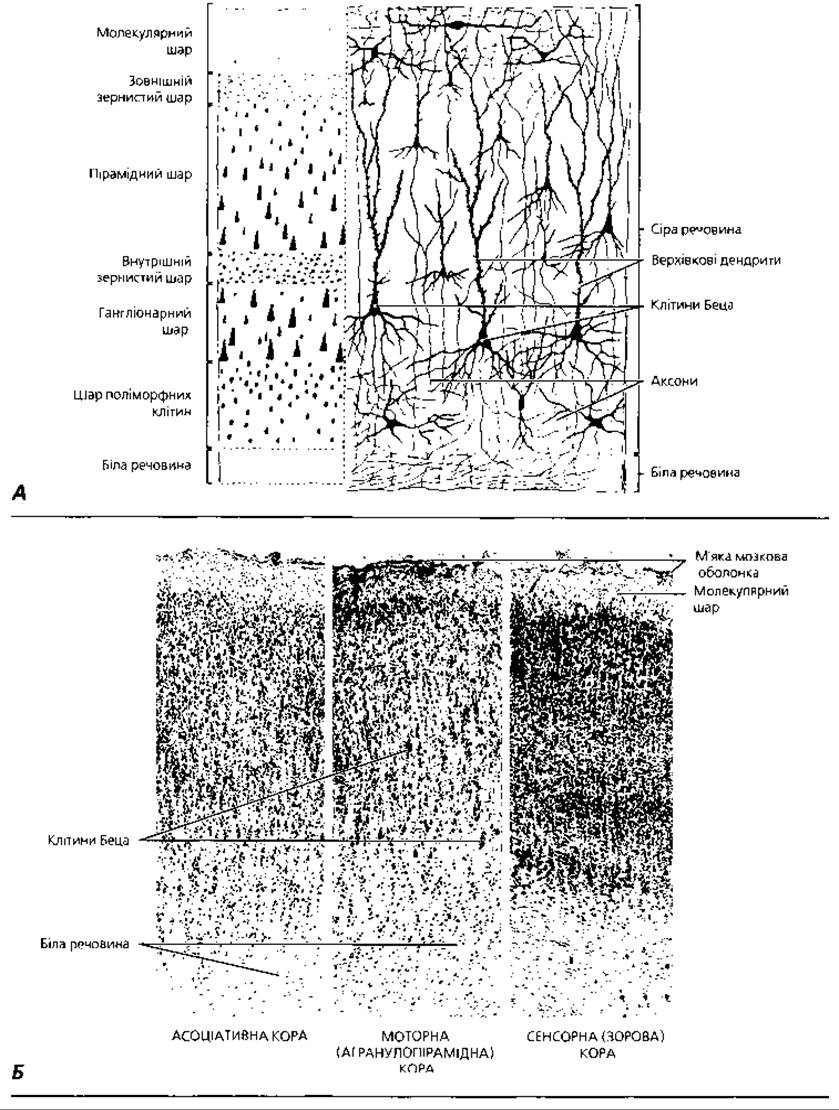
Fig. 4.117. Cytoarchitectonics of the cerebral cortex: A – schematic representation: left – layered structure, right – morphology and topography of giant pyramidal neurons (Betz cells) stained by the Golgi method; B – sagittal cortical sections from three functional areas of the cerebrum: associative, motor, and sensory (visual), x53
Beneath the cortex within the white matter of the cerebrum lies a considerable number of nerve cell aggregations known as subcortical nuclei. The processes of cortical nerve cells interact with the multipolar neurons of these subcortical nuclei, while the processes of the nuclear neurons extend beyond the cerebrum. The topography and functions of individual brain nuclei are studied in human anatomy and neurology courses.
The cerebellum is the higher center for equilibrium and motor coordination, ensuring the maintenance of Muscle tone. It consists of two hemispheres with gray matter on the surface (cerebellar cortex) and white matter and subcortical nuclei in the interior. The mass of the human cerebellum is 120–150 g, and its volume is about 160 cm3. The surface area of the cortex reaches 850 cm2, which corresponds to 50% of the cerebral cortex surface area. Due to superficial gyri and the alternation of gray and white matter, a sagittal section of the cerebellum reveals a characteristic pattern known as the arbor vitae (tree of life).
The cerebellar cortex (Fig. 4.118) has a three-layered structure comprising the molecular, ganglionic, and granular layers. The molecular layer is the most superficial, formed by The Cell bodies of basket and stellate cells. Basket cells are named for their property of sending numerous processes to the Cells of the ganglionic layer, around whose perikarya characteristic formations—known as cerebellar baskets—are formed by interwoven axonal collaterals of the basket cells. The axons of basket cells run in a horizontal (tangential) direction, always perpendicular to the gyrus above the Purkinje cells. Stellate cells of the molecular layer are subdivided into small and large types. Processes of the small cells contact the dendrites and bodies of the large Purkinje cells in the ganglionic layer. The neurocytes of the molecular layer perform an associative function by exerting an inhibitory effect on the cells of the ganglionic layer.
The second, ganglionic layer of the cerebellum is formed by a single row of large pear-shaped neurons—Purkinje cells. Their dimensions are 35×60 µm. From the tapered apex of these cells, 2–3 dendrites extend into the molecular layer, running radially and branching extensively. In a cerebellar cortical section made along the course of a gyrus, the dendrites of Purkinje cells produce a characteristic cypress-like pattern. In a section perpendicular to the gyrus, Betz cells exhibit a morphology resembling the pyramidal layer cells described above. Axons of giant pyramidal neurons project to the motor nuclei of the brainstem and spinal cord, forming numerous collaterals that exert an inhibitory effect on the cerebral cortex. Pyramidal and giant pyramidal neurons are the most Characteristic Features of histological preparations of the cerebral cortex. The multiform cell layer is formed by neurons of diverse, predominantly fusiform shapes.
Cellular layers possess specific functional specialization within the cerebral cortex. The molecular and multiform layers are predominantly associated with associative functions, the granular layers are formed by sensory neurons, and the pyramidal and ganglionic layers by motor neurons. In different cortical fields, individual cellular layers may be more or less developed. Specifically, in the precentral gyrus, which serves as the motor center of the cortex, the pyramidal, ganglionic, and multiform layers are well developed, whereas the external and internal granular layers are less prominent; such cortical structure is termed agranulopyramidal. In sensory fields where afferent pathways from the olfactory, auditory, and visual organs terminate, the granular layers reach maximal development, whereas pyramidal cells are sparse. These cortical regions are termed granular cortex.
The Structural and functional unit of the cerebral cortex (neocortex) is the cortical Column (or cortical barrel), a concept founded on the research of the prominent neuromorphologist J. Szentágothai. A cortical column can be visualized as a vertical cylinder approximately 300 µm in diameter, through which runs a cortico-cortical fiber associated with an entire complex of excitatory and inhibitory neurons. The cortico-cortical fiber is the axon of a giant pyramidal neuron from either the same hemisphere (associative fiber) or the opposite hemisphere (commissural fiber) of the brain. It forms synaptic endings across all cortical layers. The cortical column also includes two thalamocortical afferent fibers that terminate on the spinous stellate cells of cortical layer IV and the basal dendrites of pyramidal cells. Excitatory elements of the module include focal and diffuse spiny neurons, while inhibitory elements comprise axon-brush, axo-axonic, basket, and double-bouquet neurons. However, the inhibitory Influence of the latter is directed toward all types of inhibitory neurons; therefore, with respect to the pyramidal cells of the module, double-bouquet cells play an excitatory role. The axons of the module's pyramidal cells form contacts with three modules in their own hemisphere and two in the opposite hemisphere. The human cerebral cortex contains approximately 3,000,000 cortical columns.
Among the nerve fibers of the cerebrum, a distinction is made between association fibers, which connect individual cortical areas within a single hemisphere, commissural fibers, which connect the cortex of different hemispheres, and projection fibers, which connect the cortex with lower Divisions of the central nervous system. The processes of nerve cells within the cortex, including the dendrites of Purkinje cells, form numerous bush-like ramifications. Axons arise from the expanded Base of the pyriform neurons and terminate on the cells of the subcortical cerebellar nuclei. The axons of Purkinje cells form the Efferent Pathways of the cerebellum. Numerous collaterals of the pyriform neuron axons form synapses with neighboring Purkinje cells.

Fig. 4.118. Cytoarchitectonics of the cerebellar cortex: A - cellular composition and intercellular connections, 3D reconstruction: a - granule cell; b - Purkinje cell; c - basket cell; d - stellate cell; e - Golgi cell; f - mossy fiber; g - climbing fiber; B - light Microscopy of two adjacent cerebellar folia, x 30; C - morphology and topography of Purkinje cells, x 130; D - semi-schematic representation of a pyriform cell: Golgi impregnation to demonstrate the processes
The granular layer is the deepest layer of the cerebellar cortex, directly adjacent to the white matter. The granular layer contains several types of neurons: granule cells, stellate neurons (Golgi type II cells), horizontal, and spindle-shaped cells. Excitatory afferent inputs reach the cerebellar cortex via mossy and climbing fibers. The dendrites of granule cells synapse with mossy fibers, forming the so-called cerebellar glomeruli. The axons of granule cells enter the molecular layer, where they bifurcate into two branches running parallel to the surface along the cerebellar folia (known as parallel fibers), forming numerous synapses with the dendrites of pyriform, basket, and stellate neurons. Thus, excitatory signals from mossy fibers are transmitted via the axons of granule cells to many pyriform cells. The terminals of granule cell dendrites form characteristic branchings resembling bird's claws. Within the cerebellar glomeruli, there is also a significant number of synapses between the dendrites of granule cells and the axons of short-axon stellate cells. Climbing fibers terminate on Purkinje cells.
Excitatory inputs arriving at the cerebellum via mossy fibers are mediated by granule cells and cerebellar glomeruli. Inhibitory effects are exerted by basket cells and stellate cells of the molecular and granular layers; furthermore, the excitation of stellate neurons can block impulses arriving at the cerebellum via mossy fibers. Long-axon stellate cells likely provide connections between different areas of the cerebellar cortex.
Pyriform neurons are the primary functional unit of the cerebellar cortex, and The activity of all other cortical cellular elements, as well as afferent mossy and climbing fibers, is directed toward ensuring their normal function. Only the axons of pyriform neurons leave the cerebellar cortex, mediating the regulatory influence of the cerebellum on the body. It is estimated that the total number of these cells in the human cerebellum is about 15 million. Deep within the white matter lie the subcortical cerebellar nuclei—the dentate, emboliform, globose, and fastigial nuclei—which receive impulses via the axons of pyriform cells and thus function as Relay stations.
The diencephalon includes the thalamus, subthalamus, metathalamus, epithalamus, and Hypothalamus. It contains A large number of nuclei separated by layers of white matter. Ascending sensory pathways terminate in the ventral nuclei of the thalamus, from where excitation reaches the cerebral cortex. Nerve impulses travel from the cortex to the thalamus via the extrapyramidal motor pathway. Fibers of the visual pathway terminate in the caudal group of thalamic nuclei (the so-called pulvinar).
The hypothalamus houses the centers for regulating body Temperature, blood pressure, Water-salt and Lipid METABOLISM, as well as neurosecretory nuclei that belong to the central Components of the Endocrine System. Given these functions, the hypothalamus is also referred to as the autonomic brain. The STRUCTURE AND FUNCTIONS of the neurosecretory nuclei of the hypothalamus are discussed in the "Endocrine System" chapter.
The midbrain (mesencephalon) consists of the tectum (quadrigeminal region), tegmentum, substantia nigra, and cerebral peduncles. The tectum has two superior colliculi (associated with the visual analyzer) and two inferior colliculi (elements of the Auditory Analyzer). The tegmentum contains about 30 pairs of nuclei. Descending cerebrospinal and cerebellospinal tracts pass through the nuclei of the tegmentum. The cerebral peduncles are formed by myelinated fibers originating from the cerebral cortex. Neurons of the substantia nigra have The ability to accumulate melanin, a property from which the substantia nigra derives its name.
The pons (pons Varolii) includes dorsal (tegmental) and ventral parts. The dorsal part contains the nuclei of Cranial Nerves V–VIII and the reticular formation. The ventral part contains the pontine nuclei proper and the fibers of the Pyramidal Tracts.
The medulla oblongata contains the nuclei of cranial nerves—hypoglossal, accessory, vagus, and glossopharyngeal—as well as relay nuclei, the olivary nuclei. The reticular formation begins in the upper part of the spinal cord and passes through the medulla oblongata, pons, midbrain, and diencephalon. Within the reticular formation, numerous nerve fibers have different spatial orientations, forming a network-like structure. The reticular formation provides control over muscle tone and stereotypical body movements, as well as activation of the cerebral cortex.
The spinal cord (medulla spinalis) (Fig. 4.116, B, C) begins below the foramen magnum of The Skull and ends in an adult between the First and Second lumbar vertebrae, occupying about 2/3 of the volume of the vertebral canal cavity. The mass of the human spinal cord is 25-30 g. It is a rounded cord 40-45 cm long with an average diameter of 1-1.5 cm, and its cross-sectional area is about 1 cm2. At the level of the fifth to seventh cervical and third to fifth lumbar vertebrae, the spinal cord forms two enlargements—cervical and lumbar. The spinal cord is divided into segments, of which there are 31 in humans. Each segment corresponds to metamerically arranged pairs of anterior and posterior roots, ganglia, and Spinal Nerves.
In a cross-section of the spinal cord, it is visible that its central part is formed by gray matter, while the white matter is located at the periphery. The anterior median fissure and the posterior median septum divide the spinal cord into two symmetrical halves. The gray matter is shaped like an open butterfly. In each half of the spinal cord, the gray matter forms projections called horns, or columns. There are two anterior, two lateral, and two posterior horns. The anterior horns are voluminous and wide, while the posterior horns are narrow and elongated. The posterior roots enter the posterior horns, and the anterior roots of the spinal cord emerge from the anterior horns. The central canal, containing circulating CEREBROSPINAL FLUID (CSF), runs through the center of the gray matter.
Among the multipolar neurons that make up the gray matter of the spinal cord, a distinction is made between radicular, tract, and interneurons (intercalated cells). Radicular cells have axons that exit the spinal cord as part of its anterior roots. Axons of tract cells form bundles of white matter fibers that connect individual nuclei or segments of the spinal cord with each other or with the corresponding nuclei of the brain. The processes of interneurons terminate in synapses within the gray matter of the spinal cord. Neurons of the spinal cord gray matter that share common morphological features and similar functions are grouped into spinal cord nuclei. Based on the localization of neurons, their cytological characteristics, connections, and functions, B. Rexed identified ten laminae in the gray matter of the spinal cord, arranged in a rostro-caudal direction (Rexed laminae).
The anterior horns are formed by large multipolar neurons with a perikaryon size of about 100-140 μm. These are predominantly radicular motor cells. They form ventromedial, ventrolateral, dorsomedial, and central pairs of nuclei. The Medial Group of nuclei is equally well developed throughout the entire length of the spinal cord and is formed by neurons that innervate the Muscles of the Trunk. The Lateral group of nuclei is predominantly developed in the cervical and lumbar Regions of the spinal cord and is formed by neurons that innervate the muscles of the limbs.
The posterior horns are formed by The Nucleus proprius and the thoracic nucleus (Clarke's column), as well as the substantia spongiosa and substantia gelatinosa. The posterior horns are dominated by internal (intercalated) cells: association cells, whose processes terminate within their own half of the spinal cord, and commissural cells, which connect both halves of the gray matter. The interneurons of the substantia spongiosa and substantia gelatinosa, as well as scattered interneurons, provide connections between the sensory cells of the spinal ganglia and the motor cells of the anterior horns of the spinal cord. Axons of the nucleus proprius cells ascend to the cerebellum and Thalamic region, while axons of the thoracic nucleus cells reach the cerebellum.
The lateral intermediate nucleus, formed by association cells of the sympathetic reflex arc, is located in the lateral horns. The axons of the cells of the medial intermediate nucleus are located in the so-called intermediate zone of the gray matter and ascend to the cerebellum via the ventral spinocerebellar tract. Between the posterior and lateral horns, the white matter projects into the gray matter in the form of a network, forming the reticular formation. The central canal of the spinal cord, like the ventricles of the brain, is lined with ependymal glial cells, which participate in The production of cerebrospinal fluid.
The white matter of the spinal cord is divided by the horns of the gray matter into three pairs of funiculi: anterior, lateral, and posterior. The funiculi, in turn, consist of bundles of longitudinally oriented nerve fibers, or tracts, as well as neuroglial cells.
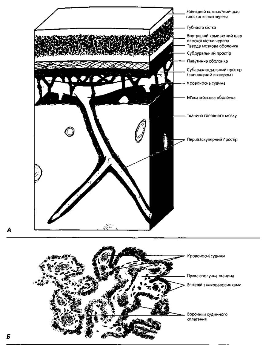
Fig. 4.119. Meninges of the central nervous system organs and cerebrospinal fluid production: A - diagram of the meninges around the brain; B - semi-schematic representation of the MICROSTRUCTURE OF THE choroid plexus of the Fourth ventricle of the human brain, which is one of the sources of cerebrospinal fluid production, x 190
Meninges of the brain. Features of cerebral blood supply. The brain and spinal cord are covered by three meninges—the pia mater, arachnoid mater, and dura mater (Fig. 4.119 A). The pia mater directly adheres to the brain tissue, separated from it by the glial limiting membrane. The pia mater is formed by loose Connective Tissue containing a significant number of blood vessels and nerve endings. The arachnoid mater is composed of loose connective tissue, separated from the pia mater by a network of Collagen and elastic fibers. The space between the pia mater and the arachnoid mater is called the subarachnoid space; it communicates with the ventricles of the brain and is filled with cerebrospinal fluid. The dura mater is formed by Dense connective tissue rich in elastic fibers. In the cranial cavity, it is fused with the periosteum of the skull bones, while in the spinal canal, it is separated from the periosteum of the vertebrae by the epidural space filled with loose connective tissue. The subdural space lies between the dura mater and the arachnoid mater. The dura mater and arachnoid mater are lined with a layer of flat gliocytes on the sides facing the subarachnoid and subdural spaces.
The organs of the central nervous system have unique features in The structure of their microcirculatory bed. Brain capillaries are characterized by a continuous endothelial lining and a well-defined basement membrane. The processes of neuroglial astrocytes accompany the capillaries along their entire length and, expanding, form a continuous layer around them that prevents neurons from direct contact with the vascular wall. This is how the blood-brain barrier is formed.
The central canal of the spinal cord, the ventricles of the brain, and the subarachnoid space are filled with cerebrospinal fluid (CSF). The latter contains water-soluble salts, a small amount of Proteins, and lymphocytes. CSF acts as a hydraulic Shock absorber for the organs of the central nervous system and also provides their immune defense. CSF is produced by the choroid plexuses of the brain ventricles (Fig. 4.119, B). The capillaries of the choroid plexuses form characteristic projections—villi—and are separated from the ventricular lumen by cuboidal cells of the ependymal Glia. The latter, together with the endothelium and thin layers of connective tissue of the villi, form the blood-CSF barrier. Reabsorption of CSF is carried out by arachnoid villi—projections of the arachnoid mater that protrude into the sinuses of the dura mater.
Development and Age-related changes of the central nervous system organs. The organs of the central nervous system develop from the neural tube, which separates from the cutaneous ectoderm during the fourth week of Embryogenesis (see chapter "Fundamentals of Human Embryogenesis"). In cross-sections of the neural tube at early Selection/3.html">Stages of development, three zones can be distinguished: the ependymal, mantle, and marginal zone (marginal veil). Ependymal glia form the lining of the central canal of the spinal cord and the ventricles of the brain. Neuroblasts of the mantle zone form the gray matter of the spinal cord, while the marginal zone forms its white matter. Spongioblasts of the neural tube serve as the source for neuroglia development.
The anterior (cranial) division of the neural tube is the source of brain development, while the spinal cord forms from the trunk (caudal) division. As it grows, the primordium of the brain forms three distinct swellings, the so-called primary brain vesicles: anterior, middle, and posterior. The three-vesicle stage is short-lived; during the sixth to seventh week of embryogenesis, it is succeeded by the five-vesicle stage. The cerebral hemispheres develop from the first brain vesicle, the diencephalon from the second, the midbrain from the third, the hindbrain (pons and cerebellum) from the fourth, and the medulla oblongata from the fifth.
The cerebral cortex is also called the neocortex because it is the latest evolutionary acquisition in phylogenesis. The Development of the cerebral cortex proceeds from the ventricular zone of the telencephalon. Neuroblasts differentiate and migrate into the cortical primordium along the vertical processes of embryonic radial glial cells. The latter undergo regression after birth. The neocortex has a predominantly six-layered structure: the first to populate the developing cortex are the neurons of the first and sixth layers, followed by the cells of the fifth, fourth, third, and second layers. All cells are aligned along the processes of radial glial cells, which thus act as organizers of cortical modules. Those cortical areas that retain the six-layered structure in the postnatal period are called the association cortex (90% of the neocortex). A smaller part of the neocortex differentiates in motor areas into the agranular pyramidal cortex, in which layers III and V are predominantly developed. In sensory areas, layers II and IV are predominantly developed; such cortex is called granular.
In addition to the neocortex, the brain also contains the archicortex (old cortex), paleocortex (ancient cortex), and intermediate cortex (periarchicortex and peripaleocortex). These types of cortex represent earlier evolutionary acquisitions compared to the neocortex; during ontogenesis, they do not pass through the six-layered stage. The archicortex is localized on the medial surfaces of the cerebral hemispheres around the corpus callosum and the inferior horn of the lateral ventricle, while the paleocortex is located on the inferior and medial surfaces of the hemispheres, between the frontal and temporal lobes. The intermediate cortex occupies a position between the neocortex and archicortex, or between the neocortex and paleocortex. The archicortex, paleocortex, and intermediate cortex are grouped under the general term of heterogenetic cortex, as opposed to the homogenetic cortex, or neocortex.
After birth, the maturation of the cerebral cortex is accompanied by an increase in the volume of neuronal perikarya, a decrease in their nucleocytoplasmic ratio, an increase in the number of synaptic contacts, and The formation of myelin sheaths around axons. A significant portion of neurons (up to 50-70%) dies via apoptosis and is phagocytosed by microglial cells. The development of a child's motor, sensory, intellectual, and communicative abilities is largely determined by the "maturation" of interneuronal connections established under the Influence of External factors and training. In The Mechanism of specific interneuronal synapse formation, adhesion molecules—so-called endogenous Lectins of nervous tissue—as well as nerve growth factors, play an important role. In adults, due to the proliferation of neuroglia and nerve fibers, and the death of some neurons, the number of neurons per unit volume of the cortex decreases.
Between the ages of 20 and 60, brain mass increases by 6 g every 10 years. With age, especially in senility, due to the progression of sclerotic changes in cerebral vessels, impaired trophics, and the death of some nerve cells, a further decrease in the number of neurons per unit volume of the brain is observed. This phenomenon is called cortical atrophy. Between the ages of 60 and 75, brain mass decreases by an average of 6% (by 50-100 g). The cortical surface area decreases by about 4%, and the brain appears to shrink. Interestingly, Betz and Purkinje cells are the first to undergo age-related involution.
Terms to remember
1. Central nervous system. 2. Brain. 3. Gray matter. 4. White matter. 5. Cerebral hemispheres. 6. Cerebral cortex. 7. Cytoarchitecture. 8. Molecular layer. 9. External granular layer. 10. Pyramidal layer. 11. Internal granular layer. 12. Ganglionic layer. 13. Polymorphic cell layer. 14. Giant pyramidal neuron (Betz cell). 15. Cerebral module. 16. Corticocortical fiber. 17. Thalamocortical fiber. 18. Association fiber. 19. Commissural fiber. 20. Projection fiber. 21. Myeloarchitecture. 22. Band of the molecular layer. 23. Band of the external granular layer. 25. Band of the ganglionic layer. 26. Subcortical nuclei of the brain. 27. Cerebellum. 28. Cerebellar cortex. 29. Molecular layer. 30. Ganglionic layer. 31. Granular layer. 32. Pear-shaped neuron (Purkinje cell). 33. Granule cells. 34. Stellate neurons, Golgi type II cells. 35. Mossy fiber. 36. Climbing fiber. 37. Cerebellar glomerulus. 38. Diencephalon. 39. Midbrain. 40. Pons. 41. Reticular formation. 42. Medulla oblongata. 43. Spinal cord. 44. Anterior median fissure. 45. Posterior median septum. 46. Anterior, lateral, and posterior horns of gray matter. 47. Radicular cell. 48. Funicular cell. 49. Interneuron. 50. Central canal. 51. Anterior, lateral, and posterior funiculi of white matter. 52. Pia mater. 53. Arachnoid mater. 54. Dura mater. 55. Blood-brain barrier. 56. Neural tube. 57. Ependymal zone. 58. Mantle zone. 59. Marginal veil. 60. Homogenetic cortex (neocortex).
The peripheral nervous system includes ganglia (spinal, cranial, and autonomic), nerve trunks (nerves), and nerve endings (Fig. 4.115).
Spinal ganglion (ganglion spinale)
(Fig. 4.120) is an accumulation of nerve cells near the junction of the anterior and posterior roots of the spinal cord. The spinal ganglion contains the perikarya of the first (sensory, afferent) neurons of the spinal reflex arcs. The spinal ganglion is covered by a connective tissue capsule, from which septa extend into the parenchyma of the organ. A characteristic morphological feature of the spinal ganglion is the orderly arrangement of neuronal perikarya and processes; the former are localized at the periphery beneath the capsule, while the latter are situated predominantly in the central part of the ganglion.
The main functional element of the spinal ganglion is the pseudounipolar neuron. This cell is characterized by a large, rounded body and a vesicular nucleus with a central Location. These cells get their name because both of their processes (axon and dendrite) originate from the same region of the perikaryon, run together for some distance, mimicking the presence of only a single process, and only then diverge in different directions. The dendrites of pseudounipolar neurons, merging into the posterior ROOT of the spinal cord, extend to the periphery to the organs they innervate. The axons of the spinal ganglion neurons form the portion of the posterior root located between the body of the ganglion and the posterior horn of the spinal cord. In addition to pseudounipolar neurons, spinal ganglia also contain small multipolar neurons that provide intraganglionic connections.
Pseudounipolar neurons are surrounded by specific neuroglial cells, so-called satellite glial cells (mantle gliocytes), which form a sort of sheath (mantle) around the perikaryon of each pseudounipolar neuron. Externally, the glial sheaths of the neurons are surrounded by layers of fine Fibrous connective tissue. The neuronal processes are covered by sheaths formed by neurolemmocytes (Schwann cells).
The STRUCTURE OF THE sensory nuclei of cranial nerves resembles that of spinal ganglia.
A nerve (nerve trunk, nervus) (Fig. 4.121) is composed of myelinated or Unmyelinated nerve fibers, as well as connective tissue elements. Individual nerve trunks may contain the cell bodies of isolated neurons and even small nerve ganglia. Externally, the peripheral nerve trunk is covered by a connective tissue sheath called the epineurium. The epineurium is rich in fibroblasts, macrophages, adipocytes, and fibrous structures. Blood vessels and nerve endings are located here. Septa (perineurium) extend from the epineurium into the nerve, dividing the peripheral nerve trunk into separate bundles of nerve fibers. The perineurium consists of longitudinally oriented thin collagen and elastic fibers and connective tissue cells. The connective tissue found within individual bundles of nerve fibers is called the endoneurium.

Fig. 4.120. Spinal (sensory) ganglion: A - topography and function: the arrow indicates the direction of the nerve impulse; B - semi-schematic representation of a whole-mount histological specimen, x100; C - light microscopy of the spinal ganglion, x300
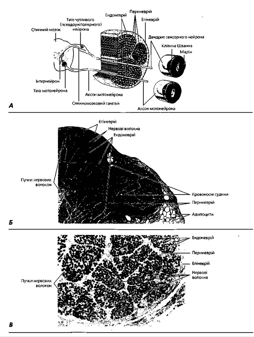
Fig. 4.121. Nerve: A - diagram of the structure; B - semi-schematic representation of a cross-section of the human tibial nerve, x76; C - light microscopy of a cross-sectioned nerve, osmium tetroxide staining, x100
Nerve endings (terminationes nervorum) are classified into receptors, effectors, and interneuronal synapses.
Receptors (receptores) are sensory endings of nerve cell dendrites adapted to perceive stimuli entering the body (Figs. 4.122, 4.123). Exteroceptors, which perceive stimuli from the external environment, and interoceptors, which receive stimuli from the body's own tissues, are distinguished. A type of interoceptor is proprioceptors—sensory nerve endings in muscles and tendons that participate in regulating movements and body position in space. Depending on The Nature of the stimuli that excite sensory nerve endings, the latter are classified into thermoreceptors (perceive temperature changes), mechanoreceptors (perceive mechanical stimuli), baroreceptors (perceive pressure changes), chemoreceptors (perceive chemical stimuli), nociceptors (perceive painful stimuli), etc.
Depending on their structure, a distinction is made between free nerve endings, which consist only of the terminal branches of a dendrite, and nerve receptors, in which the dendritic branches are surrounded by neuroglial cells. If the nerve endings are surrounded by a connective tissue capsule, they are called encapsulated; those receptors that lack a connective tissue capsule are called non-encapsulated. Receptor endings within epithelial, connective, and muscular tissues have several structural features discussed below.
Free nerve endings are characteristic of epithelium. During their formation, myelinated nerve fibers lose their myelin sheath as they approach the epithelial sheet, and their axons branch into terminal ramifications that lie between individual epithelial cells (Fig. 4.122, A). The function of free receptors, such as those in the epidermis, is associated with the perception of pain and temperature stimuli. Free nerve endings can wrap around Hair follicles in a basket-like fashion. By detecting the spatial displacement of individual hairs, they function as mechanoreceptors.
Isolated sensory epithelial cells, known as Merkel tactile cells, are localized in the basal layer of Stratified Epithelium. These electron-lucent cells with flattened nuclei contain osmiophilic granules in their Cytoplasm. Sensory nerve endings abut the basal portion of Merkel cells. This forms the so-called tactile discs (Fig. 4.112, B), which function in mechanoreception.
Sensory nerve endings within connective tissue are divided into non-encapsulated and encapsulated receptors, as well as neurotendinous spindles. In encapsulated receptors, the nerve endings are typically surrounded by neurolemmocytes and accessory elements of connective tissue origin. Among encapsulated receptors, depending on their structure, a distinction is made between Pacinian (lamellar) corpuscles, Golgi-Mazzoni corpuscles, Meissner (tactile) corpuscles, and Krause end bulbs.
The structure of the Pacinian (lamellar) corpuscle is shown in Figs. 4.122, C and 4.123, A. It is an oval-shaped structure measuring approximately 0.5x2 mm. Around the ramifications of the nerve ending, which has lost its myelin sheath, an accumulation of modified neurolemmocytes forms the inner bulb. Around the inner bulb, concentric layers of collagen fibers and flattened fibroblast-like cells form the so-called lamellae, from which the name of the lamellar corpuscle is derived. These lamellae form the outer bulb of the lamellar corpuscle. These corpuscles are abundant in the connective tissue of all internal organs, as well as in the deep layers of the dermis. They perceive changes in pressure. Golgi-Mazzoni corpuscles are smaller than Pacinian corpuscles, have a thinner capsule, and a relatively large inner bulb. They are localized in the Skin, serous, and mucous membranes, performing baroreceptive functions. A variant of dermal lamellar corpuscles is Ruffini endings (Fig. 4.122, D), which serve as receptors for detecting constant pressure. Ruffini endings are particularly numerous in the SOLE OF THE FOOT.
Meissner's tactile corpuscles (Fig. 4.122, D, 4.123, B) are located in the papillary layer of the dermis. These are oval structures measuring about 50x100 μm. Inside the tactile corpuscle, nerve endings lie in the form of a gentle spiral directed towards the skin surface. Within Meissner's corpuscle, the nerve fiber loses its myelin sheath and contacts neuroglial cells. The connective tissue capsule of Meissner's tactile corpuscle is formed by concentric layers of collagen fibers. The latter can fill the spaces between neurolemmocytes and nerve endings. Tactile corpuscles provide fine Touch sensitivity.
Krause's end-bulbs (Fig. 4.122, E) are localized in the conjunctiva of the eye, the connective tissue of the Tongue, and the external genitalia. Their characteristic feature is a very thin connective tissue capsule. Upon entering the capsule, the myelinated nerve fiber loses its myelin sheath and terminates in a bulbous expansion or may branch, forming a system of unmyelinated nerve endings. Krause's bulbs are believed to mediate cold sensitivity.
Golgi tendon organs (neurotendinous spindles) (Fig. 4.122, Ye) are formed by thick (about 15 μm in diameter) myelinated fibers which, upon approaching the collagen fibers of the tendon, lose their myelin sheath and give off numerous branches that wrap around the tendon bundles. Golgi tendon organs are considered mechanoreceptors that perceive the relative displacement of collagen fibers and changes in their position relative to adjacent tissues.
In Muscle tissue, sensory nerve endings form neuromuscular spindles (muscle spindles), which detect changes in muscle fiber length and The rate of this change (Fig. 4.122, Zh). Each spindle consists of 10-12 thin, short striated muscle fibers surrounded by an inner connective tissue capsule. These fibers are called intrafusal fibers. Externally, the connective tissue capsule is surrounded by striated muscle fibers, which form the outer capsule of the neuromuscular spindle. At the ends of the intrafusal muscle fibers, there are contractile myofibrils. The central, non-contractile portion of these fibers belongs to the receptor apparatus of the muscle spindle itself.

Fig. 4.122. Types of sensory nerve endings (schematic representation, not to scale)

Fig. 4.123. Light microscopy of sensory nerve endings: A - Pacinian corpuscle, x280; B - Meissner's corpuscles, x200; V - tactile corpuscle of the glans Penis, x800; G - tactile corpuscle of the Filiform papillae of the tongue, x600
Among the intrafusal muscle fibers, a distinction is made between nuclear bag fibers and nuclear chain fibers. Nuclear bag fibers contain a large number of nuclei in their central region. Nuclear chain fibers are half as thick and short as nuclear bag fibers. Their nuclei are arranged in a chain along the receptor region. The neuromuscular spindle has Two Types of nerve fibers. The diameter of primary fibers is 17 μm. They form the so-called annulospiral endings around both types of intrafusal muscle fibers. Annulospiral endings detect changes in muscle fiber length and the rate of this change. Secondary nerve fibers have a diameter of 8 μm. On both sides of the annulospiral ending, they form flower-spray endings that register changes in muscle fiber length.
Effectors (effectores) are formed by the terminals of nerve cell axons. There are two types of effectors: motor and secretory. Motor nerve endings in skeletal muscles are formed by the axon terminals of neurons from the motor nuclei of the anterior horns of the spinal cord or the motor nuclei of the brain. Upon approaching the muscle fiber, the myelinated nerve fiber loses its sheath, its axis cylinder branches, and, together with the Plasmalemma of the myosymplast, becomes embedded in the muscle fiber (see illustrations in the "Muscle Tissues" section). An axomuscular synapse (Neuromuscular Junction) is formed in this region, where the axolemma acts as the presynaptic membrane, and the sarcolemma of the muscle fiber serves as the postsynaptic membrane. The width of the synaptic cleft is about 50 nm. Acetylcholine accumulates within synaptic vesicles in the terminal Branches of the axon. During nerve fiber excitation, Acetylcholine is released from the synaptic vesicles, crosses the presynaptic membrane and the synaptic cleft, and, by binding to acetylcholine receptors on the surface of the postsynaptic membrane, serves as a chemical signal to excite the muscle fiber.
Motor nerve endings in Smooth muscle tissue have a somewhat simpler structure: individual nerve endings form characteristic expansions (varicosities) on the surface of smooth muscle cells, where acetylcholine or adrenaline accumulates within synaptic vesicles. Similar terminal swellings, or varicosities, in which acetylcholine predominantly accumulates, have also been described at the sites of contact between axons and secretory cells.
Interneuronal synapses (synapses interneuronales) are a special form of Intercellular junctions characteristic of nervous tissue. The classification, morphology, and function of synapses are discussed in the "Nervous Tissue" section.
The Autonomic (vegetative) nervous system regulates the activity of the digestive organs, blood pressure, perspiration and urination, body temperature, and processes related to metabolism, growth, and reproduction. The Autonomic Nervous System includes central divisions, formed by the nuclei of the brain and spinal cord, and peripheral divisions, which include nerve ganglia, trunks, and plexuses. Based on functional characteristics, the autonomic nervous system is divided into two parts—sympathetic and parasympathetic—which generally have opposing effects on the respective organs and systems of the body. In addition to these parts, the autonomic nervous system also includes the so-called metasympathetic nervous system, which comprises the intramural microganglia of internal organs, such as the digestive tract, Respiratory system, Heart, and Kidneys. These microganglia possess a significant degree of autonomy in regulating the functions of these organs, and under physiological conditions, they are not controlled by the central components of the autonomic and somatic nervous systems.
The nuclei of the central division of the autonomic nervous system are located in the midbrain, medulla oblongata, and spinal cord (specifically in the thoracic, lumbar, and sacral segments of the latter) (Fig. 4.124). The sympathetic nervous system includes the autonomic nuclei of the lateral horns of the thoracic and upper lumbar segments of the spinal cord, while the parasympathetic nervous system includes the autonomic nuclei of cranial nerves III, VII, IX, and X, as well as the nuclei of the sacral segments of the spinal cord. The nuclei of the central divisions of the autonomic nervous system are composed of multipolar associative neurons. The axons of these cells exit the central divisions within the anterior roots of the spinal cord or cranial nerves and contact neurons of the autonomic ganglia, while their dendrites synapse with the axons of pseudounipolar neurons of the spinal ganglia or associative neurons of the spinal cord. Ganglia of the autonomic nervous system are located both within organs and outside of them. Paravertebral and prevertebral sympathetic ganglia, as well as parasympathetic ganglia of the HEAD, have an extraorgan localization. Intraorgan nerve ganglia (plexuses) are located in the walls of the digestive tube, heart, Uterus, Urinary Bladder, and other organs. Paravertebral ganglia are located on both sides of THE Vertebral Column, forming sympathetic chains. Prevertebral ganglia include the celiac, superior, and inferior mesenteric ganglia, which form the celiac plexus anterior to the Abdominal Aorta and its branches.
Autonomic ganglion (ganglion autonomicum)
(Fig. 4.125) is surrounded by a connective tissue capsule, from which connective tissue septa extend into the ganglion. Ganglia of the autonomic nervous system consist of multipolar neurons, which distinguishes them from sensory spinal ganglia composed of pseudounipolar nerve cells. Another feature is that the nerve cells of autonomic ganglia are surrounded by nerve fibers, which, unlike in spinal ganglia, do not have a central localization. Each neuron of the autonomic ganglion, as well as its processes, is surrounded by neuroglial cells. The dendrites of the autonomic ganglion nerve cells branch extensively and contact the processes of neurons from the central divisions; the axons are predominantly unmyelinated and travel to the respective organs as part of postganglionic fibers. Some of the preganglionic fibers entering the ganglion terminate directly on the neuronal perikarya, forming axosomatic cholinergic synapses. The vast majority of autonomic ganglion neurons are cholinergic. Within sympathetic ganglia, small neurons with short processes have also been found; under the excitatory influence of preganglionic fibers, they release adrenaline. These cells form small groups and act as an intraganglionic inhibitory system.

Fig. 4.124. Diagram of efferent autonomic pathways and localization of autonomic ganglia: preganglionic neurons are shown as solid lines; postganglionic neurons as dashed lines; parasympathetic fibers are indicated by bold lines, and sympathetic fibers by thin lines. Sympathetic ganglia are characterized by extraorgan localization, while parasympathetic ganglia exhibit intraorgan (intramural) localization.

Fig. 4.125. Autonomic ganglia: A - semi-schematic representation of the myenteric (Auerbach's) plexus in the wall of the duodenum, x100; B - light microscopy of a sympathetic ganglion, x160; V - parasympathetic (intramural) ganglion of the Pancreas, x160
Ganglia of the Parasympathetic division of the autonomic nervous system are located either near the organ they innervate or directly within it. In addition to efferent neurons, the ganglia of intraorgan nerve plexuses contain receptor and associative cells of local reflex arcs. Based on morphological features, Three types of neurons, first described by the Russian neurohistologist A.S. Dogiel, are distinguished in intraorgan plexuses:
1) efferent neurons with short dendrites and long axons (Dogiel type I cells);
2) afferent neurons with long dendrites and short axons (Dogiel type II cells);
3) associative neurons with dendrites and axons of medium length that project to neighboring cells of the ganglion or to neighboring ganglia of the plexus (Dogiel type III cells).
The axons of nerve cells in intraorgan plexuses project to the muscular elements of the organ, on the surface of which they form varicosities with a diameter of about 0.5-2 μm.
The development of spinal ganglia and autonomic nervous system ganglia occurs in parallel with spinal cord development, originating from neural crest cells that lie in longitudinal rows between the neural tube and the surface ectoderm. A portion of these neural crest cells migrates ventrally toward the Abdominal cavity, forming the primordia of sympathetic and parasympathetic ganglia, as well as The adrenal medulla. The neural cells remaining on either side of the neural tube form ganglion plates. These plates segment, and their cellular components differentiate into neuroblasts and glioblasts, which eventually give rise to the neurons and glial cells of the spinal and paravertebral ganglia.
Terms to remember
1. Spinal ganglion. 2. Pseudounipolar neuron. 3. Satellite glial cell. 4. Nerve. 5. Epineurium. 6. Perineurium. 7. Endoneurium. 8. Nerve ending. 9. Receptor. 10. Exteroceptor. 17. Interoceptor. 12. Proprioceptor. 13. Thermoreceptor. 14. Mechanoreceptor. 15. Baroreceptor. 16. Nociceptor. 17. Free nerve ending. 18. Merkel tactile cell. 19. Encapsulated nerve ending. 20. Pacinian (lamellar) corpuscle. 21. Meissner's (tactile) corpuscle. 22. Krause end bulb. 23. Unencapsulated nerve ending. 24. Golgi tendon organ (neurotendinous spindle). 25. Ruffini corpuscle. 26. Neuomuscular spindle (muscle spindle). 27. Intrafusal muscle fiber. 28. Nuclear bag fiber. 29. Nuclear chain fiber. 30. Annulospiral (primary) nerve ending. 31. Flower-spray (secondary) nerve ending. 32. Effector. 33. Axomuscular synapse. 34. Autonomic nervous system. 35. Sympathetic nervous system. 36. Parasympathetic nervous system. 37. Metasympathetic nervous system. 38. Autonomic ganglion. 49. Neural crest. 40. Ganglion plate.
Last update: 09/08/2026
Editorial and Educational Adaptation: This material has been compiled based on the primary/original source text. The project team performed an editorial review, corrected technical inaccuracies, structured sections, and adapted the content for an educational format.
What was processed:
- elimination of formatting defects (OCR errors, structural breaks, corrupted characters);
- editorial organization of content;
- standardization of terminology in accordance with academic sources;
- verification of factual statements against the original source text.
All mentions of the author, publication year, and origin of the primary text have been preserved in accordance with the source.