Human Anatomy - Kotsan I. Y. 2009
Brain
Thalamic region
The thalamic region includes the thalamus, metathalamus, and epithalamus.
The thalamus, or dorsal thalamus, is a paired egg-shaped Structure located on either side of the Third ventricle. The thickened posterior end of the thalamus is called the pulvinar. Anteriorly, the thalamus narrows and terminates in the anterior tubercle (tuberculum anterius thalamicum). The anterior, inferior, lateral, and almost the entire posterior surface (except for the pulvinar) of the thalamus are fused with the Brain tissue of adjacent regions. Only two surfaces of the thalamus are free: the medial surface, which forms the lateral wall of the third ventricle, and the superior surface, which contributes to the floor of the central part of the lateral ventricle. The superior surface of the thalamus is separated from the medial surface by a thin white medullary stria of the thalamus (stria medullaris thalamica). The medial surfaces of the right and left thalami are connected to each other by the interthalamic adhesion (adhesio interthalamica). The lateral surface of the thalamus is adjacent to the internal capsule.
The thalamus consists of Gray matter containing distinct clusters of Nerve Cells (thalamic nuclei). These clusters are separated by thin layers of White matter. There are about 40 nuclei performing various Functions. The main nuclei of the thalamus are the anterior nuclei (nuclei anteriores), posterior nuclei (nuclei posteriores), medial nuclei (nuclei mediales), and ventrolateral nuclei (nuclei ventrolaterales). The processes of nerve cells belonging to second-order (Relay) Neurons of all sensory pathways (except for olfactory, gustatory, and auditory) synapse with the nerve Cells of the thalamus. Consequently, the thalamus essentially serves as a subcortical sensory center. The processes of thalamic neurons project partly to the nuclei of the corpus striatum of the Telencephalon (therefore, the thalamus is considered a sensory center of the extrapyramidal system) and partly to the Cerebral Cortex via thalamocortical tracts (fasciculi thalamocorticales).
Inferior to the thalamus lies the so-called subthalamic region (regio subthalamica), which extends inferiorly into the tegmentum of the Midbrain. This is a small area of brain tissue located ventral to the thalamus and separated from it (on the side of the third ventricle) by the hypothalamic sulcus (sulcus hypothalamicus). The red Nucleus and substantia nigra of the midbrain extend into the subthalamic region and terminate there. Lateral to the substantia nigra lies the subthalamic nucleus (nucleus subthalamicus).
The metathalamus — the retrothalamic region — is formed by the paired medial and lateral geniculate bodies located posterior to each thalamus (Fig. 247). The medial geniculate body (corpus geniculatum mediale) is situated posterior to the pulvinar of the thalamus. The lateral geniculate body (corpus geniculatum laterale) is located near the inferolateral surface of the thalamus, lateral to its pulvinar. The lateral geniculate bodies, together with the superior colliculi of the midbrain, serve as subcortical visual centers. The medial geniculate bodies and inferior colliculi of the midbrain form the subcortical auditory centers. The geniculate bodies are connected to the colliculi of the midbrain by the brachia of the superior and inferior colliculi (brachium colliculi superioris et brachium colliculi inferioris).
Class="center">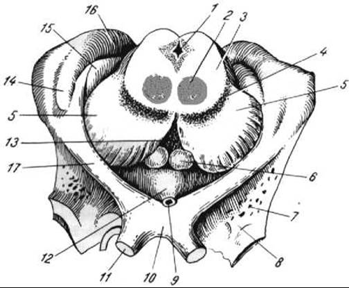
Fig. 247. Metathalamus and Hypothalamus
1 — cerebral aqueduct; 2 — red nucleus; 3 — tegmentum; 4 — substantia nigra; 5 — cerebral peduncle; 6 — mamillary body; 7 — anterior perforated substance; 8 — olfactory trigone; 9 — infundibulum; 10 — optic chiasm; 11 — Optic nerve; 12 — tuber cinereum; 13 — posterior perforated substance; 14 — lateral geniculate body; 15 — medial geniculate body; 16 — pulvinar of the thalamus; 17 — optic tract
The epithalamus — the suprathalamic region — is located dorsal and posterior to the thalamus. It consists of the pineal body, habenulae, and habenular commissure.
The pineal body, or epiphysis (corpus pineale, epiphysis), lies on the tectum of the midbrain between the superior colliculi of this plate. This organ belongs to the Endocrine glands (see "Endocrine Glands"). The pineal body is attached to the thalamus by the habenulae.
The habenula is a paired elongated structure that connects the thalamus to the pineal body and represents the posterior continuation of the medullary stria of the thalamus. In the posterior part of the habenulae, There is a triangular plate of brain tissue called the trigonum habenulae. Deep within the habenular trigone lie the medial and lateral habenular nuclei (nuclei habenulae medialis et lateralis), which belong to the structures of the rhinencephalon. The habenular trigones are interconnected by the habenular commissure (commissura habenularum), which passes anterior to the pineal body. Anterior to the habenular commissure is the epithalamic (posterior) commissure (commissura epithalamica [posterior]). It consists of a small number of commissural nerve fibers connecting adjacent areas of the cerebral hemispheres. Between the epithalamic commissure and the habenular commissure, a shallow blind pocket — the pineal recess — extends into the anterosuperior part of the pineal body and its base.
Last update: 08/08/2026
Editorial and Educational Adaptation: This material has been compiled based on the primary/original source text. The project team performed an editorial review, corrected technical inaccuracies, structured sections, and adapted the content for an educational format.
What was processed:
- elimination of formatting defects (OCR errors, structural breaks, corrupted characters);
- editorial organization of content;
- standardization of terminology in accordance with academic sources;
- verification of factual statements against the original source text.
All mentions of the author, publication year, and origin of the primary text have been preserved in accordance with the source.