Human Histology - O.D. Lutsyk 2003
Special Histology
Sense Organs
Sensory Organs are specialized organs through which The Nervous System receives stimuli from the external and internal environments and perceives them as sensations. Information coming from the sensory organs is the source of our perception of the surrounding world. There are five sensory organs: Touch, taste, smell, Hearing and Balance (equilibrium), and Vision. According to I.P. Pavlov's definition, sensory organs are the peripheral parts of analyzers. Analyzers are complex neurodynamic systems, the afferent divisions of reflex arcs that connect the Central Nervous System with the external and internal environments. Each analyzer consists of a peripheral part where stimuli are perceived (which are the sensory organs themselves); an intermediate part, comprising pathways and subcortical structures that transmit nerve impulses; and a central part, which is the Cerebral Cortex, where the final analysis and Synthesis of the perceived sensation take place.
Classification of sensory organs. Based on their origin and Structure, sensory organs are divided into three main types. The first type includes the organs of vision and smell; their receptor Cells, called neurosensory or primary sensory cells, develop from the embryonic neural plate. The second type includes the Organs of taste, hearing, and equilibrium, whose receptive elements are specialized epithelial cells (sensory epithelial cells). From these cells, the transformed stimulus is transmitted to Nerve Cells. Such sensory organs are called secondary sensory organs. In Embryogenesis, these organs develop from special ectodermal thickenings known as placodes. The third type of sensory organs, which lack a distinct organ structure, includes sensory (encapsulated and unencapsulated) nerve endings, as well as individual cells that serve as the peripheral PARTS OF THE corresponding analyzers (pressure, touch).
The structure and histophysiology of the organs of vision, hearing, and equilibrium are discussed below. A detailed description of other sensory organs is provided in other chapters of the textbook: The ORGAN OF TASTE in the chapter "Oral Cavity. Tongue"; the Organ of Smell in the chapter "Respiratory system. Nasal cavity"; and the Organ of Touch in the chapter "Peripheral Nervous System. Receptors".
The Organ of Vision, as defined by V. P. Filatov, is the most precious of all sensory organs. It provides us with 60-80% of all information about the surrounding world. The eye (oculus) is the peripheral part of the visual analyzer. The eye consists of the Eyeball and accessory structures, which include the eyelids, the Muscles of the eyeball, and the Lacrimal Apparatus.
The eyeball (bulbus oculi) (Fig. 4.126) is roughly spherical, with a diameter of 24 mm. It is located in the anterior part of the eye socket, or Orbit. Between THE EYE AND the bony wall of the orbit lie fat, Connective Tissue, ligaments, muscles, and the lacrimal gland. The eye is suspended by ligaments so that voluntary muscles located in the orbit can move it down, up, and from side to side. The wall of the eyeball is formed by three tunics — outer, middle, and inner. The outer, fibrous tunic consists of two parts — the opaque white of the eye, the sclera, which surrounds the eyeball and makes up 5/6 of its surface, and the transparent cornea, which covers the anterior pole of the eyeball (1/6 of the surface). The sclera transitions into the cornea gradually — first the inner and middle layers, then the outer layers. The transition zone is called the limbus. The middle (vascular) tunic consists of three parts — the choroid proper, the ciliary body, and the iris. The inner (sensory) tunic is called the retina. It is divided into three parts: the optic, iridial, and ciliary parts. The optic part of the retina is the most complex in structure and the most important in function. The iridial and ciliary parts are actually the inner layer of the ciliary body and iris, which together are also referred to as the blind retina (pars caeca retinae).
Class="center">
Fig. 4.126. Eyeball: A — diagram of the structure showing the main structural components and optical axes: horizontal section of the human right eye; B — 3D reconstruction of the left eye: horizontal projection
Inside the eyeball are the lens, the vitreous body, and cavities — the Chambers of the eye, filled with the so-called aqueous humor. A distinction is made between the anterior chamber, which occupies the space between the cornea and the iris, and the posterior chamber — the space between the iris, the ciliary zonule, and the lens.
The eyeball has three main functional apparatuses:
1) the dioptric, or refractive apparatus, which includes all transparent (optical) media through which light passes before reaching the retina; it comprises the cornea, the aqueous humor of the anterior chamber, the lens, and the vitreous body;
2) the accommodative apparatus, which provides Changes in the shape and refractive power of the lens to focus images of objects on the retina, as well as the adaptation of the eye to light of varying intensity; it includes the ciliary body with the ciliary zonule and the iris;
3) the receptor, or photosensory apparatus, which includes the optic part of the retina.
The sclera (sclera) (Figs. 4.126, 4.127) is a Cytology/practical/45.html">Dense connective tissue tunic that performs protective and supportive Functions. The thickness of the posterior part of the sclera is 0.3-0.4 mm, and near the cornea, it is 0.6 mm. The sclera is composed of lamellae built of Collagen fibers oriented parallel to The surface of the eye, with fibroblasts and elastic fibers located between them. Numerous melanocytes are localized in the deep layers of the sclera near the exit of the Optic nerve. In the transition zone to the cornea, the sclera contains small, irregularly shaped cavities that communicate with each other to form the scleral venous sinus (or the so-called canal of Schlemm). In the angle between the cornea and the iris, with which the inner surface of the sclera contacts, lies the pectinate ligament. This region, together with the scleral venous sinus, provides the outflow of fluid from the anterior chamber of the eye. The anterior surface of the sclera is covered by the conjunctiva. The sclera is connected to the adjacent Tissues of the orbit by loose, highly vascular episcleral tissue.

Fig. 4.127. Angle of the eye: A — diagram of the anterior part of the eyeball: the arrows in the left half show the direction of aqueous humor flow, the right half illustrates the structural details of the ciliary body; B — semi-schematic rendering of a human eye angle specimen, x 15
The cornea (cornea) (Figs. 4.126, 4.127) is a continuation of the sclera. Due to its unique Structure and Chemical composition, the cornea is transparent. Its thickness is 0.8-0.9 mm in the center and 1.1 mm at the periphery. The refractive index is 1.37. Five layers are distinguished in the cornea.
The first, outer layer is formed by a stratified squamous non-keratinized epithelium up to 50 μm thick, containing numerous nerve endings that mediate the corneal reflex. Its surface is moistened by secretions from the lacrimal and conjunctival glands. It has a high regenerative capacity and is permeable to liquids and gases. The anterior corneal epithelium is continuous with the stratified squamous epithelium of the conjunctiva. The basement membrane of this epithelium consists of electron-lucent and electron-dense layers.
The anterior limiting lamina (Bowman's membrane) is located beneath the basement membrane. Under the Light Microscope, it appears homogeneous, while under the Electron microscope, it has a fibrillar structure, with collagen fibrils measuring 20-30 nm in diameter. The thickness of Bowman's membrane is 8-14 μm.
The substantia propria of the cornea (corneal stroma) consists of 200-250 thin connective tissue lamellae that regularly alternate and cross each other at an angle. Each lamella is formed by bundles of collagen fibers. Between and within these lamellae are flat cells with long, branching processes, which are a type of fibroblast (keratocytes). The cells and lamellae are embedded in an amorphous ground substance rich in keratan sulfates, which ensure the transparency of the cornea. Blood Vessels are absent here. In the region of the iridocorneal angle, the substantia propria of the cornea transitions into the opaque sclera.
The posterior limiting lamina (Descemet's membrane) is 10-14 μm thick. It consists of collagen fibers embedded in an amorphous ground substance.
The posterior corneal epithelium (corneal endothelium) is a simple squamous epithelium with cells 5 μm high and 18-20 μm wide, facing the anterior chamber of the eye.
The choroid (choroidea) of the middle tunic of the eyeball is located in its posterior part, between the sclera and the optic part of the retina, providing nourishment to the latter. In the choroid, starting from the outside, there are four layers:
1) the suprachoroid lamina borders the sclera and is formed by loose connective tissue containing A large number of melanocytes;
2) the vascular lamina consists of Arteries and Veins, between which is loose Connective tissue with a large number of pigment cells; bundles of smooth myocytes are also located here;
3) the vascular-capillary layer contains hemocapillaries, including those of a sinusoid type, interspersed with fibroblasts;
4) the basal membrane (Bruch's membrane) is 1-4 µm thick and located between the choroid and the retinal pigment epithelium; it consists of three layers: elastic, fibrous, and the basal membrane proper.
The ciliary body (corpus ciliare) (Figs. 4.126, 4.127) is a forward continuation of the vascular tunic (choroid). It is divided into two parts: an inner ciliary crown and an outer ciliary ring. Ciliary processes extend from the ciliary crown, and from these processes arise the zonular fibers (suspensory ligament), which anchor the lens to the ciliary body. The bulk of the ciliary body, aside from the processes, is formed by the ciliary Muscle, which is the active component of the eye's accommodation apparatus. The ciliary Muscle consists of smooth myocytes arranged in three directions: meridian, radial, and circular. Loose connective tissue containing melanocytes is found in bundles between the smooth muscle cells. Contraction of the ciliary muscle relaxes the zonular fibers, releasing tension on the lens capsule; As a result, the lens becomes more convex due to its elasticity, thereby increasing its refractive power.
The ciliary body and its processes are covered by the ciliary part of the retina, which in this region comprises two layers: a single layer of pigmented cuboidal epithelial cells and a single layer of non-pigmented columnar epithelium. The non-pigmented epithelium is lined on the inside by the vitreous ciliary membrane. Together with the capillaries of the ciliary processes, these epithelial cells are responsible for producing the aqueous humor that fills the eye chambers.
The iris (Figs. 4.126, 4.127) extends anteriorly from the ciliary body as its direct continuation. It appears as a pigmented disc with a centrally located aperture of variable size—the pupil—which separates the anterior and posterior chambers of the eye. The iris features a ciliary margin, where it attaches to the ciliary body, and a pupillary margin; the boundary between them lies 1.5 mm from the edge of the pupil (along the so-called ora serrata). The iris consists of five layers:
1) the anterior epithelium, formed by flat, polygonal cells, which is a continuation of the corneal posterior surface epithelium;
2) the outer limiting layer, a connective tissue containing ground substance, numerous fibroblasts, and pigment cells; varying numbers and locations of melanocytes determine eye color;
3) the vascular layer, containing numerous blood vessels surrounded by loose connective tissue with melanocytes; two smooth muscles are located in this layer—the sphincter pupillae and the dilator pupillae; the sphincter is situated at the pupillary margin, while the dilator lies near the ciliary margin of the iris;
4) the inner limiting layer, structurally similar to the outer limiting layer;
5) the posterior pigmented epithelium, which is a continuation of the two-layered retinal epithelium that also covers the ciliary body.
The iris functions as the Diaphragm of the eye, regulating The amount of light falling on the retina via the aforementioned muscles.
The retina (Figs. 4.126, 4.127, 4.128, 4.129). Due to the presence of photoreceptor cells, the optic part of the retina acts as the light-sensitive tunic. It extends across the Fundus of the eyeball up to the ora serrata, where it transitions into the non-visual (blind) retina that covers the posterior surface of the ciliary body and the iris. The choroid lies adjacent to the optic part of the retina.

Fig. 4.128. STRUCTURE OF THE retina: A - horizontal section of the right eye demonstrating the topography and morphological Features of the macula and blind spot; B - schematic diagram of the Cellular Organization of the optic retina (ten layers) showing three-neuron chains; C - comparative Morphology of a cone and a rod

Fig. 4.129. Light Cell/15.html">Microscopy of the retina, sagittal section: A - general view of the optic retina (ten layers), × 350; B - region of the macula lutea (fovea centralis) of the retina, × 275; C - region of the blind spot (optic disc), × 25
The retina is composed of ten layers: the pigment layer, photoreceptor layer, outer limiting layer, outer nuclear layer, outer plexiform layer, inner nuclear layer, inner plexiform layer, ganglion cell layer, nerve fiber layer, and inner limiting layer (Fig. 4.128, B). All these retinal layers, except for the pigment layer, are formed by Neurons. Three of these neuronal types are primary and arranged in radial chains, while two additional types integrate into the chain at two levels as interneurons. Additionally, the retina contains specialized cells known as radial glial cells. The neurons are organized such that their cell bodies form the outer and inner nuclear layers and the ganglion cell layer, whereas their processes and synaptic contacts form the outer and inner plexiform layers and the nerve fiber layer. Characteristically, the dendrites of all three neurons in the chain are directed outward, while their axons are directed toward the interior of the eyeball.
The pigment layer is the outermost layer of the retina, consisting of a single layer of pigmented epithelial cells approximately 8 µm in height. These cells rest on the basal membrane of the choroid and remain attached to the choroid even during retinal detachment. The outer part of a pigment cell contains one or two nuclei, while eight to ten cytoplasmic processes extend from its inner surface. The Cytoplasm contains melanin granules in the form of melanosomes, which migrate into the processes under intense illumination and return to The Cell body in the dark.
The first neuron of the retina is a photosensitive bipolar cell, whose modified dendrites are called rods (one cell type) and cones (the other cell type). Light rays are perceived by these photoreceptor cells. The rods and cones form the second photosensitive layer of the retina, interspersed among the processes of the pigment epithelial cells. The nuclear regions of these photosensory neurons constitute the outer nuclear layer, and their axons contribute to The formation of the outer plexiform layer of the retina.
Rods and cones (Fig. 4.128, C) consist of outer and inner segments connected by a connecting cilium. The outer segment of a rod is cylindrical and contains a large stack (up to 1,000) of flat membrane discs 140 nm thick and 2 µm wide. The disc membrane contains the visual pigment rhodopsin, which is composed of the protein opsin and retinal, an aldehyde of vitamin A. The outer segment of a cone is conical, wider, and shorter than that of a rod; it contains half-discs formed by the invagination of the Plasmalemma, with one end of each half-disc closed and the other open. The membrane of cone half-discs contains a different visual pigment, iodopsin. The human retina possesses three distinct types of cone cells, sensitive to the blue, green, and red Regions of the spectrum. The outer segments of rods and cones are modified dendrites.
The connecting cilium linking the outer and inner segments of rods and cones originates from a basal body in the inner segment. The inner segment contains numerous Mitochondria, Endoplasmic reticulum, and enzyme systems that support METABOLISM/26.html">Energy Metabolism and The Biosynthesis of vital cell components. The inner segment of a cone differs from that of a rod by the presence of an ellipsoid—a lipid droplet surrounded by a dense cluster of closely packed mitochondria.
The human retina contains approximately 130 million rod cells and 6-7 million cone cells. Rods function as the apparatus for black-and-white vision in dim light (scotopic vision), whereas cones mediate color daylight vision (photopic vision). Vision begins when a quantum of light interacts with rhodopsin, the photosensory protein embedded in the membrane of the outer segment of retinal rods. The rhodopsin molecule consists of the transmembrane polypeptide opsin and the chromophore 11-cis-retinal. Upon absorbing a photon of light, cis-retinal isomerizes to trans-retinal and dissociates from opsin, causing the latter to undergo a conformational change. This, in turn, leads to the closure of The Plasma Membrane sodium channels, its hyperpolarization, and the generation of an Action Potential. Color Vision is provided by the Three types of cones in the retina. The photosensory protein iodopsin in the first type of cones is sensitive to wavelengths around 700 nm (red light); the second type contains a protein sensitive to 550 nm (green); and the third type responds to 400-450 nm (blue-violet spectrum). The discs undergo continuous regeneration. Every day, up to 80 membrane discs are formed in a rod cell at night and in a cone cell during the day. Worn-out discs are shed and phagocytosed by pigment epithelial cells, each of which engulfs about 2-3 thousand discs per day. Due to the presence of retinol-binding protein in their cytoplasm, pigment cells help retain vitamin A under intense illumination and supply it to photoreceptor cells to regenerate rhodopsin. Besides their phagocytic function, pigment cells facilitate light and dark adaptation of visual cells. Each rod outer segment is surrounded by 2 to 7 processes of pigment cells, whereas a cone is enclosed by 30-40 processes. During bright light, melanosomes move into the pigment cell processes, causing rods to elongate and become shielded, while cones contract and receive optimal illumination. In twilight, when melanosomes return to the pigment cell bodies, cones elongate and are shielded, while rods shorten and resume their function.
Vitamin A deficiency causes nyctalopia (night blindness), a pathology that drew researchers' attention to the crucial role of vitamin A in rod function. It was later discovered that vitamin A deficiency also leads to cone degeneration. Vitamin therapy helps restore normal retinal function if initiated before receptor destruction begins. Partial color blindness (the inability to distinguish certain colors) was first described in the late 18th century by the English physicist John Dalton, who suffered from this visual defect himself. Daltonism is caused by the absence of genes encoding specific peptide chains of cone opsins on the unpaired male X chromosome.
The second neuron in the retinal three-neuron chain is known as the bipolar cell. Its cell body is located in the inner nuclear layer; its dendrite forms synapses with the axons of rod and receptor cells in the outer plexiform layer (several rod cells synapse with a single bipolar neuron, whereas cones connect in a 1:1 ratio); and its axon is situated in the inner plexiform layer, where it synapses with the dendrite of the third neuron.
In addition to bipolar cells, the inner nuclear layer of the retina contains Two Types of associative (intercalated) neurons: horizontal and amacrine cells. Horizontal cells send their processes into the outer plexiform layer, where they synapse with the axons of photoreceptor neurons. Their excitation causes a temporary blockade of impulses from photoreceptors, thereby enhancing image contrast. Amacrine cells lack axons. Based on the branching pattern and length of their processes, several types of these neurons are distinguished. Taking into account their morphology and the Neuropeptides they produce, amacrine cells are divided into six subtypes. Amacrine cells project their processes into the inner plexiform layer, where they perform a function similar to that of horizontal cells, albeit at the level of the synapse between the second and third neurons.
The third neuron of the retina is called the ganglion neuron and is multipolar. It is the largest neuron of the retina and has a well-developed chromatophilic substance. The bodies of ganglion cells form the ganglion cell layer of the retina; their dendrites are located in the inner plexiform layer, forming synapses with the axons of bipolar neurons, while their axons form the nerve fiber layer. The retinal nerve fibers run radially and converge, like spokes in a wheel, at a single point where they form the optic disc, or the so-called blind spot of the retina. From here, they exit the eyeball as the optic nerve. In the region of the blind spot, the retina consists only of the nerve fiber layer. All other layers are absent here, so this area does not perceive light stimuli.
Lateral to the blind spot, at the posterior pole of the optical axis of the eyeball, there is another specific area on the retina, the so-called macula lutea (yellow spot). It is round or oval, with a diameter of about 2 mm. In the center of the macula, There is a depression called the fovea centralis. This is the site of the sharpest vision. In this region, all inner layers of the retina (up to the outer nuclear layer) are absent, as if pushed aside. The outer nuclear layer is composed mainly of the cell bodies of cone cells, which are larger and elongated. The axons of photosensory cells in this area must run laterally in a horizontal direction to meet the bipolar cells, resulting in the formation of an additional fibrous layer.
The presence of the fovea centralis in the retina is due to the fact that the human eye belongs to the so-called inverted eye type, in which the visual elements—cones and rods—are oriented in the direction opposite to the incoming light, and light rays must pass through the entire thickness of the retina before they reach the photosensory layer. In the fovea centralis, this obstacle is removed, and light falls directly on the cone cells.
The retinal neuroglia is represented by specialized, fiber-like cells called radial gliocytes, or Müller cells (fibers). They are arranged radially throughout the thickness of the retina, extending from the outer to the inner limiting membrane. Their nuclear regions are located in the center of the inner nuclear layer, and their inner processes form the inner limiting membrane, which separates the retina from the vitreous body. The outer limiting membrane is formed at the boundary between the photosensory and outer nuclear layers due to the tight junction of the peripheral ends of the radial gliocytes.
Visual information Processing in the retina involves the formation of three sequential images: in photoreceptor, bipolar, and ganglion cells. During the Formation of the second image, the signal is modified by horizontal cells, and during the third, by amacrine cells. Changes during transmission through the subcortical visual centers are minor, and the third image reaches the occipital areas of the cerebral cortex.
The lens (lens) (Figs. 4.126, 4.127) is a transparent biconvex structure connected to the ciliary body by the fibers of the ciliary zonule. Due to this, the lens changes its shape during the contraction of the ciliary muscle and is thus a passive part of the accommodative apparatus of the eye. Together with the cornea and the vitreous body, the lens is the primary light-refracting medium; its refractive index is 1.42, and its radius of curvature is 6-10 mm.
The lens is covered by a transparent capsule, the thickness of which is 11-18 µm. On the anterior surface beneath the capsule, there is a simple squamous epithelium. In the equatorial region, the epithelial cells become taller and form the germinative zone of the lens, which supplies new cells to its anterior and posterior surfaces. These cells differentiate into lens fibers.
The lens substance proper constitutes its main mass and consists of lens fibers, which are modified epithelial cells. The central and transitional fibers lack nuclei and together form the dense lens Nucleus. The lens cortex is formed by the main fibers, which contain nuclei. The fibers are shaped like hexagonal prisms containing the protein crystallin. The fibers are cemented together by a special substance that has the same refractive index. At the poles of the lens, where the ends of the fibers meet, characteristic star-like figures with three or more rays are formed.
The vitreous body (corpus vitreum) (Fig. 4.126) is a transparent mass of gelatinous substance that fills the cavity between the lens and the retina. In fixed specimens, the vitreous body has a reticular structure. A canal—the remnant of the embryonic hyaloid Vascular System of the eye—runs through the vitreous body (from the optic disc to the posterior surface of the lens). The vitreous body contains the protein vitrein and hyaluronic acid. Its refractive index is 1.33.
Accessory apparatus of the eye. The conjunctiva (tunica conjunctiva) (Fig. 4.126) is a thin, transparent mucous membrane that covers the sclera and lines the eyelids. The conjunctiva consists of a lamina propria covered by epithelium. The lamina propria is composed of loose connective tissue containing lymphoid aggregates. It also contains the mucous tubuloalveolar conjunctival glands (glands of Krause). The conjunctival epithelium is stratified squamous or cuboidal, lacking the middle spinous layer in most parts, and contains scattered goblet cells that produce mucus.
Eyelids (palpebrae) (Fig. 4.130). They include an anterior cutaneous surface and a posterior conjunctival surface, which is continuous with the conjunctiva of the eye. Inside the eyelid, closer to the posterior surface, lies the tarsal plate made of dense Fibrous connective tissue—the so-called tarsal Cartilage. Closer to the anterior surface, within the thickness of the eyelid, lies the orbicularis oculi muscle. The cutaneous surface of the eyelid is covered with thin Skin containing vellus Hair and Sebaceous Glands. Eyelashes are arranged in 2-3 rows along the margin of the eyelid. The ducts of several sebaceous glands (glands of Zeis) open into the follicle of the eyelash ROOT. Concurrently, the ducts of the ciliary glands of Moll, which are modified Sweat Glands with straight terminal portions, open there. Branched sebaceous Meibomian glands are located within the tarsal plate and open at the margin of the eyelid.
The lacrimal apparatus of the eye (Fig. 4.B0) consists of the lacrimal glands, the lacrimal sac, and the nasolacrimal duct. The lacrimal glands are formed by several groups of compound tubuloalveolar glands, which are serous by type of secretion. The secretion of the lacrimal glands contains 1.5% sodium chloride, 0.5% albumin, and mucus, as well as a bactericidal substance—Lysozyme. The walls of the lacrimal sac and the nasolacrimal duct are lined with pseudostratified or Stratified Epithelium resting on loose connective tissue. Small, branched tubular glands open into the lacrimal sac.
Development of the organ of vision. The retina and the optic nerve of the eye develop from the neural tube; the corneal epithelium and the lens develop from the ectoderm; the corneal stroma proper, sclera, choroid, and vitreous body develop from the mesenchyme. Development of the eyeball begins with the formation of outgrowths of the neural tube—optic vesicles, which maintain connection with the embryonic Brain via hollow optic stalks. Vessels grow along the stalk into the optic vesicle. The anterior part of the optic vesicle invaginates, whereby it takes the shape of a double-walled optic cup. The ectoderm located opposite the opening of the optic cup, the so-called lens placode, thickens, invaginates into the optic cup, and then pinches off, giving rise to the lens. Ectodermal changes occur under The Influence of differentiation inductors produced by the optic vesicle. The inner wall of the optic cup differentiates into the retina, and the outer wall into the pigment epithelium of the retina. The muscles of the iris develop from the marginal regions of the optic cup, meaning they have a neural origin.

Fig. 4.130. Accessory Organs of the eye: A—diagram of the arrangement of extraocular muscles; B—diagram of the arrangement of the muscles of the eyeball; C—semischematic representation of a histological section of the upper eyelid of a 2.5-year-old child, sagittal section, x 15
Terms to remember
1. Sense Organs. 2. Analyzer. 3. Neurosensory cells. 4. Sensory epithelial cells. 5. Receptor nerve endings. 6. Organ of vision. 7. Eyeball. 8. Sclera. 9. Cornea. 10. Corneoscleral limbus. 11. Choroid proper. 12. Ciliary body. 13. Iris. 14. Pupil. 15. Retina. 16. Optic part of the retina. 17. Non-visual (blind) part of the retina. 18. Anterior chamber of the eye. 19. Posterior chamber of the eye. 20. Dioptric apparatus of the eye. 21. Accommodative apparatus of the eye. 22. Photosensory apparatus of the eye. 23. Photosensory bipolar neuron. 24. Rods and cones. 25. Rhodopsin. 26. Iodopsin. 27. Bipolar associative neuron. 28. Horizontal cell. 29. Amacrine cell. 30. Ganglion neuron. 31. Blind spot. 32. Macula lutea (yellow spot). 33. Fovea centralis. 34. Radial gliocytes (Müller cells). 35. Lens. 36. Lens capsule. 37. Crystallin. 38. Vitreous body. 39. Vitrein. 40. Eyelid. 41. Cutaneous surface of the eyelid. 42. Conjunctival surface of the eyelid. 43. Eyelashes. 44. Sebaceous Glands of the eyelid (Zeis). 45. Sweat glands (Moll). 46. Tarsal glands (Meibomian). 47. Lacrimal glands. 48. Optic vesicle. 49. Optic cup.
Organ of Hearing and Equilibrium
The Organ of Hearing and Equilibrium (Vestibulocochlear Organ) consists of the outer, middle, and Inner ear (Fig. 4.131), which functions to perceive auditory, gravitational, and vibrational stimuli, as well as linear and angular accelerations. The receptive elements of the organ of hearing and equilibrium are localized in the inner ear, while the outer and Middle ear serve as the transmitting apparatus of the hearing organ.
The outer ear (auris externa) (Fig. 4.131, A, B) consists of the auricle (pinna), the external acoustic meatus, and the tympanic membrane. The auricle is a complexly shaped plate of elastic cartilage covered with thin skin containing vellus hair, sebaceous glands, and a small number of sweat glands. It helps determine the source of sound. The external acoustic meatus is a tube 2.5-3 cm long. The framework of the wall of the external acoustic meatus is formed by elastic cartilage closer to the surface, and by bone deeper in the canal. Its surface is lined with thin skin containing hair and sebaceous glands. Deeper lie modified apocrine sweat glands, the so-called ceruminous glands, which produce earwax. They open independently onto the surface of the external acoustic meatus or into the ducts of the sebaceous glands.
The tympanic membrane lies at the boundary with the middle ear cavity and forms its lateral wall. This thin, elastic membrane, 0.1 mm thick, is unevenly tensioned and has no natural frequency of vibration. This is essential for transmitting sound vibrations coming from the external environment. The core of the tympanic membrane is the lamina propria, which consists of two layers of collagen fibers (an outer radial and an inner circular layer) and fibroblasts lying between the fibers. Externally, the tympanic membrane is covered by epidermis 50-60 µm thick, and internally, from the side of the middle ear, by a mucous membrane (20-40 µm) lined with simple squamous epithelium.
The middle ear (auris media) (Fig. 4.131, A, B, 4.134, A, B) consists of the tympanic cavity, auditory ossicles, and the auditory (Eustachian) tube. The tympanic cavity measures 15x2 mm; in shape, it is a low cylinder standing on its edge. Six walls are distinguished in the tympanic cavity—anterior, posterior, superior, inferior (bony), lateral (tympanic membrane), and medial. The latter wall is also bony but has two openings, the so-called windows. The upper, oval window is closed by the Base of the stapes, the vibrations of which are transmitted to the perilymph of the scala vestibuli of the cochlea. The lower, round window is closed by a fibrous membrane—the secondary tympanic membrane—and leads into the scala tympani.
The auditory ossicles—the malleus, incus, and stapes—are located in the tympanic cavity. The malleus has a HEAD connected by a neck to the manubrium (handle). The latter is attached to the tympanic membrane. The head of the malleus is mobile and articulates with the incus, which in turn connects to the stapes.

Fig. 4.131. The ear: A—topography and Components of the human right ear; B—representation of the structures of the outer, middle, and inner ear: for ease of visualization, the muscles of the middle ear have been removed; C—three-dimensional reconstruction of the membranous labyrinth of the left ear with the localization and SCHEMATIC STRUCTURE OF its sensory areas
The stapes consists of two crura and a footplate that closes the oval window by attaching to its margin via a thin annular ligament. Thus, the auditory ossicles form a mobile chain extending along the tympanic cavity from the lateral to the medial wall, behind which lies the inner ear. The internal walls of the tympanic cavity, as well as the surfaces of the auditory ossicles, are lined with a simple squamous epithelium (with patches of cuboidal or columnar epithelium).
The auditory (Eustachian) tube connects the tympanic cavity with the nasopharynx and helps equalize air pressure between the middle ear cavity and the external atmosphere. It is 35-40 mm long with a lumen diameter of 1-2 mm. The portion closer to the tympanic cavity is supported by bone, whereas the remaining part consists of cartilage. The internal surface of the auditory tube is lined with a mucous membrane featuring a pseudostratified ciliated epithelium, identical to that of the respiratory tract. In the inferior segment of the auditory tube, a submucosa is present beneath the mucous membrane. Its connective tissue is rich in lymphocytes and contains mucous glands. Around the pharyngeal opening of the tube, the tubal tonsil is located (see chapter "Digestive System. Tonsils").
The inner ear (auris interna) (Figs. 4.131, 4.134, A) is situated within the petrous part of the Temporal bone; due to its complex architecture, it is referred to as the labyrinth. It comprises the bony labyrinth and the membranous labyrinth enclosed within it. Composed of fibrous tissue, the membranous labyrinth generally mirrors the shape of the bony labyrinth and is suspended within it so that a fluid-filled space, the perilymph, remains between the two labyrinths. The membranous labyrinth is anchored to the periosteum of the bony labyrinth wall only in select areas. The interior of the membranous labyrinth also contains a fluid, though with a slightly distinct chemical composition known as endolymph.
The bony labyrinth consists of three parts: the vestibule, three semicircular canals, and the cochlea. The vestibule forms the central part of the labyrinth. It is an oval cavity communicating posteriorly via five openings with the semicircular canals and anteriorly via a broader opening with the cochlear canal. A bony crest divides the vestibular cavity into two recesses: a posterior and an anterior one. The semicircular canals are arched in shape and lie in three mutually perpendicular planes: the anterior (superior) in the sagittal plane, the posterior in the frontal plane, and the lateral in the horizontal plane. Each canal terminates in two limbs (crura), one of which expands before entering the vestibule to form the ampulla. There are three ampullae: anterior, posterior, and lateral.
The cochlea is a bony canal coiled like a snail shell, making 2.5 turns around a central axis, the modiolus (Figs. 4.131, B; 4.132, A). The diameter of the canal varies: it is 6 mm at the base, 4 mm in the middle, and 2 mm near the apex. The total length of the cochlea is 35 mm. The axis of the cochlea is positioned horizontally. The walls of the canal closer to the axis are termed internal, and the opposite ones are external. The walls lying closer to the apex of the cochlea are upper, and those near the base are lower. The internal wall of the bony cochlear canal features a bony ridge known as the spiral lamina. Internally, the bony wall is lined with periosteum. In the region of the spiral lamina, the thickened periosteum forms a prominence—the spiral limbus—which is divided by a spiral cleft into two Lips: an upper vestibular lip and a lower tympanic lip. The margin of the lower lip features a single row of openings through which nerve fibers pass toward the sensory Cells of the hearing organ. The external wall of the bony canal is also uneven and bears a periosteal thickening called the spiral ligament.

Fig. 4.132. Structure of the spiral organ: A - schematic representation of an axial section of the cochlea; B - transverse section of the lower part of the cochlear canal; C - diagram of The structure of the spiral organ
Like the bony labyrinth, the membranous labyrinth consists of three parts: within the bony vestibule, There are two membranous structures—the elliptical sac (utricle) and the spherical sac (saccule). These sacs are connected by a narrow duct within the bony recesses of the vestibule. The second part of the membranous labyrinth comprises three semicircular ducts housed within the bony semicircular canals. The semicircular ducts open via five apertures into the utricle. The third part of the membranous labyrinth is the cochlear duct, which communicates with the saccule via a narrow membranous canal.
The wall of the membranous labyrinth contains specialized areas where the dendrites of neurons from the vestibular and cochlear nerve ganglia terminate on sensory epithelial cells. There are six such areas in the labyrinth: three are located in the ampullae of the semicircular ducts and are termed ampullary crests (cristae ampullares), two are situated in the maculae of the sacs (utricle and saccule), and one is located in the cochlear duct (Figs. 4.131, B), known as the spiral organ (of Corti). Functionally, the Divisions of the bony and membranous labyrinths with their sensory areas are categorized as follows: the vestibule with the utricle, saccule, and their respective maculae, along with the semicircular canals with their ducts and ampullary crests, constitute the vestibular apparatus (the Organ of Equilibrium, vibration, and gravitational sensitivity); the bony cochlea with the cochlear duct and the enclosed spiral organ forms the peripheral division of the Auditory Analyzer.
The cochlear duct is a spiral canal with a triangular cross-section, 3.5 cm in length, which ends blindly near the apex of the bony cochlea and is fused with it in the region of the spiral ligament and limbus (Fig. 4.132). The cavity of the bony cochlear canal is divided by the cochlear duct into three scalae: upper, middle, and lower. The upper and lower scalae are designated as: the upper scala vestibuli and the lower scala tympani. These scalae are filled with perilymph and communicate with each other at the apex of the cochlea through an opening known as the helicotrema. Perilymph is derived mainly from Blood Plasma and structurally resembles intercellular fluid. The middle scala is the endolymph-filled cochlear duct.
In an axial section of the cochlea, the cochlear duct appears triangular, with upper, outer, and lower walls. The upper wall is stretched between the upper margin of the spiral ligament and the base of the vestibular lip of the limbus. It is called the vestibular (Reissner's) membrane. With a thickness of 3 µm, it is constructed from a thin fibrillar connective tissue plate covered by a simple squamous epithelium on the endolymphatic side and an endothelium on the perilymphatic side. The outer wall of the cochlear duct is formed by the stria vascularis, which overlies the spiral ligament. The stria vascularis is composed of a stratified epithelium containing flat, light basal cells and tall, dark prismatic cells with abundant mitochondria; blood capillaries run between these cells. Notably, the stria vascularis is the only site in the body where an epithelium contains blood vessels. The cells of the stria vascularis are characterized by high Na⁺-K⁺-ATPase activity and a unique electrogenic K⁺ pump that maintains an 85 mV potential difference between the cochlear duct on one side and the scala vestibuli and scala tympani on the other. The stria vascularis produces endolymph, which differs from perilymph by its high concentration of potassium ions and low concentration of sodium ions.
The lower wall of the cochlear duct is formed by the basilar membrane, which spirals along the entire length of the cochlear duct, stretched between the tympanic lip of the limbus and the PROJECTION OF THE spiral ligament. This wall consists of three layers: the basement membrane, a layer of collagen fibers, and a covering layer. The epithelial cells of the spiral organ rest directly on the basement membrane. Underlying the basement membrane is a layer of fine collagen fibers known as auditory strings (fibers). These auditory strings vary in length along the course of the duct—longer ones are located at the apex of the cochlea (reaching up to 500 µm in length), while shorter ones are found near its base (about 100 µm long). The fibers consist of fine fibrils 30 nm in diameter that anastomose with each other via thinner bundles. The fibers are embedded in a homogeneous ground substance. The covering layer, which lines the basilar membrane on the side of the scala tympani, is formed by a single layer of flat epithelial cells.
Structure of the spiral organ. The spiral organ is an epithelial strip approximately 3.5 cm long, measuring up to 0.5 mm in width at the base of the cochlea and 0.05 mm at its apex. The spiral organ is composed of two main cell types: hair (sensory) cells and supporting (auxiliary) cells. Topographically, all these cells are divided into outer and internal groups, separated by the inner tunnel. The supporting cells include pillar cells (outer and inner), phalangeal cells (outer and internal), border cells (outer and internal), and outer supporting cells. All these cellular elements rest with their bases directly on the basement membrane; their cytoplasm contains tonofibrils, and their apical parts, expanded into plate-like structures, join together to form a continuous membrane known as the reticular lamina.
The pillar cells are arranged in two rows. They feature an elongated, slightly curved body and an expanded base resting on the basement membrane. The outer and internal pillar cells are positioned such that their bases are apart while their apices contact one another, creating a triangular space known as the inner tunnel. Lateral to the outer pillar cells lie three to five rows of outer phalangeal cells (Deiters' cells). These cells are prismatic in shape, with a nucleus located in the basal region surrounded by bundles of tonofibrils. The upper third of these cells contains a cup-shaped depression that cradles the bases of the outer Hair cells. The narrow apical process of the phalangeal cells (the phalanx) reaches the surface of the spiral organ, where it expands into a flat cuticular plate (Fig. 4.133, C).
Lateral to the outer phalangeal cells lie several rows of outer border cells (Hensen's cells). These are tall cells of diverse SHAPES AND SIZES whose height gradually decreases in the lateral direction, with nuclei positioned at varying levels. Their apices bear numerous microvilli, and their cytoplasm is rich in Glycogen, presumably reflecting a trophic function. Outward from Hensen's cells are the outer supporting cells (Claudius' cells), which gradually transition into the epithelium of the stria vascularis. Bottcher's cells lie beneath Claudius' cells.
Medial to the internal pillar cells is a single row of internal phalangeal cells, which are similar in structure to the outer phalangeal cells described above. Medial to the internal phalangeal cells are the internal border cells, which gradually transition into the epithelium of the spiral sulcus located between the vestibular and tympanic lips of the limbus.
The hair cells of the spiral organ are divided into outer and internal groups. Both types rest upon their respective phalangeal cells within specialized depressions of the latter, forming corresponding parallel rows (Fig. 4.134, B). Thus, the sensory cells do not contact the basement membrane directly, but their apices reach the surface of the spiral organ. Hair cells form tight junctions with adjacent phalangeal cells, establishing a barrier that prevents endolymph from reaching the bases of the hair cells. Meanwhile, perilymph percolates through the basilar lamina to fill the inner tunnel and the clefts between the bases of the internal and outer hair cells.
The internal hair cells, numbering about 3,500 in humans, are flask-shaped with broadened bases and are arranged in a single row. The surface of their slightly convex apices bears 30 to 60 tall microvilli known as stereocilia (Figs. 4.133, G; 4.134, G). These are rigid cylindrical structures, 3 µm long and 0.3 µm in diameter, tapered at their bases—a design that allows them to bend and then spring back to their original position. The core of each stereocilium consists of parallel Actin filaments associated with Various Forms of Myosin. On the surface of the hair cell, stereocilia of varying lengths are arranged in a precise graded height order, resembling organ pipes. The apical part of the cell is covered by a cuticular plate pierced by the stereocilia. Extremely fine filaments, termed tip links, connect the tip of each stereocilium to the lateral surface of an adjacent taller stereocilium. Mechanically sensitive cation channels are located near these contact points. When shorter stereocilia are deflected away from taller ones, the open time of these channels decreases; conversely, deflecting shorter stereocilia in the opposite direction increases their open time. The tension of each channel is regulated by an "adaptation motor" composed of myosin located in the taller stereocilium.

Fig. 4.133. Details of the micromorphology of the spiral organ: A - horizontal section of the cochlear canal and the macula of the saccule, x 90; B - light microscopy of the spiral organ, x 180; C - scanning electron micrograph of outer hair and phalangeal cells, x 2400
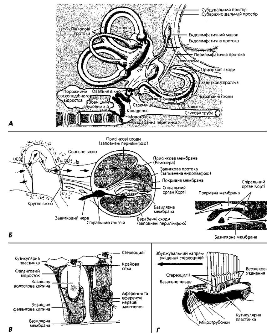
Fig. 4.134. Histophysiology of sound perception: A - pathway of sound waves in the outer, middle, and inner ear, with arrows indicating the direction of perilymph displacement; B - diagram of hair cell excitation resulting from vibrations of the vestibular, basilar, and tectorial membranes, with arrows showing the direction of sound wave propagation; C - interrelationships between outer hair cells and phalangeal cells of the spiral organ; D - structure of the apical portion of a hair cell
Aminoglycoside Antibiotics (such as streptomycin and gentamicin) block the Ion Channels of the tip links and can induce degeneration of hair cells, leading to deafness. Hearing loss can also result from Mutations in myosins that form part of the stereocilia and the "adaptation motor".
The outer hair cells are cylindrical in shape with rounded bases and are arranged in 3 to 5 rows. Humans possess 12,000 to 20,000 of these cells. Like the internal hair cells, they bear a cuticular plate with stereocilia on their apical surface, which form a V-shaped brush of several rows (Fig. 4.134). It is estimated that 90-95% of spiral ganglion neurons provide innervation to the internal hair cells, whereas only 5-10% innervate the outer hair cells; thus, a single afferent fiber terminates on multiple outer hair cells. Efferent fibers running within the auditory nerve predominantly innervate the outer hair cells. The total number of afferent and efferent nerve fibers comprising the auditory nerve is approximately 28,000.
Suspended freely above the spiral organ is the tectorial membrane. This is a gelatinous, ribbon-like spiral structure that continues from the epithelium of the vestibular lip of the limbus. It extends along the spiral organ, positioned directly above the apices of its hair cells. The tips of the stereocilia of the outer hair cells are embedded in the tectorial membrane, whereas the stereocilia of the internal hair cells do not reach it. The tectorial membrane is composed of fine, radially oriented collagen fibers embedded in a transparent amorphous matrix rich in glycosaminoglycans and alpha-tectorin. Mutations in the Gene encoding this protein can lead to hereditary deafness.
Histophysiology of the organ of hearing (Fig. 4.134). Sound vibrations strike the tympanic membrane and are transmitted via the auditory ossicle chain to the base of the stapes. Acting like a piston in the oval window, the footplate of the stapes transmits these vibrations to the perilymph of the scala vestibuli in the cochlea, generating a series of so-called traveling waves. Through the helicotrema at the cochlear apex, these waves pass into the perilymph of the scala tympani. The dissipation of vibrations occurs via the round window, which bulges into the tympanic cavity as the stapes footplate presses inward into the oval window. As a wave propagates along the cochlea, its amplitude reaches a maximum at a specific point and then rapidly decays. The distance from the stapes footplate to the point along the cochlea where the wave reaches its peak amplitude is inversely proportional to the frequency of the sound waves.
The bony walls of the vestibular scala are rigid and practically unaffected by perilymph vibrations. In contrast, the basilar membrane is readily displaced by traveling waves, with the point of maximum displacement determined by the wave frequency. As previously noted, the stereocilia of the outer hair cells are in contact with the tectorial membrane. As traveling waves pass through, both membranes are displaced, and their relative movement causes the stereocilia to bend. Although the hairs of the inner hair cells do not reach the tectorial membrane, they are likewise deflected by the movement of endolymph within the space between the tectorial membrane and the apices of the hair cells.
Deformation of the stereocilia in hair cells alters the permeability of their cation channels. Because these cells are bathed in endolymph rich in potassium ions, $ ext{K}^+$ ions enter the hair cells and trigger membrane depolarization. This influx of potassium ions prompts the release of a neurotransmitter, which in turn depolarizes the membranes of afferent neurons innervating the hair cells. Glutamate is believed to function as this neurotransmitter.
Inner hair cells are the primary sensory cells of the spiral organ responsible for generating action potentials in the afferent nerve fibers of the Vestibulocochlear nerve. The outer hair cells are innervated predominantly by cholinergic nerve fibers originating from the superior olivary nuclei. Upon hyperpolarization induced by acetylcholine, these cells elongate, whereas depolarization causes them to shorten. Thus, The primary function of outer hair cells is to amplify and sharpen the tuning peaks of basilar membrane vibration, although the finer mechanisms of this process remain incompletely understood.
Due to the varying lengths of the auditory strings, different regions of the spiral organ respond via Resonance to sound vibrations of different frequencies. High-frequency waves stimulate longer auditory strings in the basal turn of the cochlea, whereas low-frequency waves stimulate strings near the cochlear apex, thereby exciting the hair cells of the corresponding Location. This spatial segregation of stimuli is subsequently mapped and processed in the auditory cortex of the cerebrum.
The vestibular division of the membranous labyrinth comprises the utricle, the saccule, and three semicircular ducts (Figs. 4.131; 4.134, 4.135, 4.136). The walls of these structures are lined by a simple squamous epithelium resting on a basement membrane, beneath which lies a layer of dense, finely fibrous connective tissue. In the regions of the ampullary crests of the semicircular ducts and the maculae of the utricle and saccule, the connective tissue stroma thickens to form elevations, and the epithelium becomes columnar. The wall of the membranous labyrinth is anchored to the bony labyrinth by connective tissue strands, and in one area it directly fuses with the wall of the bony canal.

Fig. 4.135. Maculae of the utricle and saccule: A - localization of sensory areas of the vestibular apparatus in the membranous labyrinth of the inner ear; B - structural diagram of the utricular macula; C - structural diagram of a vestibular hair cell; D - mechanism of stimulation of hair cells in the maculae of the utricle and saccule; E - scanning electron micrograph of human otoliths; F - scanning electron micrograph of the stereocilia of a hair cell in the saccular macula following removal of the otolithic membrane, × 6000
The maculae of the utricle and saccule consist of an epithelium resting on a basement membrane, comprising hair cells and supporting cells. The apical surfaces of the hair cells face the labyrinthine lumen, while their bases contact nerve endings without reaching the basement membrane. The human saccular macula contains approximately 18,000 hair cells, whereas the utricular macula contains 33,000. Hair cells are classified into two types. Type I cells are pear-shaped, featuring a broad, rounded base embraced by nerve endings that form a chalice-like calyx. Type II cells are columnar in shape with punctate nerve endings at their base. Their apical surfaces bear a cuticular plate from which 30 to 150 stereocilia and a single kinocilium project. Stereocilia are tall, specialized microvilli structurally similar to those of the spiral organ hair cells, whereas the kinocilium is a modified cilium containing nine peripheral microtubule doublets and one central doublet. The kinocilium is the longest projection, terminating in a bulbous expansion (Fig. 4.135).
Supporting cells rest directly on the basement membrane, are characterized by dark oval nuclei, and contain abundant mitochondria. Their apical surfaces feature numerous microvilli. The surfaces of the utricular and saccular maculae are covered by a gelatinous otolithic membrane containing otoliths, or statoconia, composed of calcium carbonate crystals.
The utricular macula functions as a receptor for linear acceleration and gravity, whereas the saccular macula detects gravity and vibration. During relevant head and body movements, the otolithic membrane—acting analogously to a flat stone—tends to slide relative to the macula, deflecting the hairs of the sensory cells and thereby generating nerve impulses.
The ampullary crests appear as transverse folds within the ampullae of the semicircular ducts (Figs. 4.131, 4.135, 4.136). They are formed by hair cells and supporting epithelial cells, whose structure, classification, and innervation closely resemble those of the maculae described above. The apical regions of these cells are covered by a non-cavitated, bell-shaped gelatinous cupula approximately 1 mm in height. The ampullary crests serve as receptors for angular acceleration. When angular acceleration occurs in the plane of a given semicircular canal, endolymph inertia causes it to shift in the direction opposite to the rotation. This fluid movement deforms the cupula, bending the embedded hair bundles. Once a constant rotational velocity is reached, the endolymph moves at the same speed as the bony walls of the semicircular canals, allowing the cupula to return to its resting position. Upon deceleration, the cupula is once again deformed by the endolymph, which due to inertia continues moving faster than the rest of the body. Deflection of the stereocilia toward the kinocilium results in excitation, whereas deflection in the opposite direction leads to inhibition of the hair cells (Fig. 4.136, B).
Development of the inner ear. The membranous labyrinth develops from an ectodermal thickening lateral to the Hindbrain anlage known as the otic placode. This placode invaginates and subsequently detaches from the surface ectoderm to sink into the mesenchyme, forming the otic vesicle (otocyst). The vesicle is lined by a pseudostratified epithelium that secretes the endolymph filling its lumen. Simultaneously, the otic vesicle comes into contact with the embryonic statoacoustic ganglion, which subsequently divides into the vestibular and cochlear ganglia. As development proceeds, the vesicle changes shape, dividing into two main regions: the first gives rise to the utricle and three semicircular ducts, while the second forms the saccule and cochlear duct. Where the vestibular ganglion contacts the otic vesicle, the vesicle wall thickens to form a sensory placode, which later subdivides into upper and lower parts. The upper part gives rise to the utricular macula and ampullary crests, whereas the lower part gives rise to the saccular macula and the spiral organ. Perilymphatic spaces form through the resorption of surrounding mesenchyme, followed by ossification and the final shaping of the bony labyrinth.

Fig. 4.136. Histophysiology of the vestibular apparatus: A - structural diagram of an ampullary crest; B - mechanism of Excitation and Inhibition of vestibular hair cells; C - mechanism of perception of head and body angular acceleration by the receptor cells of the ampullary crests
Terms for Memorization
1. External ear. 2. Middle ear. 3. Inner ear. 4. Auricle (pinna). 5. External acoustic meatus. 6. Tympanic membrane. 7. Ceruminous glands. 8. Tympanic cavity. 9. Oval window. 10. Round window. 11. Auditory ossicles. 12. Malleus. 13. Incus. 14. Stapes. 15. Secondary tympanic membrane. 16. Pharyngotympanic (auditory) tube. 17. Tubal tonsil. 18. Bony labyrinth. 19. Membranous labyrinth. 20. Perilymph. 21. Endolymph. 22. Vestibule. 23. Semicircular canals: anterior (superior), posterior, and lateral. 24. Cochlea. 25. Ampullae: anterior, posterior, lateral. 26. Spiral osseous lamina. 27. Spiral limbus. 28. Vestibular lip of the limbus. 29. Tympanic lip of the limbus. 30. Spiral sulcus. 31. Spiral ligament. 32. Utricle. 33. Saccule. 34. Semicircular ducts: anterior (superior), posterior, lateral. 35. Cochlear duct (scala media). 36. Ampullary crests. 37. Macula of the utricle. 38. Macula of the saccule. 39. Spiral organ (organ of Corti). 40. Vestibular apparatus. 41. Vestibular membrane (Reissner's membrane). 42. Stria vascularis. 43. Basilar membrane (lamina). 44. Scala vestibuli. 45. Scala tympani. 46. Outer cells of the spiral organ. 47. Inner cells of the spiral organ. 48. Hair cells. 49. Pillar cells (rods). 50. Pharyngeal (phalangeal) cells. 51. Supporting cells. 52. Border cells. 53. Reticular lamina (membrane). 54. Inner tunnel. 55. Tectorial membrane. 56. Otolithic membrane. 57. Gelatinous cupula.
Last update: 09/08/2026
Editorial and Educational Adaptation: This material has been compiled based on the primary/original source text. The project team performed an editorial review, corrected technical inaccuracies, structured sections, and adapted the content for an educational format.
What was processed:
- elimination of formatting defects (OCR errors, structural breaks, corrupted characters);
- editorial organization of content;
- standardization of terminology in accordance with academic sources;
- verification of factual statements against the original source text.
All mentions of the author, publication year, and origin of the primary text have been preserved in accordance with the source.