Human Histology - O.D. Lutsyk 2003
Systemic Histology
Female Reproductive System
The FEMALE Reproductive System, much like the male system, performs two primary Functions: generative, which involves The production of germ Cells, and endocrine, which entails the synthesis of Sex Hormones. Both of these functions are interconnected and create the necessary conditions for reproduction. Additionally, the female reproductive system fulfills two more functions: it ensures the INTRAUTERINE DEVELOPMENT OF the fetus and the secretion of milk. The Organs of the female reproductive system (Fig. 4.104) include the Ovaries (female sex glands or female Gonads), the oviducts (Fallopian tubes), the Uterus, the Vagina, the external genitalia, and the Mammary Glands.
The Ovary (ovarium) (Figs. 4.104-4.107) is the female gonad responsible for producing Female Germ Cells (oocytes) and sex hormones (estrogens and progesterone). It is a paired organ located near the lateral wall of the lesser pelvis. Oval in shape, it typically measures 4x2x1.5 cm, with the right ovary being slightly larger than the left. The weight of an adult woman's ovary is 5-7 g, compared to up to 0.5 g in a newborn girl and 2-3 g in elderly women. In a newborn, the ovary is spindle-shaped with a smooth surface; in an adult, it becomes rounded with an uneven, bumpy surface. The ovary is attached to the broad ligament of the uterus by a peritoneal fold (mesovarium).
Externally, the ovary is covered by a surface epithelium derived from the coelomic epithelium. This consists of a single layer of cuboidal cells with a height of approximately 18 µm. Beneath the epithelium lies the tunica albuginea, a 100 µm-thick capsule composed of Collagen and elastic fibers along with a small number of smooth myocytes. The tunica albuginea is poorly developed in newborn girls and typically forms during the third to fourth year of life. The Internal Structure of the ovary is divided into a medulla and a cortex.
The medulla is formed by a Connective Tissue stroma containing numerous elastic fibers, many large Blood Vessels, nerve fibers, and nerve endings, as well as the so-called ovarian rete (rete ovarii), which is present in 93% of women.
The cortex surrounds the medulla in a horseshoe-like shape and is absent only at the hilum of the ovary. It consists of a stroma and a parenchyma. The stroma is formed by connective tissue containing collagen fibers and a small amount of elastic tissue. This connective tissue is rich in smooth-Muscle-like fibroblasts known as interstitial cells, which are capable of hormone production. The parenchyma comprises primordial, primary, secondary (vesicular), and mature follicles (tertiary follicles or Graafian follicles), corpora lutea, corpora albicantia, atretic follicles, and atretic bodies.
A primordial follicle consists of a primary oocyte (oocyte of the first (I) order) with a diameter of 15-25 µm, arrested in the diplotene stage of meiotic prophase, surrounded by a single layer of flat follicular cells up to 9 µm in diameter. Primordial follicles are roughly spherical, measure about 50 µm in diameter, and are located in the peripheral region of the cortex. Their formation begins during the third month of human Embryogenesis.
Primary follicles are larger than primordial ones; their follicular epithelium becomes cuboidal and arranges itself in one or several layers. The volume of the oocyte also increases. It is in these follicles that the zona pellucida first becomes discernible (see below).
Secondary (preantral) follicles are surrounded by a multi-layered follicular epithelium, within which a fluid-filled cavity—the antrum—begins to form. The fluid is produced by the follicular cells and contains Female Sex Hormones, namely estrogens. These follicles start to develop during Puberty.
Class="center">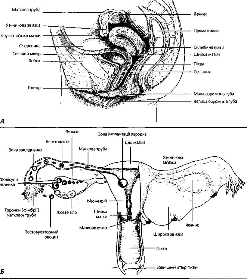
Fig. 4.104. General structural layout of the female reproductive system: A – sagittal projection; B – frontal projection illustrating events of the first week of embryogenesis
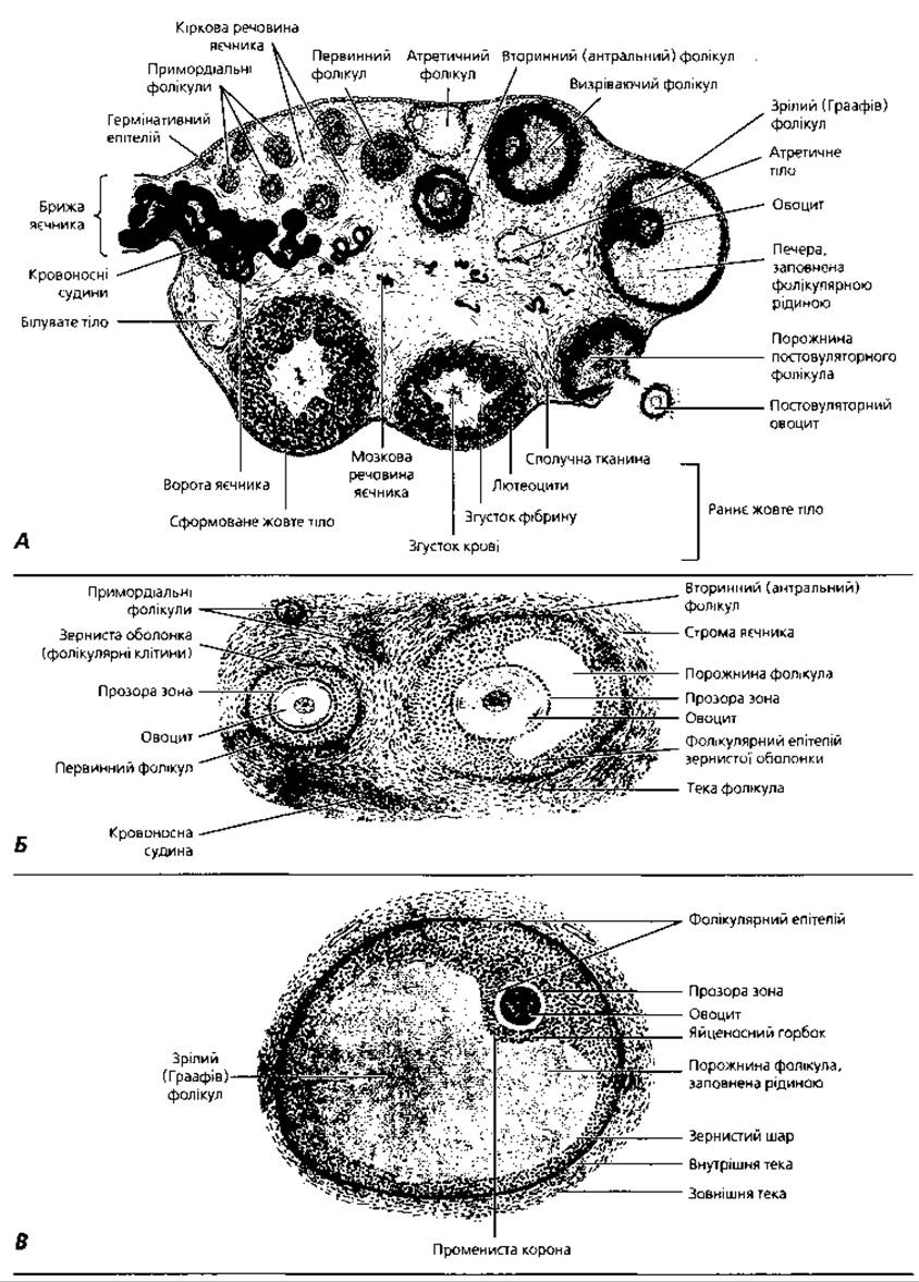
Fig. 4.105. The ovary: A – schematic representation; B, C – details of the microstructure of follicles at various stages of maturity
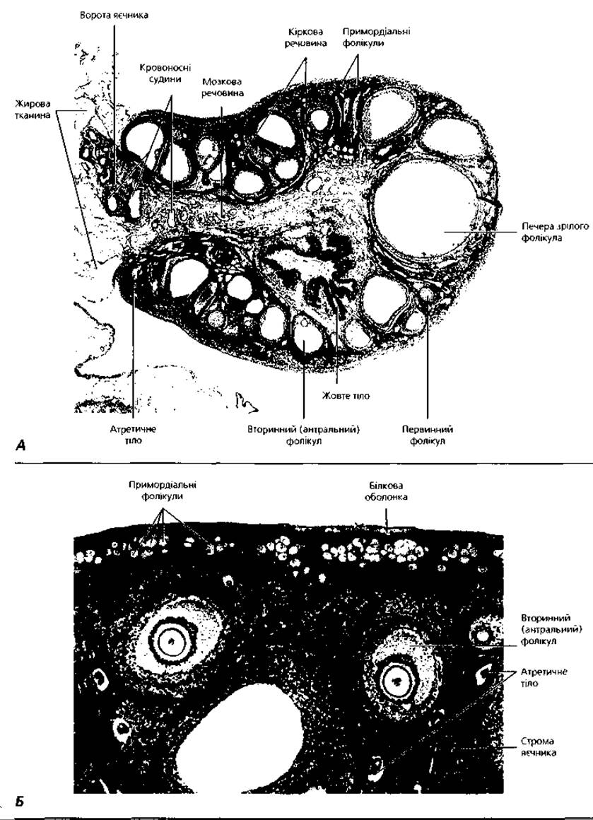
Fig. 4.106. Light Cell/15.html">Microscopy of the ovary: A – whole mount preparation, x 7; B – cortical substance, x 90
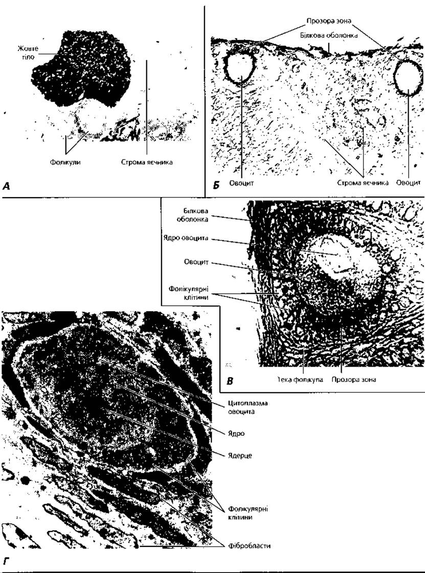
Fig. 4.107. Details of ovarian micromorphology: A – corpus luteum, selective histochemical reaction with peanut agglutinin, x60; B – demonstration of the zona pellucida of primary follicles using wheat germ agglutinin reaction, x100; C – primary follicle, Cytology/cytology/16.html">Early stages of zona pellucida biogenesis revealed by concanavalin A reaction, x500; D – transmission Electron microscopy of a primary follicle, x4000
A tertiary, antral, or mature (Graafian) follicle is characterized by a fully formed, large cavity that occupies the majority of its volume. The process of transforming primordial follicles into primary, secondary, and mature ones is known as folliculogenesis (follicular growth). This growth is driven by pituitary gonadotropic hormones—follicle-stimulating hormone (FSH) and a small amount of luteinizing hormone (LH)—although the Initial Stages of follicular growth are gonadotropin-independent. Several follicles grow simultaneously, but under normal conditions in humans and primates, many large antral follicles degenerate prior to ovulation. Out of twenty large antral follicles, typically only two develop into mature follicles (Graafian vesicles), of which one usually degenerates upon reaching a diameter of 1 cm while the other undergoes ovulation. Follicular growth is accompanied by the growth of the oocyte. In all mammals, the oocyte reaches its maximum size (130-140 µm) within follicles whose walls consist of multiple layers of follicular cells, even before the follicular cavity has formed. Evidence suggests that oocytes produce specific substances that stimulate the proliferation of follicular cells and follicular growth. In humans, the number of follicular cells is about 50 in primary follicles, rising to roughly 50 million just before ovulation.
During growth, the oocyte gradually becomes enveloped by protective structures—the zona pellucida and the corona radiata—while the follicular epithelium forms the stratum granulosum (granulosa layer) of the follicle, and an internal cavity, the antrum, develops. Along the inner wall of the follicle, the granular layer forms a protrusion known as the cumulus oophorus, which houses the oocyte surrounded by multiple layers of follicular cells. The surrounding connective tissue organizes into the follicular theca, comprising a basement membrane along with internal and external layers. The theca interna contains blood vessels, collagen fibers, abundant nerve fibers, and specialized cells called theca cells (thecocytes). The theca externa is composed of Dense connective tissue. The growth phase culminates in The formation of a mature ovarian follicle with the aforementioned structure. It migrates toward The surface of the ovary, its wall thins, and due to increasing follicular fluid pressure, it ruptures—a process known as ovulation.
Ovulation is the rupture of the mature follicular wall and the ovarian surface, resulting in the release of the oocyte. Prior to ovulation, the oocyte, together with the Cells of the corona radiata, detaches from the cumulus oophorus and floats freely within the follicular fluid. In the region where the follicle bulges against the ovarian surface, the theca, tunica albuginea, and surface epithelium undergo marked thinning and softening under METABOLISM/18.html">The Influence of Enzymes produced by follicular cells and migrating leukocytes. This localized area is termed the stigma. Approximately 30 minutes before ovulation, Blood Circulation in the stigma region ceases, leading to local tissue necrosis. The stigma protrudes above the ovarian surface as a translucent bulge. Following its rupture, the oocyte—surrounded by cumulus cells and a cloud of viscous follicular fluid—enters the lumen of the oviduct. Typically, the oocyte is captured immediately by the fallopian tube because the fimbriae of its ampullary region embrace the ovary during ovulation. At this juncture, the oocyte is arrested in metaphase of the second meiotic division. Ovulation is triggered by pituitary luteinizing hormone. The granulosa layer of growing follicles produces estrogens (estradiol, estrone, and estriol). The thecocytes synthesize small amounts of estrogens and testosterone (an androgen). Within the follicular cells, testosterone is converted into estradiol (an estrogen) via the enzyme aromatase (estrogen synthetase). Aromatase synthesis in the ovary is induced by follitropin. Estrogens drive The Development of female secondary sex characteristics (widening of the pelvis, growth of the mammary glands, uterus, and oviducts, female-pattern Hair distribution, and the onset of menstruation), as well as Changes in the reproductive tract During the first half of the Menstrual cycle (the regeneration and proliferation phases).
Following ovulation, the remnants of the mature follicle (the granulosa and theca) give rise to a temporary endocrine structure known as the corpus luteum. The corpus luteum undergoes several developmental stages. Initially, at the moment of follicular rupture, Hemorrhage occurs from damaged Vessels of the theca, and blood accumulates in the center of the future corpus luteum. This blood clot is rapidly organized, eventually forming a connective tissue scar. Cells of the follicular granulosa layer begin to proliferate and are invaded by a dense network of blood capillaries; this phase is known as proliferation and vascularization. Subsequently, the granulosa cells accumulate a yellow pigment, lutein, and transform into glandular luteal cells, or granulosa lutein cells. Another source of luteal cells is the theca interna, which gives rise to theca lutein cells. This second phase is termed the stage of glandular metamorphosis. During the third phase—full maturity (florescence)—the cells of the corpus luteum begin to secrete the hormone progesterone. Driven by progesterone, the secretory phase of the menstrual cycle unfolds; this hormone prepares the uterus for implantation and is essential for maintaining a normal Pregnancy through the first three to four months.
If pregnancy does not occur, the florid stage of the corpus luteum lasts for 12-14 days; such a structure reaches 1.5-2 cm in diameter and is referred to as a cyclic or menstrual corpus luteum. Should pregnancy ensue, the florid stage extends to 11-12 weeks, with the corpus luteum expanding up to 5 cm in diameter, and is designated as the corpus luteum of pregnancy. The final stage of the corpus luteum's lifecycle is regression (involuting stage). During this phase, the luteal cells degenerate, and the connective tissue of the central scar proliferates. This results in the formation of a corpus albicans, which persists in the ovary for up to five years before gradually being resorbed and replaced by a fibrous scar.
Atretic follicles and atretic bodies arise because not all follicles that initiate growth reach maturity. A proportion of these undergo reduction and regression—a process known as atresia. During atresia, the oocyte degenerates first, while its zona pellucida—shrunken, thickened, and hyalinized—persists for a longer period in the center of the atretic body. This characteristic distinguishes it from the corpus luteum, which features a central connective tissue scar. Following the demise of the oocyte in an atretic follicle, cells of the theca interna proliferate and synthesize estrogens. Consequently, follicular atresia is necessary not only to eliminate surplus germ cells but also to supply the Organism with estrogens. The atresia process is mediated by gonadocrinin, a protein hormone (analogous to testicular inhibin) produced concurrently with estrogens by the granulosa layer of growing and mature follicles. Another protein hormone produced by the ovary is relaxin, which promotes the relaxation of the Pubic Symphysis and the dilation of the cervical canal during childbirth. Furthermore, large follicles secrete inhibin, which suppresses follicle-stimulating hormone (FSH) production, as well as Prostaglandins.
Hilar cells, located in the ovarian medulla near the hilum and morphologically similar to testicular Leydig cells, produce androgens. Their hyperplasia can lead to masculinization.
Characteristics of oogenesis. Oogenesis—the developmental process of female germ cells—comprises three periods: multiplication, growth, and maturation (Fig. 4.108).
The multiplication period takes place in the fetal ovary from the second to the fifth month of embryogenesis and involves the mitotic proliferation of oogonia. Oogonia originate from primordial germ cells (gonocytes) of extragonadal origin that migrate into the gonadal ridge, interact with follicular epithelial cells, and differentiate into oogonia. Unlike gonocytes, oogonia exhibit high mitotic activity. As a result of this proliferation, the number of oogonia in a single ovary reaches 1 to 5 million. Concurrently with multiplication, massive cell death of oogonia occurs via apoptosis, causing their numbers to decrease significantly by birth. Following their final mitotic division, oogonia differentiate into preleptotene oocytes and enter the next phase of oogenesis—the growth period.
The growth period in human oogenesis begins in the third month of embryonic development and consists in the Formation of primary oocytes (oocytes I), in whose nuclei a complex reorganization occurs as preparation for the reduction of chromosome number. At this time, the size of the oocyte itself increases, it becomes surrounded by follicular cells, and follicles are formed. This process is called the minor growth phase. Primary oocytes enter prophase of Meiosis and, just as in primary spermatocytes (see section "Male Reproductive System"), pass through the stages of leptotene, zygotene (second to seventh months of embryogenesis), pachytene, and diplotene (sixth to ninth months). However, unlike male meiosis, in oogenesis prophase is not followed by metaphase; instead, meiosis is blocked, and the oocytes enter dictyotene for a long period—a unique phase characteristic only of oogenesis.
The arrest of the primary oocyte in the diplotene of prophase of the 1st meiotic division is mediated by the so-called oocyte maturation inhibitor (OMI). In dictyotene, the Chromosomes of the primary oocyte decondense and become invisible until the end of the growth period. In humans and other mammals, oocytes enter dictyotene during the prenatal period or immediately after birth and remain in this state for decades (from 10-13 to 45-50 years). In humans, different generations of oocytes undergo prophase at different stages of prenatal ontogeny. Thus, in the third month of embryonic development, about 1% of oocytes reach the dictyotene stage, by the fourth month their number is already 20%, and by the eighth month, it reaches 90%.
With the onset of sexual maturity, oocytes enter the process of further growth (the so-called major growth phase). During this, the size of the oocyte increases, yolk accumulates in its Cytoplasm, and the oocyte becomes surrounded by the zona pellucida and corona radiata. The zona pellucida exhibits oxyphilic properties and is clearly visible under a Light Microscope. It is formed by a complex network of Glycoproteins and Proteoglycans. Microvilli of follicular cells penetrate the zona pellucida and form Gap Junctions with the oocyte Plasmalemma. Outside the zona pellucida are the follicular cells of the corona radiata, which merge without a sharp boundary into the cells of the cumulus oophorus (Fig. 4.105, B).

Fig. 4.108. Summary of folliculogenesis processes: FSH - follicle-stimulating hormone; LH - luteinizing hormone; hCG - human chorionic gonadotropin
The maturation phase of oogenesis begins in mature follicles immediately before ovulation, when oocytes resume meiosis, starting from the metaphase of the first maturation division. Meiotic resumption is triggered by luteinizing hormone (LH). After the First Division, two cells are formed: one large—the secondary oocyte (oocyte II), which retains almost all of the cytoplasm, and one small—the first polar body (polocyte I). Each of these cells receives 23 dyads from the chromosome Complement of the primary oocyte. The second maturation division begins immediately after the first, but is arrested at the metaphase stage and is completed only after sperm penetration through the plasmalemma of the secondary oocyte. As a result of the second meiotic division, a small cell—the second polar body (polocyte II)—and a large cell—the mature egg (ovum)—are formed; both cells receive 23 monads. The polar, or reduction, bodies contain about 1% of the egg's cytoplasm. At the metaphase stage of the second maturation division, the oocyte is released from the ovary due to ovulation, and maturation is completed in the fallopian tubes after Fertilization.
The uterine (Fallopian) tube (tuba uterina, salpinx) (Figs. 4.104, 4.109, 4.110) is a paired tubular organ that starts from the Fundus of the uterus, runs within the broad ligament to the lateral wall of the lesser pelvis, and ends near the ovaries. The length of the Uterine tube is 10-12 cm; the lumen diameter is 6-10 mm in the ampullary part, 3 mm at the level of the isthmus, and 0.5-1 mm in the uterine part. The wall of the uterine tube consists of three layers: mucosa, muscularis, and serosa. The mucosa consists of the epithelium and lamina propria. The mucosal epithelium is simple columnar, containing ciliated and secretory cells. The lamina propria is composed of loose connective tissue. The mucosa of the uterine tube forms numerous tall folds, which in the ampullary part are called fimbriae.
The muscularis consists of two layers of smooth muscle cells—an inner circular and an outer longitudinal layer. In the uterine part of the tube, the inner layer becomes longitudinal, and the outer layer becomes circular. The serosa consists of a lamina propria made of loose connective tissue and the overlying mesothelium, similar to all other serous membranes.
The Functions of the uterine tubes include transporting Gametes to the site of fertilization and the embryo to the uterus, providing conditions for sperm capacitation, and creating an environment favorable for fertilization. The first 4-5 days of embryonic life take place in the uterine tube. Transport of the embryo into the uterus occurs due to the peristalsis of the muscularis, as well as the beating of epithelial cell cilia.

Fig. 4.109. Uterine (Fallopian) tube: A - semi-schematic representation of a cross-section of the ampullary part, x12; B - light microscopy of the ampullary part of the uterine tube of a 23-year-old woman, x30; C - region of the isthmus, x30; D - uterine part of the tube, x30

Fig. 4.110. Uterine tube; A - fimbriae of the mucosa, x280; B - epithelial lining of the mucosa, x1,200; C - scanning electron microscopy of the surface of the epithelial lining of the uterine tube, x7,000

Fig. 4.111. Uterus: A - semi-schematic representation, x20; B - dynamics of morphological changes in the endometrium during the menstrual cycle, x30
The uterus (Figs. 4.104, 4.111, 4.113, A) is a flattened, pear-shaped organ whose function is to gestate the fetus. The uterus is located in the geometric center of the lesser pelvis, between the Urinary Bladder and the rectum. In the non-pregnant state, the mass of the uterus is 70-100 g, and its dimensions are 8x5x4 cm. The wall of the organ consists of three layers: the endometrium (mucosa), myometrium (muscularis), and perimetrium (serosa).
The endometrium in girls under 10 years of age is about 0.15 mm thick, and in sexually mature women, it is up to 2-3 mm. The endometrium does not form folds, and the uterine lumen appears as a slit. The endometrium consists of two layers—the epithelium and the lamina propria. The endometrial epithelium is simple tall columnar (cell height 20-30 µm), consisting of ciliated and secretory cells. The epithelial layer forms tubular invaginations into the lamina propria, forming uterine glands. The lamina propria of the mucosa is composed of loose connective tissue.
The myometrium in young girls contains few muscle cells. In sexually mature women, it is well-developed, formed by smooth myocytes with numerous processes. The smooth myocytes of the myometrium form three layers: the sub-mucosal, with an oblique-longitudinal orientation of myocytes; the vascular, with a predominantly circular orientation of muscle cells; and the supra-vascular, with an oblique-longitudinal orientation of myocytes. The perimetrium is formed of loose connective tissue covered by mesothelium.
The mucosa of the cervix has several distinctive features. Similar to the vagina, its vaginal surface is covered with stratified squamous epithelium. The cervical canal is lined with simple columnar epithelium that produces mucus. The mucosa of the cervical canal forms folds and two longitudinal ridges. In addition, there are numerous branched glands that also produce mucus. The muscularis of the cervix is formed by a well-developed circular layer of smooth muscle cells, which forms the so-called uterine sphincter.
The vagina (Figs. 4.104, 4.113, B, C) is a fibromuscular tube 7-9 cm long and 2-3 cm in diameter, located in the lesser pelvis between the Urethra and the rectum. Three layers are distinguished in the vaginal wall: mucosa, muscularis, and adventitia. The mucosa has two layers—the epithelium and the lamina propria. The vaginal epithelium is stratified squamous non-keratinized, in which basal, intermediate, and superficial layers are distinguished. The latter is also called the functional layer because it undergoes rhythmic changes during the menstrual cycle. Cells of the superficial layers of the epithelium contain keratohyalin granules rich in Glycogen. The breakdown of glycogen by microbes leads to the formation of lactic acid, so the vaginal fluid has an acidic pH, which prevents infection. There are no glands in the vaginal wall. The lamina propria of the mucosa forms papillae that invaginate into the epithelium and is infiltrated with lymphocytes. Elastic fibers of the lamina propria form superficial and deep networks.
The muscularis of the vagina is formed by longitudinal bundles of smooth myocytes, with a small number of circularly arranged muscle elements between them. The adventitia is composed of loose connective tissue that connects the vagina to adjacent organs.
The female external genitalia include the Vestibule of the vagina, the Labia minora, the Labia Majora, and the Clitoris. The vestibule of the vagina is lined with stratified squamous epithelium. Two greater vestibular glands (Bartholin's glands), which are tubuloalveolar in shape and produce mucus, open here.
The labia minora are folds of mucosa covered by Stratified squamous keratinized pigmented epithelium. Their core consists of loose connective tissue rich in elastic fibers and blood vessels, containing numerous Sebaceous Glands. The labia majora are Skin folds with a significant amount of adipose tissue, sebaceous, and Sweat Glands. The outer surface of the labia majora is covered with hair.
The clitoris corresponds in Development and Structure to the dorsal part of the Penis. It consists of two corpora cavernosa and a glans covered by stratified squamous keratinized epithelium.
The mammary glands (mammae) are, by origin, modified sweat glands. The regulation of Mammary Gland Function is mainly carried out by two hormones: prolactin (an adenohypophyseal hormone), which stimulates The Biosynthesis of milk components by lactocytes, and oxytocin (a hormone of the paraventricular nuclei of the Hypothalamus), which promotes milk ejection. In turn, prolactin secretion is activated by hypothalamic prolactoliberin and inhibited by prolactostatin. A detailed Description of the STRUCTURE OF THE mammary glands is provided in the section "Skin and its derivatives".
Ovarian-Menstrual Cycle. The cyclic changes occurring in the internal (functional) layer of the endometrium, manifested by monthly uterine bleedings known as menstruation, are termed the menstrual cycle (Figs. 4.106, 4.107). The menstrual cycle encompasses not only the functional layer of the endometrium but the entire female organism as well, depending on cyclic changes within the ovary and its secretion of estrogens and progesterone (the ovarian cycle). Consequently, these monthly cyclic changes in the female body are referred to as the ovarian-menstrual cycle. In animals, the analog of menstrual cycles is known as estrous cycles.
The duration of the menstrual cycle is calculated from the first day of the previous menstruation to the first day of the next. In most women, the cycle lasts 28 days and comprises several distinct phases.
During the desquamation or menstrual phase (days 1–4 of the cycle), the functional layer of the endometrium is shed. The deep portion of the endometrium remaining after desquamation is called the basal layer. The Blood vessels of the endometrium have a distinctive structure, including spiral and straight Arteries. The former supply blood to the functional layer of the endometrium, while the latter supply the basal layer. Just before menstruation, as a result of declining progesterone levels and the absence of estrogen influence, the spiral arteries undergo spasm, reducing blood flow to the Superficial layer of the endometrium (leading to its ischemia) and causing necrotic changes. The necrotic portion of the endometrium is shed, and the vessels bleed until the end of the fourth day. Blood loss during menstruation ranges from 50 to 200 ml. Menstrual blood does not clot and contains an Abundance of lymphocytes.
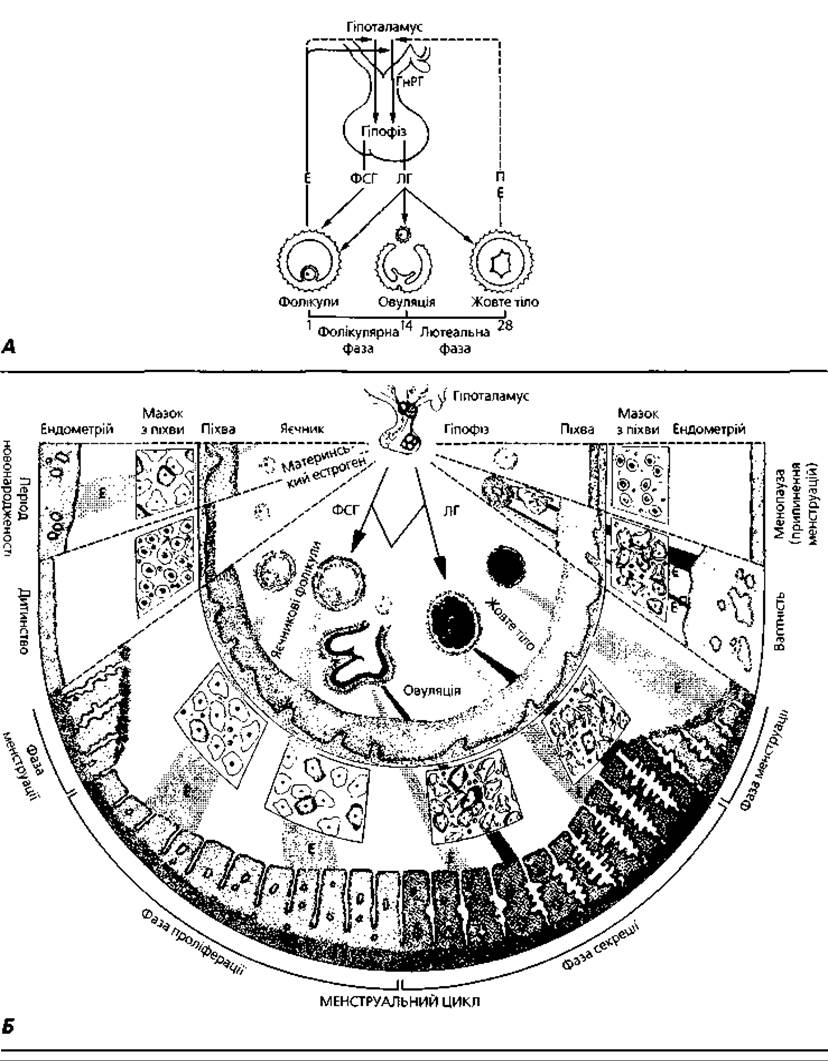
Fig. 4.112. Hormonal Regulation of the female reproductive system: A - diagram of the interaction among the hypothalamus, pituitary, and ovaries, with solid arrows indicating stimulation and dashed arrows indicating negative feedback; B - ovarian-menstrual cycle and functional changes in FEMALE REPRODUCTIVE ORGANS under the influence of Hypothalamic and pituitary hormones: GnRH - gonadotropin-releasing hormone; LH - luteinizing hormone; FSH - follicle-stimulating hormone; E - estrogen; P - progesterone
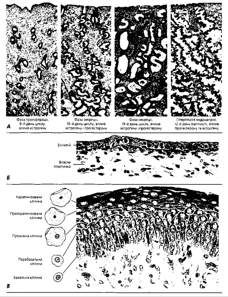
Fig. 4.113. Hormonal influence on the Morphology of the endometrium and vaginal mucosa: A - light microscopy of the endometrium in various Phases of the menstrual cycle and during pregnancy; B - vaginal mucosa in the absence of estrogen influence; C - vaginal mucosa stimulated by estrogen
The proliferative phase (follicular, postmenstrual) spans days 5–14 of the cycle. It begins with follicular growth and the production of estrogens by the follicles. Estrogens drive the regeneration of the functional layer of the endometrium. Renewal of the mucosal epithelial lining occurs through the proliferation of epithelial cells from the bases of the uterine glands that survive the shedding of the functional layer. The thickness of the endometrium increases two- to threefold in this phase, reaching 2–3 mm. Due to enhanced proliferation, epithelial cells often overlap one another. Secretory cells produce a small amount of watery mucus, interspersed with small groups of ciliated cells. The uterine glands are narrow and straight. The stroma contains a small amount of ground substance, and leukocytes are rarely encountered. This phase, like the preceding one, is maintained by the action of estrogens. Ovulation occurs in the ovary at the end of this phase.
The secretory phase (luteal, premenstrual) spans days 15–28 of the cycle. The endometrium thickens to twice its thickness in the previous phase—not through cell multiplication as in the postmenstrual phase, but as a result of edema, accumulation of secretion within the glands, and an increase in the volume of stromal cells. The uterine glands become tortuous and continue to secrete large amounts of fluid, and their cells accumulate significant amounts of glycogen. During the secretory phase, two zones can be distinguished in the functional layer of the endometrium: a superficial compact zone and a deep spongy zone (where widened glands impart a spongy appearance). The Formation of the superficial and deep zones reflects the process of endometrial preparation for embryo reception, i.e., implantation. The secretory phase is driven by progesterone, which is produced by the corpus luteum formed at the site of the postovulatory follicle under the influence of pituitary lutropin. Progesterone production is also stimulated by prolactin. Progesterone helps stabilize the edematous endometrium and prevents its shedding. If pregnancy does not occur and the corpus luteum degenerates, the drop in progesterone levels leads to the shedding of the functional layer of the endometrium and the onset of the menstrual phase. In the absence of progesterone influence, the growth of ovarian follicles is unblocked, and they begin to produce estrogens. Estrogens then stimulate the regeneration and proliferation of the functional layer of the endometrium, thereby repeating the cycle.
The cyclical functioning of the female reproductive system is driven by the specific secretory patterns of luteinizing hormone (lutropin) by the Pituitary Gland. In the male organism, both follicle-stimulating hormone (follitropin) and luteinizing hormone are produced continuously and at steady levels, whereas in females, lutropin is released periodically when the pituitary discharges an elevated surge of the hormone sufficient for ovulation and corpus luteum development (the so-called ovulatory quota of lutropin). Hypothalamic regulation of this adenohypophyseal function is mediated by two centers. One of these (the lower center) is localized in the tuberal nuclei of the mediobasal hypothalamus. It stimulates the anterior pituitary to maintain continuous tonic secretion of both gonadotropins. Under these conditions, The amount of lutropin produced is sufficient only to support estrogen secretion by the ovaries and testosterone by the Testes. The second (higher, or ovulatory) center is localized in the preoptic area of the mediobasal hypothalamus. It modulates The activity of the lower center, prompting the latter to stimulate the pituitary to release a large quantity (the ovulatory quota) of lutropin into the bloodstream.
In the absence of androgen influence, the higher center retains the capacity to periodically excite the activity of the lower center, a characteristic feature of the female organism. In the male embryo, the ovulatory center becomes masculinized due to the production of Male Sex Hormones. By the end of the Prenatal period of development, the hypothalamic ovulatory center loses The ability to undergo male-type differentiation.
Development of the female reproductive system. The reproductive system in individuals of both sexes initially develops according to a unified plan and in close association with the development of the Urinary System. The gonadal primordia arise in the embryo during the fourth week of development as thickenings of the coelomic epithelium On the surface of the primary Kidney; these thickenings are termed the genital ridges. Parallel to the mesonephric (Wolffian) duct, the paramesonephric (Mullerian) duct separates and develops. The upper PARTS OF THE paramesonephric ducts form the fallopian tubes, while their lower parts form the uterus and vagina. During the development of the female reproductive system, the mesonephric ducts regress, transforming into the vestigial ducts of the epoophoron. In addition to the coelomic epithelium and mesenchyme, a separate source for the development of the gonads consists of the primordial germ cells—gonocytoblasts, which are of extrazonal origin and are first detected in the primitive streak and the region of the allantoic ROOT, subsequently migrating into the endoderm of the yolk sac, the endoderm of the midgut, and the dorsal mesentery; from there, via the coelomic lining and germinal epithelium, they reach the genital ridges, i.e., the gonadal primordia. Within the female gonadal primordium, the gonocytes differentiate into oogonia. Differentiation of the ovaries occurs later than that of the testes and becomes morphologically evident only by the end of the 7th–8th week of embryogenesis.
Terms for Memorization
1. Ovaries. 2. Fallopian tubes. 3. Uterus. 4. Vagina. 5. Mammary glands. 6. Surface epithelium. 7. Tunica albuginea. 8. Ovarian cortex. 9. Ovarian medulla. 10. Interstitial cells. 11. Primordial follicle. 12. Primary follicle. 13. Secondary (vesicular, antral) follicle. 14. Mature (tertiary, Graafian) follicle. 15. Corpus luteum. 16. Corpus albicans. 17. Atretic follicle. 18. Follicular antrum. 19. Granulosa layer of the follicle. 20. Cumulus oophorus. 21. Zona pellucida. 22. Corona radiata. 23. Theca interna. 24. Theca externa. 25. Thecocytes. 26. Ovulation. 27. Estrogens. 28. Granulosa lutein cells. 29. Theca lutein cells. 30. Cyclic (menstrual) corpus luteum. 31. Corpus luteum of pregnancy. 32. Progesterone. 33. Follicular atresia. 34. Gonadokinin. 35. Relaxin. 36. Oogenesis. 37. Gonocytes. 38. Oogonia. 39. Preleptotene oocytes. 40. Dictyotene stage. 41. Primary oocyte. 42. Secondary oocyte. 43. First polar body (polocyte I). 44. Mature ovum. 45. Second polar body (polocyte II). 46. Endometrium. 47. Uterine glands. 48. Functional layer. 49. Basal layer. 50. Myometrium. 51. Perimetrium. 52. Cervix. 53. Cervical glands. 54. Vaginal vestibule. 55. Vestibular glands (Bartholin's glands). 56. Labia minora. 57. Labia majora. 58. Clitoris. 59. Ovarian-menstrual cycle. 60. Desquamation (menstrual) phase. 61. Straight and spiral arteries. 62. Proliferative (follicular, postmenstrual) phase. 63. Ovulatory quota of lutropin. 64. Mesonephric (Wolffian) duct. 65. Paramesonephric (Mullerian) duct.
Last update: 09/08/2026
Editorial and Educational Adaptation: This material has been compiled based on the primary/original source text. The project team performed an editorial review, corrected technical inaccuracies, structured sections, and adapted the content for an educational format.
What was processed:
- elimination of formatting defects (OCR errors, structural breaks, corrupted characters);
- editorial organization of content;
- standardization of terminology in accordance with academic sources;
- verification of factual statements against the original source text.
All mentions of the author, publication year, and origin of the primary text have been preserved in accordance with the source.