Human Histology - O.D. Lutsyk 2003
Systemic Histology
Male Reproductive System
The Male Reproductive System (Fig. 4.92) includes the male Gonads—the Testes, the reproductive tract (epididymides, vasa deferentia, ejaculatory ducts, and Urethra), and accessory Organs (Seminal Vesicles, Prostate Gland, Bulbourethral Glands, and Penis). The primary function of the Male reproductive organs is reproductive (generative), which consists in The production of Male Germ Cells—spermatozoa. The Organs of the male reproductive system contain endocrine cells; therefore, In addition to their reproductive function, they also perform an endocrine function.
The Testis (testicle, testis) is located in the Scrotum. It is a paired oval-shaped organ (Figs. 4.92, 4.93). In a sexually mature male, its weight is 18–25 g, and its dimensions are 4x3x2.5 cm. Spermatozoa are formed and Male Sex Hormones are produced in the testis; it is also the Water/144.html">Origin of the reproductive tract, through which spermatozoa travel to complete their maturation. Externally, the testis is surrounded by a Connective Tissue capsule covered with mesothelium—the tunica albuginea. At the posterior border of the testis, the tunica albuginea thickens to form the Mediastinum testis. Connective tissue septa extend inward from the tunica albuginea, dividing the parenchyma of the organ into lobules. The lobule is the Structural and functional unit of the testis, with each testis containing 250–300 lobules.
Each lobule contains one to four tightly packed, convoluted tubes called seminiferous tubules (Figs. 4.93, A, 4.94, 4.95, A). The length of a seminiferous tubule ranges from 30 to 70 cm, and its diameter is 150–250 μm. The seminiferous tubules begin and end near the mediastinum testis, where they transition into straight seminiferous tubules (tubuli recti). There are 300 to 450 straight tubules in each testis. Within the mediastinum, the straight tubules merge to form the rete testis. From the latter, 10–15 efferent ductules emerge and empty into the duct of the Epididymis.
Class="center">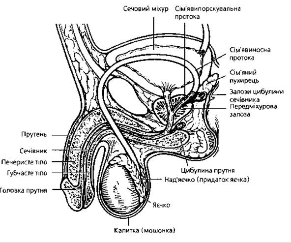
Fig. 4.92. General Organization OF THE male reproductive system
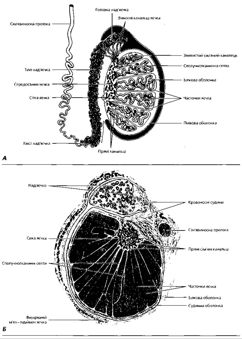
Fig. 4.93. Testis and epididymis: A—diagram of structural organization; B—semi-schematic representation of a whole histological section of the testis of a 2.5-year-old boy, midsagittal section, x 7
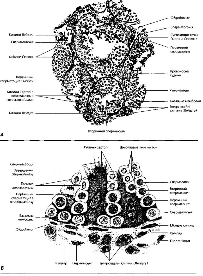
Fig. 4.94. Structure of seminiferous tubules: A—semi-schematic representation of a human testis histological specimen, x150; B—diagram of a wall fragment of a seminiferous tubule with adjacent interstitial endocrine Cells of the testis (Leydig cells)
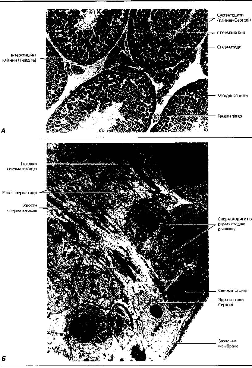
Fig. 4.95. Seminiferous tubules: A—light Cell/15.html">Microscopy, x400; B—transmission Electron microscopy, x1700
The wall of the seminiferous tubule consists of three layers: basal, myoid, and fibrous. The basal layer is formed by a network of Collagen fibers separated from the internal Contents of the tubule by the basement membrane. The myoid layer consists of myoid cells, whose periodic contractions facilitate The transport of spermatozoa from the seminiferous tubules. The fibrous layer, closer to the myoid layer, consists of a basement membrane and a plexus of collagen fibers; the outer part of the fibrous layer is formed by cells of the fibroblastic Lineage.
The connective tissue surrounding the seminiferous tubules is permeated by a dense network of lymphatic and Blood capillaries that supply spermatogenic cells with nutrients. The combination of these Structural elements of the seminiferous tubule wall, endothelial cells, and the parabasal layer of the blood Capillary Wall constitutes the blood-testis barrier. The latter provides selective permeability for various chemical compounds into the seminiferous tubule. Blood capillaries are accompanied by layers of connective tissue containing interstitial endocrine cells of the testis (Leydig cells). The function of the latter is to produce the male sex hormone, testosterone. Leydig cells are round or polygonal, with acidophilic Cytoplasm and a well-developed smooth Endoplasmic reticulum. Mitochondria contain characteristic tubular and vesicular cristae. The cytoplasm of these testicular endocrine cells contains inclusions of Glycogen and Glycoproteins (the latter appearing as rods or ribbons), with a significant number of vacuoles found at the periphery.
The internal contents of the seminiferous tubule consist of two cell populations: supporting cells (sustentacular cells, or Sertoli cells) and spermatogenic cells at various stages of maturity. Sertoli cells have an irregular conical shape, with their bases resting on the basement membrane. The nuclear envelope of Sertoli cells forms numerous invaginations. The cytoplasm of these cells contains a well-developed smooth endoplasmic reticulum, elements of the Golgi complex, and inclusions of crystalloids, CARBOHYDRATES, and Lipids. Maturing spermatogenic cells are embedded in the recesses of the lateral surfaces of Sertoli cells. Tight junctions form between adjacent Sertoli cells, serving as the primary component of the blood-testis barrier and dividing the interior of the seminiferous tubules into two compartments: an outer basal compartment and an inner adluminal compartment.
The basal compartment contains spermatogonia and preleptotene spermatocytes—cells that are genetically identical to other cells of the body. They receive nutrients directly from the microvasculature. Trophic support for the spermatogenic cells of the adluminal compartment (Primary and secondary spermatocytes, spermatids, and spermatozoa, i.e., cells that have undergone Meiosis and become genetically distinct from the body's somatic cells) is provided by the Sertoli cells. The latter create a microenvironment for the maturing germ cells, isolating them from toxins and autoimmune reactions. Sertoli cells can phagocytose defective germ cells and their fragments formed during spermiogenesis, and they also produce BIOLOGICALLY ACTIVE SUBSTANCES (androgen-binding protein, inhibin, transferrin, Insulin-like growth factor, and spermatogonial proliferation stimulator) that regulate Spermatogenesis.
The process of male germ Cell Formation (spermatogenesis) takes place in the seminiferous tubules through the following sequence of cell stages: spermatogonium, primary and secondary spermatocytes, spermatid, and spermatozoon (Fig. 4.96). As the germ cells mature, they gradually migrate from the basement membrane toward the lumen of the seminiferous tubule. Spermatogenesis is divided into four successive phases: proliferation, growth, maturation, and formation (spermiogenesis).
The proliferation phase corresponds to spermatogonia—cells occupying the most peripheral position within the seminiferous tubule. Among spermatogonia, there are subpopulations of slowly dividing stem cells and progenitor cells (which proliferate much faster, roughly one division every 75 days). Spermatogonia are cells with large, prominent nuclei and an irregular round or polygonal shape that divide by mitosis. Spermatogonia contain a diploid set of Chromosomes, and their proliferation is controlled by pituitary follicle-stimulating hormone.
Under METABOLISM/18.html">The Influence of testosterone, some spermatogonia enter the growth phase and differentiate into primary spermatocytes (spermatocytes I). During this process, they migrate to the adluminal compartment of the seminiferous tubule, increase in volume, and undergo meiotic prophase. The latter consists of 5 stages: leptotene, zygotene, pachytene, diplotene, and diakinesis. Before the onset of prophase, DNA Replication occurs in the primary spermatocyte during the S phase of the Cell Cycle, and the spermatocyte becomes tetraploid, the so-called preleptotene stage. At the leptotene stage, chromosomes resembling thin threads become visible in The Nucleus of spermatocyte I. At the zygotene stage, homologous chromosomes pair up, conjugating along their length to form bivalents, or dyads. During this time, homologous chromosomes exchange genetic material (Crossing Over), which ensures genetic variation across generations. At the pachytene stage, the chromosomes thicken and shorten due to further Condensation, remaining in close contact along their entire length. At this stage, under an Electron microscope, paired ribbon-like structures called synaptonemal complexes can be seen at the contact sites of homologous chromosomes. In humans, 23 synaptonemal complexes are formed. During diplotene, the chromosomes forming each bivalent pull apart, remaining connected only at the sites of chiasmata. At the same time, it becomes apparent that each chromosome of the bivalent is composed of two chromatids, which is why this stage is also called the tetrad stage (four chromatids are visible in place of each pair of homologs). In humans, 23 tetrads are formed. At the diakinesis stage, the chromosomes thicken even further and move apart.
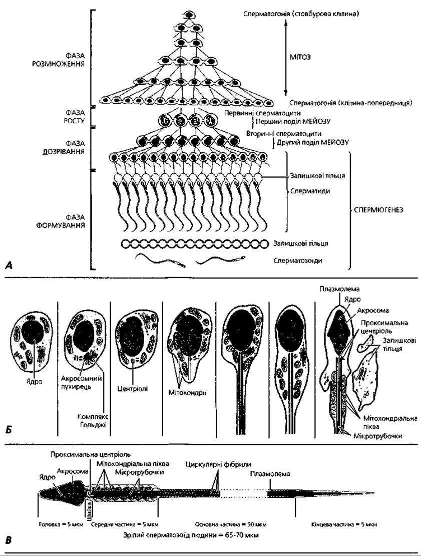
Fig. 4.96. Spermatogenesis and spermiogenesis: A—diagram of successive stages of spermatogenesis: until the Separation of mature spermatozoa from residual bodies, all spermatogenic cells are connected by cytoplasmic bridges and form a syncytium; B—spermiogenesis: Transformation of a spermatid into a spermatozoon; C—Morphology of a mature spermatozoon
After the growth phase is complete, the primary spermatocyte enters meiotic metaphase, transitioning into the next phase of spermatogenesis—maturation. As a result of the first (reduction) meiotic division, a secondary spermatocyte (spermatocyte II) is formed. During the anaphase of this division, the duplicated chromosomes of each bivalent, or dyads, migrate to opposite poles. Each cell receives 23 dyads. Spermatocyte II is smaller and located closer to the lumen of the seminiferous tubule compared to spermatocyte I. Prior to the second meiotic division, no DNA Synthesis or replication of chromosomal material occurs in spermatocyte II. The second (equational) meiotic division results in The formation of a spermatid—a cell with a haploid set of chromosomes, each consisting of a single chromatid. During the anaphase of this division, the chromatids of each chromosome, or monads, migrate to opposite poles. Thus, each cell receives 23 monads.
Spermatids are small cells that, depending on their stage of development, can be polygonal, irregularly round, or elongated. They are located near the lumen of the seminiferous tubule. At the end of the maturation phase, the nuclei of spermatids condense and become oval. Consequently, at the end of the maturation phase, as a result of meiotic division, four spermatids of two types are formed from each original spermatogonium: the first type carries an X chromosome and, upon fusion with an egg cell, can form a female embryo; the second type carries a Y chromosome, from which a male Organism can develop upon fusion with an egg cell. It should be noted that individual cell forms arising at successive stages of spermatogenesis remain connected to each other by cytoplasmic bridges, forming a characteristic multicellular structure—a syncytium (Fig. 4.96 A), and only the spermatozoa that detach into the lumen of the seminiferous tubules become completely free cells.
During the formation phase (or spermiogenesis), spermatids transform into spermatozoa (Fig. 4.96, B; 4.97). This process involves a series of Changes in the nucleus and cytoplasm of The Cell:
1) Chromatin condenses due to the replacement of Histones with non-histone Proteins; the nucleus decreases in size and becomes pear-shaped;
2) elements of the Golgi complex transform into the acrosome—a flat membrane-bound vesicle containing lytic Enzymes that covers the anterior surface of the nucleus like a cap;
3) both centrioles move toward the posterior pole of the nucleus. The proximal centriole is positioned in a fossa formed by the nucleus, while the distal centriole gives rise to the axoneme of the tail, thereby forming the flagellum;
4) as the tail develops, specific cytoskeletal elements are formed. These include 9 longitudinally arranged segmented columns around the centrioles (connecting piece), which are distally associated with 9 dense fibers located around the axoneme microtubules in the middle piece. In the principal piece, a fibrous sheath is formed, consisting of longitudinal columns connected by Ribs;
5) mitochondria acquire a spiral orientation, arrange themselves around the dense fibers in the middle piece, and abut one another to form the mitochondrial sheath;
6) excess cytoplasm containing Organelles and lipid inclusions detaches from the spermatozoon and is shed into the lumen of the tubule as so-called residual bodies.
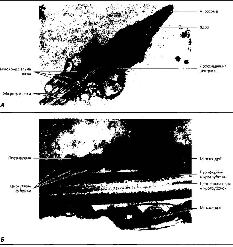
Fig. 4.97. Electron micrograph of a human spermatozoon; A - HEAD, neck, and middle piece of the tail, x 16,000; B - sperm tail at the transition from the middle piece to the principal piece, x 55,000
The activity of spermiogenesis is driven by the expression of the creme Gene, which encodes the CREME protein (cAMP-responsive element modulator). The latter is a Transcription factor that initiates the transformation of spermatids into spermatozoa. It should be noted that spermatogenesis proceeds in a wave-like fashion along the tubule, with certain phases predominating in specific regions, forming characteristic cellular associations. The process of spermatogonia transforming into mature spermatozoa takes about 75 days and is regulated by the Endocrine System (Fig. 4.98).
The straight seminiferous tubules connect the convoluted seminiferous tubules to the rete testis. They mark the beginning of the male genital ducts (Figs. 4.93, A, 4.99, 4.100). The wall of the straight tubules (as well as other PARTS OF THE genital ducts) consists of three layers: mucosa, muscularis, and adventitia. The mucosa of the straight seminiferous tubules is lined with a simple columnar epithelium, that of the rete testis tubules with a simple cuboidal epithelium, and that of the efferent ductules of the testis with a simple columnar epithelium, where ciliated cells alternate with secretory cells. Notably, the epithelial lining of the rete testis contains macrophages that phagocytose defective spermatozoa. The muscular layer is composed of circular bundles of smooth myocytes. The adventitia is formed by loose connective tissue.
The epididymis is an organ weighing about 3 g, located adjacent to and closely applied to the testis. It has an elongated shape, measuring 4x1x0.5 cm. Within the epididymis lies a tightly coiled tube—the duct of the epididymis (Figs. 4.93, A, 4.99, A). The latter ensures the transport of sperm, and the final differentiation of spermatozoa is completed within its lumen. Under the influence of androgens, the epididymis produces protein substances that regulate the proper packaging of Polysaccharides and enzymes in the acrosome; they also modify the surface glycopolymers (glycocalyx) of spermatozoa, which plays an important role in Fertilization. Furthermore, the secretory products of the epithelial cells lining the duct of the epididymis dilute the semen. The lumen of the epididymal duct serves as a reservoir for sperm.
Anatomically, the epididymis is divided into a head, body, and tail. The head of the epididymis is formed by the efferent ductules, which merge to form the duct of the epididymis, located in its body and tail. Sperm transport occurs in the direction from the head of the epididymis to its tail. Connective tissue septa divide the parenchyma of the epididymis into lobules. The wall of the epididymal duct consists of three layers: mucosa, muscularis, and adventitia. The mucosa is lined by a Cytology/practical/33.html">Pseudostratified columnar epithelium containing Two Types of cells:
1) principal cells—tall columnar epithelial cells with long, atypical microvilli (up to 40-80 µm in length) on their apical surface, known as stereocilia;
2) basal cells, located in the spaces between the basal portions of the principal cells. The muscular layer of the epididymal duct is composed of circular bundles of smooth myocytes, and the adventitia consists of loose Fibrous connective tissue.
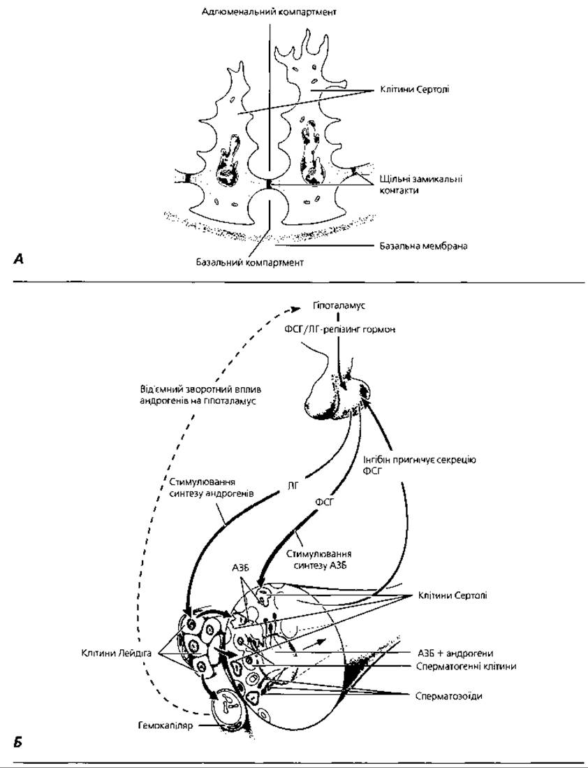
Fig. 4.98. Hormonal Regulation of spermatogenesis: A - basal and adluminal compartments formed by sustentacular cells for maturing spermatogenic cells; B - interaction of the Hypothalamus, Pituitary Gland, endocrine cells, and sustentacular cells in The regulation of spermatogenesis: FSH - follicle-stimulating hormone; LH - luteinizing hormone; ABP - androgen-binding protein
The Ductus deferens (vas deferens) (Figs. 4.93, A, 4.99, B, 4.100, C, 4.101, C) is a paired organ, representing a tube about 45 cm long with a lumen of 0.2-0.5 mm. The function of the ductus deferens is sperm transport: from the distal part of the ductus deferens, through the ejaculatory duct, semen is discharged into the urethra, and from there into the female reproductive tract. Contraction of the wall of the ductus deferens, due to the substantial development of muscular elements, drives ejaculation.
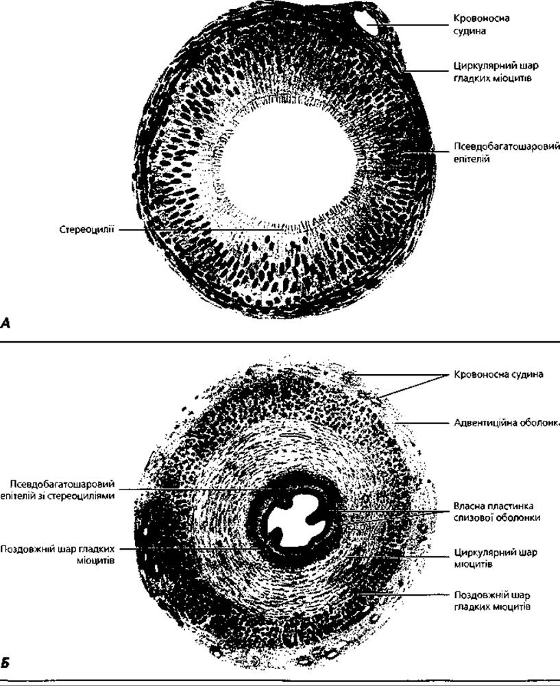
Fig. 4.99. Male genital ducts, semi-schematic representation: A - duct of the epididymis, x 200; B - ductus deferens, x 15
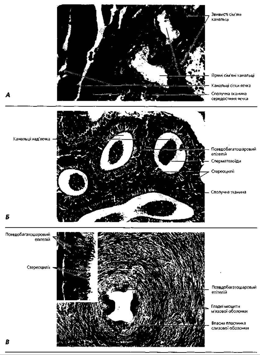
Fig. 4.100. Light microscopy of the male genital ducts: A - comparative morphology of convoluted and straight seminiferous tubules and the rete testis, x 110; B - ductules of the epididymis, x 200; C - ductus deferens, x 16 (inset on the left - x 400)
The wall of the ductus deferens consists of three layers: mucosa, muscularis, and adventitia. The mucosal epithelium is pseudostratified columnar, identical to that described in the epididymis. The muscularis contains three layers of smooth myocytes: inner and outer longitudinal, and middle circular. The adventitia is composed of loose connective tissue that gradually merges with the tissue of the Spermatic Cord. The latter contains Arteries, numerous Veins, and striated Muscle fibers of the cremaster muscle. The distal portion of the ductus deferens forms an ampullary dilation.
The ejaculatory duct (ductus ejaculatorius) (Figs. 4.92, 4.101, A) is a paired segment of the male genital tract, located below the junction of the Seminal Vesicle duct with the ampulla of the ductus deferens. The ejaculatory duct passes through the substance of the prostate gland and opens into the prostatic urethra. Its wall consists of three layers: mucosa, muscularis, and adventitia. The mucosa of the ejaculatory duct forms numerous folds. It is lined by a pseudostratified epithelium, which contains tall columnar cells with stereocilia on their apical surface and low basal cells. It is believed that stimulation of the stereocilia of the epithelial lining of the ductus deferens and ejaculatory ducts during sperm passage is one of the triggers of orgasm—the pleasurable sensation that occurs at the moment of ejaculation. A characteristic feature of the muscularis of the ejaculatory duct is its much weaker development of muscular elements compared to the ductus deferens. The connective tissue of the adventitia of the ejaculatory duct merges with the prostatic stroma.
The urethra (Figs. 4.92, 4.103) is a tubular structure 22-25 cm long that runs inside the corpus spongiosum of the penis. It is divided into three parts: the posterior prostatic part, the middle membranous part, and the anterior spongy part. The wall of the urethra is formed by three layers: mucosa, submucosa, and muscularis. The mucosal epithelium varies in structure across all three parts of the urethra. Specifically, the prostatic part is lined with transitional epithelium, the membranous part with pseudostratified columnar epithelium, and the spongy part with stratified squamous epithelium, which may show signs of cornification. The pseudostratified epithelium of the membranous urethra contains numerous goblet cells and occasional endocrine cells. The epithelium rests on the loose fibrous connective tissue of the lamina propria, which contains a dense network of venous vessels. The submucosa is composed of loose connective tissue containing a network of wide venous sinuses. Numerous small mucous glands, known as glands of Littre, are located in the urethral wall. The muscular layer of the urethra is formed by bundles of smooth myocytes, which are particularly well-developed in the prostatic part (where inner longitudinal and outer circular layers are distinguished) and gradually thin out toward the spongy part.

Fig. 4.101. Accessory Glands of the male reproductive system, semi-schematic representation; A - seminal vesicles; B - prostate gland, x 120; C - Bulbourethral gland, x 120
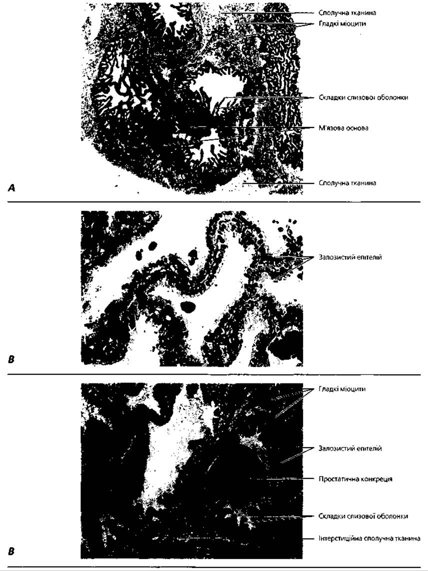
Fig. 4.102. Light microscopy of the accessory glands of the male reproductive system: A - seminal vesicle, x16; B - Glandular Epithelium of the seminal vesicle, x300; C - prostate gland, x120
The seminal vesicle (vesicula seminalis) (Figs. 4.92, 4.101) is a paired glandular organ located lateral to the ductus deferens and superior to the prostate gland. It has an oblong shape with dimensions of 5x2x1 cm. The excretory ducts of the seminal vesicles empty into the distal part of the ductus deferens just above the point where the latter transitions into the ejaculatory duct. The secretion of the seminal vesicles is rich in fructose, a monosaccharide used by spermatozoa to maintain metabolism. In addition, the secretory products of the seminal vesicles liquefy semen and create an alkaline environment within it, which helps enhance sperm motility in the female reproductive tract.
The wall of the seminal vesicle consists of three layers: mucosal, muscular, and adventitial. The mucosa is lined by pseudostratified columnar epithelium and forms numerous folds. The lamina propria is composed of connective tissue rich in elastic fibers, containing the terminal portions of mucosal alveolar glands. The muscular layer is formed by bundles of smooth myocytes running in two mutually perpendicular directions: an inner circular and an outer longitudinal layer. The adventitia of the seminal vesicle is built of Dense Fibrous Connective tissue rich in elastic fibers.
The prostate gland (prostata) (Figs. 4.92; 4.101, A, C; 4.102) is a fibromuscular and glandular organ weighing about 20 g, which surrounds the seminal pathways like a sleeve at the level of the ejaculatory duct and the proximal part of the urethra. Chestnut-shaped, the organ measures 4x3x2 cm. The Significance of the prostate is related to its endocrine and exocrine Functions. As an endocrine organ, it secretes a group of biologically active substances into the blood—Prostaglandins—with various physiological effects. The latter influence male sex hormone production and spermatogenesis, stimulate nerve growth, and promote smooth myocyte contraction. As an exocrine gland, the prostate produces a mucous secretion that liquefies semen and increases sperm motility. The contraction of its muscular elements facilitates ejaculation.
The prostate gland is surrounded by a connective tissue capsule. The parenchyma of the organ is composed of separate mucosal alveolar glands, whose excretory ducts empty into the prostatic urethra. The first group of glands is arranged in a ring within the urethral mucosa, the second is located in the connective tissue surrounding the urethra, and the third forms the actual parenchyma of the prostate. The terminal secretory units of the prostate are formed by two types of epitheliocytes: tall columnar cells with a mucous type of secretion, and intercalated (basal) cells located between the bases of the secretory cells. Secretory products are released into the lumen of the prostatic acini in the form of membrane-bound vesicles with a diameter of 125-150 µm, known as prostasomes. The function of basal cells is not yet fully understood; however, they are believed to perform an endocrine or paracrine role by regulating the interactions between the stromal and parenchymal elements of the gland.
The fibromuscular elements of the prostate consist of loose connective tissue and radially oriented bundles of smooth myocytes that divide it into lobules. The contraction of smooth myocytes at the moment of ejaculation facilitates the expulsion of secretion from the prostatic glands. In the region where the ductus deferens transitions into the urethra, the Tissues of the prostate form a thickening known as the seminal colliculus (verumontanum), the erection of which prevents the reflux of semen into the Urinary Bladder. Posterior to the seminal colliculus lies the prostatic utricle, which opens onto The surface of the seminal colliculus.
The bulbourethral glands (glandulae bulbo-urethrales) (Fig. 4.92) are complex tubuloalveolar glands whose excretory ducts empty into the proximal part of the urethra. The function of their secretion is to liquefy semen. The terminal secretory units of the bulbourethral glands are composed of flat, cuboidal, or columnar mucous cells, in whose cytoplasm mucus droplets and characteristic rod-shaped inclusions are found. The terminal secretory units of the glands are surrounded by loose connective tissue and bundles of smooth myocytes.
The penis (penis) (Figs. 4.92; 4.103) is a copulatory organ that ensures the delivery of semen into the female reproductive tract and also serves for urination. In the flaccid state, the length of the penis is 6-11 cm. The organ is formed by two corpora cavernosa and one corpus spongiosum, which provide rigidity (erection) when filled with blood. The urethra runs through the lower corpus spongiosum. Externally, the penis is covered by Skin, beneath which lies the tunica albuinea. The latter is formed by Dense connective tissue rich in elastic fibers and smooth myocytes. The glans penis is built of dense connective tissue containing a dense network of anastomosing veins. The glans is surrounded by thin skin forming a double fold, on the inner surface of which the ducts of specific sebaceous preputial glands open. The penile arteries are termed helicine because they coil in a spiral fashion in the flaccid state. The walls of the penile arteries and veins are rich in muscular elements, the contraction of which blocks the outflow of blood from the organ, ensuring its rigidity during erection. The vascular spaces of the corpora cavernosa and corpus spongiosum are located between the arteries and veins and have thin walls lined with endothelium.
Development and Age-related changes of the male reproductive system. Primordial germ cells—gonocytoblasts—are first detected in the primitive streak of the embryo and the adjacent mesoderm. In the third week of Embryogenesis, they migrate to the endoderm of the yolk sac, where they undergo intensive proliferation. At the same time (during the third and fourth weeks), the genital ridges—thickenings of the coelomic epithelium On the surface of the pronephros/mesonephros—form in the embryo's body. Gonocytoblasts migrate from the yolk sac wall initially into the hindgut wall, from where they are carried by the blood vessel system into the genital ridges. This stage, which in humans lasts from the third to the sixth week of embryogenesis, is common to both male and female embryos and is therefore called the indifferent stage. During The Development of the male gonad, the coelomic epithelium of the genital ridges gives rise to the sustenticular cells (Sertoli cells) of the testis, the mesenchymal cells of the primitive Kidney give rise to its endocrinocytes (Leydig cells), and the gonocytoblasts give rise to gonocytes and spermatogenic cells.
During male-type Differentiation of the reproductive system primordia, cords of epithelial cells—the sex cords—grow from the genital ridges into the mesonephros, with gonocytes localizing within them. The sex cords gradually transform into seminiferous tubules, where gonocytes differentiate into spermatogonia at around the 22nd week of embryogenesis. In the postnatal period, proliferation of spermatogonia continues within the primordia of the convoluted seminiferous tubules, whereas in the straight tubules and the rete testis, these cells regress. The efferent ductules of the testis develop through the remodeling of the mesonephric tubules. The mesonephric duct gives rise to the epididymal duct, the ductus deferens, and the ejaculatory duct.
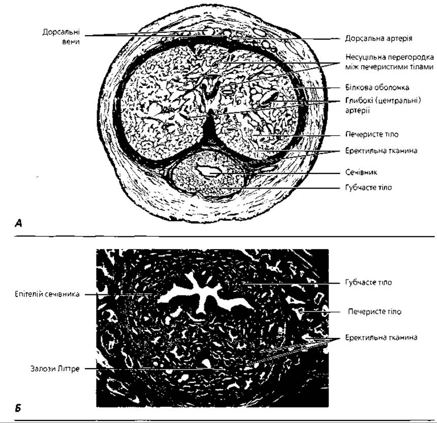
Fig. 4.103. Penis: A - semi-schematic representation of a cross-section of the distal part of the penis of a 21-year-old male, x 2.5, B - light micrograph of the corpus spongiosum with the urethra, cross-section, x 25
The endocrine function of the testes is established earlier in ontogeny than their reproductive function. Genetic information encoded on the Y chromosome directs the synthesis of a series of hormones that determine the Development of the reproductive system along the male pathway. Thus, as early as the sixth week of embryogenesis, the indifferent gonad begins to produce inhibin, which induces the regression of the paramesonephric duct and the male-type differentiation of the gonadal primordia, as well as the hypothalamus and the Pineal Gland. During the eighth to tenth weeks of embryogenesis, the testis begins to produce testosterone. Around the fifth month of fetal development, the epithelial cells of the rete testis produce inhibin, which suppresses the secretion of pituitary follicle-stimulating hormone, slowing down proliferation and causing the death of some gonocytes. Consequently, spermatogenesis is arrested until the onset of Puberty.
The ESTABLISHMENT OF THE spermatogenic function of the testis is completed only with the onset of puberty. Thus, starting from the ninth year of life, the first primary spermatocytes appear in the testicular tubules. Between the ages of 10 and 15, sustentocytes reach full development. In the convoluted seminiferous tubules, in addition to primary spermatocytes, secondary spermatocytes and spermatids also appear; the seminiferous tubules become tortuous. During the same period (12–14 years), the number of interstitial endocrinocytes in the interstitial connective tissue increases, and their hormonal activity drives the rapid growth of the epididymis and the ductus deferens.
Age-related involution of the testis is observed between 50 and 80 years of age. It manifests as a decline in the spermatogenic and endocrine Functions of the organ, accompanied by the proliferation of connective tissue. Age-related changes in spermatogenesis include meiotic abnormalities, the appearance of immature germ cells in the semen, atrophic changes and diverticula in the seminiferous tubules, and lipid accumulation in testicular sustentocytes. Notably, even in advanced age, isolated convoluted seminiferous tubules with normal Structure and function persist in the testes. The decline in male sex hormone production leads to Atrophy of the external genitalia and the prostate gland.
The prostate gland begins to form as an organ at the 10th to 12th week of embryonic development as epithelial outgrowths of the urethral primordium into the surrounding mesenchyme. During the first half of embryogenesis, the proliferation of epithelial cords and the development of the organ's glandular structures predominate, while the second half is dominated by the development of its fibromuscular elements. The prostatic utricle is formed as a result of the regression of most of the paramesonephric duct. The seminal vesicles arise as outgrowths of the urogenital sinus.
The prostate gland reaches its peak development between 20 and 35 years of age. At this time, its secretory elements predominate over connective tissue, and the epithelial cells of the prostatic glands become tall columnar and actively secrete. Between 35 and 60 years of age, age-related involution of the prostate gland is observed: the height of the epithelial cells in the secretory endpieces decreases, individual lobules of the gland atrophy, and the connective tissue proliferates and becomes denser. Prostatic concretions (corpora amylacea, consisting of desquamated epithelium and condensed secretory products) accumulate in the lumen of the glands, and their number increases significantly in old age.
Terms to remember
1. Testis. 2. Tunica albuginea. 3. Mediastinum testis. 4. Lobule of the testis. 5. Convoluted seminiferous tubule. 6. Basal layer. 7. Myoid layer. 8. Fibrous layer. 9. Blood-testis barrier. 10. Interstitial endocrinocyte of the testis (Leydig cell). 11. Testosterone. 12. Sustentocyte of the testis (Sertoli cell). 13. Androgen-binding protein. 14. Inhibins. 15. Spermatogonial proliferation stimulator. 16. Spermatogenesis. 17. Spermatogonium. 18. Primary spermatocyte. 19. Secondary spermatocyte. 20. Spermatid. 21. Spermatozoon. 22. Proliferation phase of spermatogenesis. 23. Growth phase. 24. Maturation phase. 25. Formation phase. 26. Straight seminiferous tubule. 27. Tubule of the rete testis. 28. Efferent ductule of the testis. 29. Epididymis. 30. Lobule of the epididymis. 31. Duct of the epididymis. 32. Principal cell. 33. Basal cell. 34. Ductus deferens (vas deferens). 35. Ejaculation. 36. Ejaculatory duct. 37. Urethra. 38. Prostatic urethra. 39. Membranous urethra. 40. Spongy (cavernous) urethra. 41. Seminal vesicle. 42. Prostate gland (prostate). 43. Prostaglandins. 44. Bulbourethral gland. 45. Penis. 46. Preputial glands. 47. Glands of Littre. 48. Gonocytoblast. 49. Genital ridge. 50. Sex cord. 51. Mesonephros (primary kidney). 52. Mesonephric duct.
Last update: 09/08/2026
Editorial and Educational Adaptation: This material has been compiled based on the primary/original source text. The project team performed an editorial review, corrected technical inaccuracies, structured sections, and adapted the content for an educational format.
What was processed:
- elimination of formatting defects (OCR errors, structural breaks, corrupted characters);
- editorial organization of content;
- standardization of terminology in accordance with academic sources;
- verification of factual statements against the original source text.
All mentions of the author, publication year, and origin of the primary text have been preserved in accordance with the source.