Human Histology - O.D. Lutsyk 2003
Systemic Histology
Urinary System
The Urinary System includes the Kidneys, which are the urine-producing Organs, as well as the Ureters, Urinary Bladder, and Urethra, which make up the Urinary Tract (Fig. 4.81).
The Kidney (ren) is an organ in which urine is continuously formed. It is the primary organ that rids the body of metabolic end products: 80% of all waste products are excreted in the urine due to kidney function. It is well known that impairment of excretory processes leads to death much faster than the disruption of normal nutrient intake. The primary and vital function of the kidneys is excretion. In addition, the kidneys participate in regulating Blood OSMOTIC PRESSURE, maintaining acid-base balance, and also perform an endocrine function.
The kidney is a paired, bean-shaped parenchymal organ located in the retroperitoneal space on both sides of the lumbar spine. The mass of each kidney is 100-300 g, and its dimensions are 11x6x4 cm. The kidney is covered by a fibrous capsule 100-200 μm thick, and also has a serous membrane anteriorly. External to the fibrous capsule, especially in the hilum region and on the posterior surface, lies a layer of adipose tissue—the perinephric fat (adipose capsule of the kidney). In section, two layers can be distinguished in the kidney: the renal cortex (located in a continuous layer beneath the capsule, dark red, granular) and the renal medulla (inner layer, light-colored, striated). The cortex extends into the medulla as the so-called renal columns of Bertin, which divide the medulla into 8–12 pyramid-shaped regions known as renal pyramids. The broad bases of the renal pyramids face The surface of the organ, while their apices point toward the hilum; the apices of the pyramids form the renal papillae, which project freely into the renal calyces. A renal pyramid with its overlying cortex is called a renal lobe.
Class="center">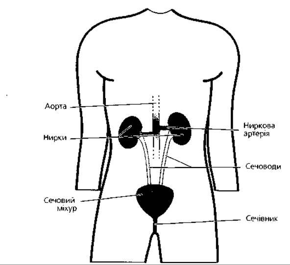
Fig. 4.81. General structural plan of the urinary system.
The renal medulla, in turn, extends into the cortex as thin bundles called medullary rays of Ferrein. A medullary ray together with the surrounding cortical tissue forms a cortical lobule. The lobules are demarcated by interlobular Arteries and Veins (see below, Fig. 4.89, B, C). The renal stroma consists of loose Fibrous Connective Tissue rich in reticular Cells and reticular fibers. The renal parenchyma is formed by renal corpuscles and epithelial renal tubules, which include convoluted and straight tubules. The former, together with the corpuscles, form the cortex, while the latter form the medulla.
The nephron (Fig. 4.82) is the Structural and functional unit of the kidney. It consists of a system of convoluted and straight epithelial tubules originating from each renal corpuscle. The length of a single nephron ranges from 18 to 50 mm, and the total length of all nephrons is about 100 km. The number of nephrons in both kidneys is 2–2.5 million. The nephron includes Bowman's capsule, the proximal convoluted and straight tubules, the thin tubule (with descending and ascending limbs), and the distal straight and convoluted tubules. The thin tubule and the distal straight tubule form the Loop of Henle. The glomerulus and Bowman's capsule form the renal (Malpighian) corpuscle.
Depending on their Location and structural features, nephrons are classified into cortical and juxtamedullary. Among cortical nephrons, a distinction is made between superficial (short) nephrons, which are entirely located within the cortex (accounting for 1%), and midcortical (intermediate) nephrons, whose loops descend into the outer zone of the medulla (accounting for 80%). Juxtamedullary nephrons, which make up about 20%, have very long loops that extend deep into the medulla, while their renal corpuscles, proximal, and distal segments are located in the cortex near the corticomedullary junction. Nephrons empty into collecting ducts (tubules). The latter begin in the cortex and, together with the straight tubules of cortical nephrons, form part of the medullary rays. The collecting ducts then pass into the medulla and, at the apices of the pyramids, merge into the papillary ducts.
Histophysiology of Urine Formation. The renal corpuscle, where the nephron begins, is spherical, with a diameter of 100 to 240 μm, and consists of a capsule and a glomerulus (Figs. 4.82, 4.83, 4.84). The capsule is shaped like a double-walled cup, consisting of visceral (inner) and parietal (outer) layers that enclose a slit-like cavity—the so-called urinary space. The glomerulus consists of 50–100 capillary loops, which are Branches of the afferent arteriole.
The capillaries merge to form the efferent arteriole, which has a smaller diameter than the afferent arteriole, thereby ensuring high pressure (over 50 mmHg) in the glomerular capillaries. This is a prerequisite for The first phase of urine formation, which occurs via filtration of Blood Plasma through the filtration barrier. About 1000 liters of blood pass through the glomerular capillaries daily, meaning that the entire blood volume of The Human Body passes through the kidneys approximately 200 times a day, or once every 5–10 minutes.
The endothelial Cells of the glomerular capillaries have numerous fenestrae up to 0.1 μm in diameter and rest on the inner surface of a thick, three-layered glomerular basement membrane. Its outer surface is covered by the epithelium of the visceral layer of the capsule. In the three-layered basement membrane, the outer and inner layers are electron-lucent and less dense; between them lies the electron-dense middle layer, which contains a microfibrillar meshwork with a pore diameter of up to 7 nm.
The glomerular capillaries are enveloped by the visceral layer of the capsule along almost their entire length, like fingers in a glove. The visceral layer of the nephron capsule is formed by large (up to 30 μm), irregularly shaped epithelial cells called podocytes. Several broad primary processes, or cytotrabeculae, extend from the basal surface of these cells, giving rise to numerous small secondary processes called cytopodia (pedicels). The latter, with their slightly expanded bases (the so-called attachment feet), contact the three-layered basement membrane described above. Between the pedicels, which form numerous interdigitations, lie narrow filtration slits (Fig. 4.83, C, D) bridged by slit diaphragms.
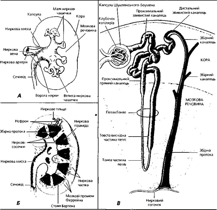
Fig. 4.82. Kidney: A - general structural plan; B - principle of structural Organization; C - Structure OF THE nephron and its connection with the glomerulus and collecting duct
All the aforementioned components—the fenestrated endothelium of the glomerular capillaries, the podocytes of the visceral layer of the capsule, the filtration slits, and the three-layered basement membrane—make up the filtration barrier (renal filter), through which blood is filtered and primary urine is formed, accumulating in the capsular space. The renal filter has selective permeability, retaining everything larger than the Pores in the meshwork of the Middle layer of the basement membrane. Normally, formed elements of blood and high-molecular-weight Plasma Proteins, such as IMMUNOGLOBULINS and fibrinogen, do not pass through the filtration barrier. If the filter is damaged by a pathological process in the kidneys, these blood components can be detected in the patient's urine. In the glomeruli of the renal corpuscles, where there are no podocytes of the visceral layer between the capillaries, another Cell type is present—the so-called mesangial cells (mesangiocytes). They produce the Extracellular matrix, together with which they form the mesangium of the glomeruli. Some mesangial cells are macrophages involved in immune defense and the clearance of the renal filter. Evidence suggests that mesangial cells participate in the physiological regeneration of the basement membrane of the filtration barrier, as well as in the endocrine Functions of the kidney. The parietal (outer) layer of Bowman's capsule is formed by a single layer of simple squamous or cuboidal cells surrounded by a basement membrane. The epithelium of the parietal layer of the capsule is continuous with the epithelium of the proximal tubule of the nephron.
The proximal segment of the nephron (Figs. 4.82, 4.83, 4.85, 4.86) consists of two parts: a long convoluted part and a short straight part. The length of this tubule is about 15 mm, and its diameter is 50-60 μm. The convoluted part of the proximal segment begins at the urinary pole of the renal corpuscle and then, after winding, returns toward its renal corpuscle; the short straight part descends and transitions into the thin tubule of the nephron. The wall of the proximal segment is formed by a single layer of simple columnar or cuboidal cells resting on the basement membrane. The apical surface of these cells features a brush border formed by microvilli, while the basal part exhibits basal striation, which arises from the orderly arrangement of rod-shaped Mitochondria oriented with their long axes perpendicular to the basement membrane and located between deep invaginations of the basal Plasmalemma. The Cytoplasm of the cells in this segment of the nephron is acidophilic (oxyphilic) and contains inclusions of urates, Lipids, pigments, as well as a significant number of pinocytotic vesicles and Lysosomes. Cell boundaries are poorly visible, and the lumen of the tubule appears as a barely outlined slit. Reabsorption—the back-absorption of proteins, glucose, electrolytes, and Water from the primary urine into the blood—takes place in the proximal segment. The Mechanism of this process is determined by the structural CHARACTERISTICS OF THE cells in this segment. The brush border, which has high alkaline phosphatase activity, facilitates the complete reabsorption of glucose. Through pinocytosis, the cells absorb proteins from the primary urine and break them down into Amino Acids, which are then released into the blood. Mitochondria facilitate the active reabsorption of certain electrolytes, while the folds of the cytolemma are of great importance in the mechanism of passive water reabsorption.
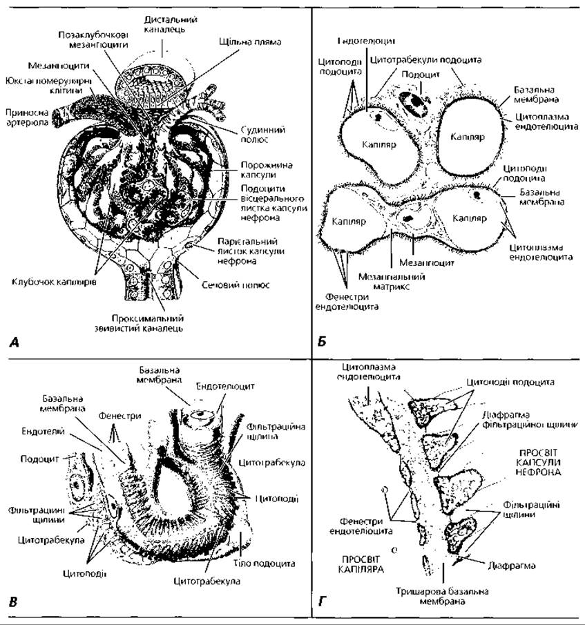
Fig. 4.83. ULTRASTRUCTURE OF THE renal corpuscle: A - renal corpuscle with adjacent segments of proximal and distal tubules; B - relationship of glomerular capillaries with podocytes and mesangial cells; C - three-dimensional reconstruction of the filtration barrier; D - structural details of the filtration barrier
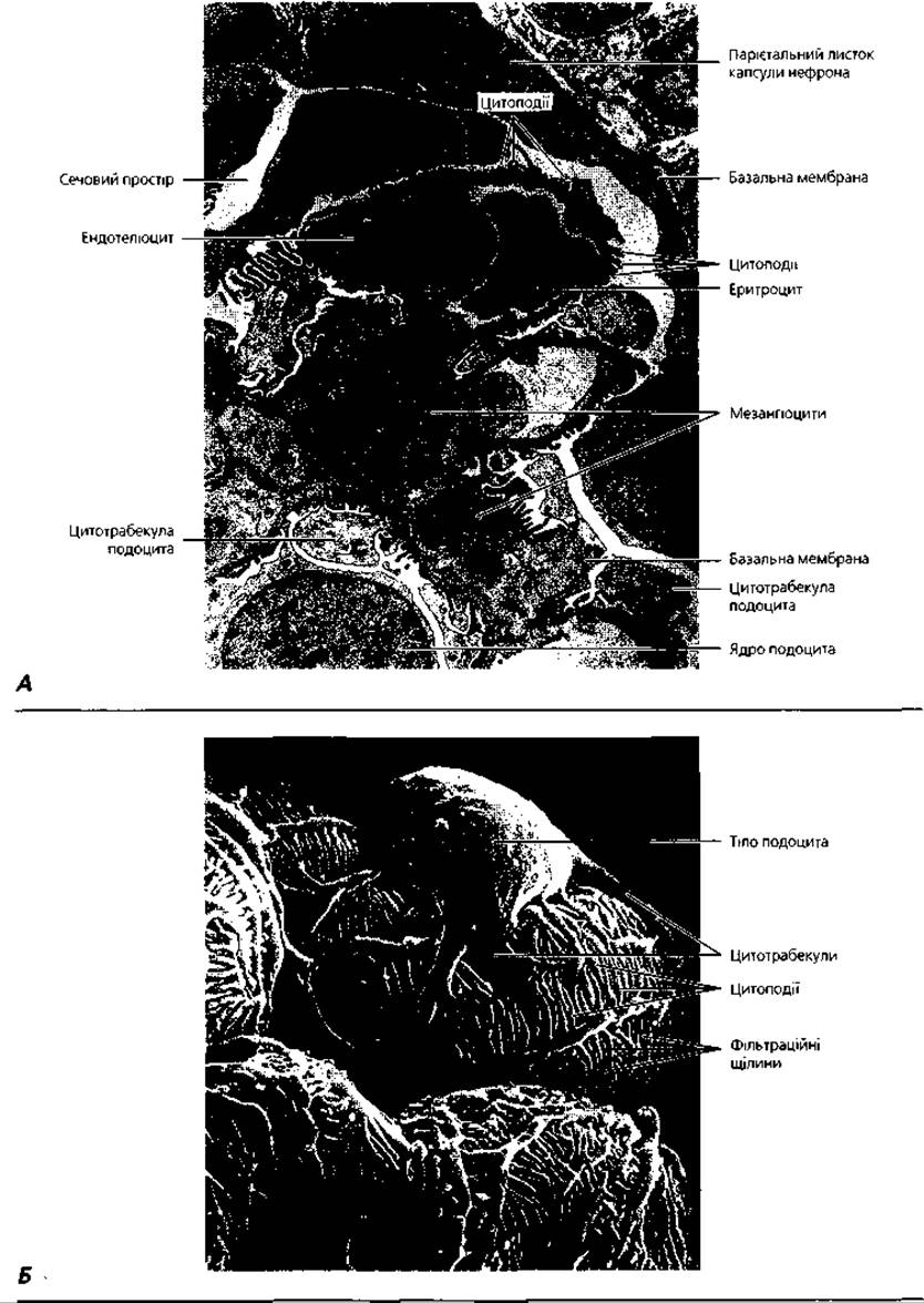
Fig. 4.84. Electron Microscopy of the renal corpuscle: A - transmission electron microscopy, x 4500; B - scanning electron microscopy of a podocyte, x 6200
The thin tubule of the nephron has a diameter of 13-15 μm. Its wall is formed by a single layer of simple squamous epithelial cells with light cytoplasm poor in Organelles. The nucleated portions of the cells bulge into the lumen of the tubule. In cortical nephrons, the thin tubule has only a descending limb, whereas in juxtamedullary nephrons, there is also a long ascending limb that transitions into the distal straight tubule. As it passes through the thin tubule, the primary urine loses water, which exits through the tubule walls into the interstitium (the connective tissue between the tubules) due to the high concentration of sodium chloride there. The cells of the straight and adjacent PARTS OF THE convoluted distal tubules actively transport sodium ions from the urine into the interstitium, thereby creating the necessary osmotic pressure gradient between the urine and the interstitial fluid. As a result, the fluid, which was hypertonic in the lower part of the thin tubule, becomes hypotonic in these segments, while the osmotic pressure in the interstitium increases. The latter drives passive water reabsorption in the terminal part of the convoluted distal tubules and in the collecting ducts.
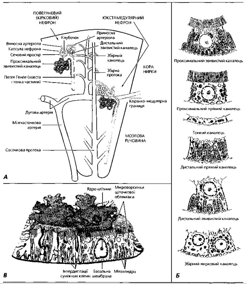
Fig. 4.85. Structural details of nephrons: A - Two Types of nephrons; B - ULTRASTRUCTURAL FEATURES OF cells in different segments of the nephron; C - three-dimensional reconstruction of a portion of the proximal convoluted tubule wall
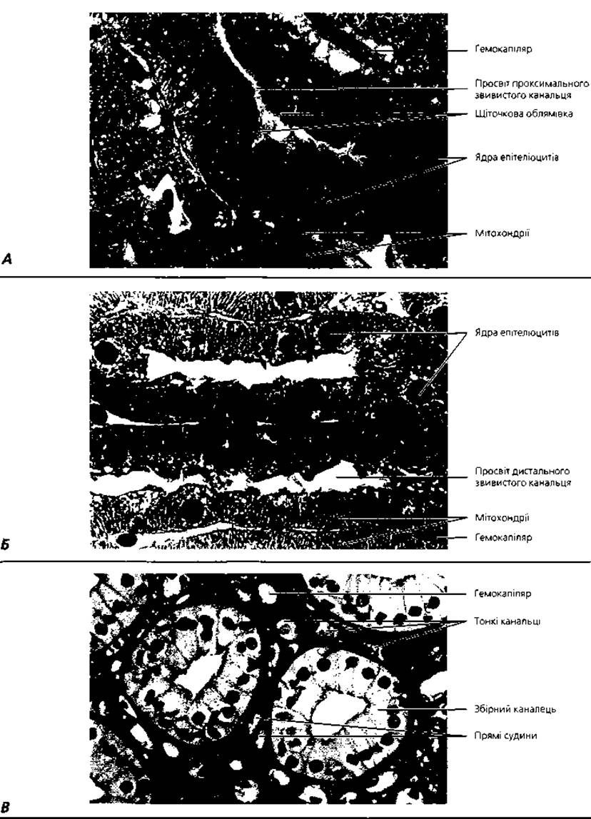
Fig. 4.86. Light microscopy of renal tubules: A - proximal convoluted tubule, x 1000; B - distal convoluted tubules, x 1000; C - thin and collecting tubules, x 400
The distal straight tubule has a diameter of 30 μm, and its epithelium is similar to that of the Distal convoluted tubule. The diameter of the latter is 30-40 μm. Its epithelium is low columnar, resting on a well-defined basement membrane. Unlike the proximal nephron segment, the cells of the distal segment lack a brush border, but the invaginations of the plasmolemma in the basal part are deeper, containing large, elongated mitochondria. Because the distal convoluted tubule is shorter than the proximal one, its cross-sections are observed less frequently in histological sections than those of the proximal segment. The distal tubules lie near their renal corpuscle because the nephron loops back to it. The cells of the distal segment have a lighter cytoplasm and more distinct boundaries compared to the epithelium of the proximal tubule.
Collecting renal tubules (Figs. 4.86, 4.87) do not belong directly to the nephron; they drain several nephrons. In the upper cortical part, they are lined with simple cuboidal epithelium, and in the lower part, with simple columnar epithelium. Light and dark cells are distinguished within the epithelium. Light cells are poor in organelles, and their cytoplasm forms internal folds. The function of these cells is the passive reabsorption of water, which occurs under METABOLISM/18.html">The Influence of antidiuretic hormone, which also acts on the cells of the distal segment. The ultrastructure of dark cells resembles that of the parietal cells of gastric glands that secrete H+ ions. The secretory function of dark cells is believed to ensure urine acidification.
Thus, the complex process of urine formation occurs in the nephrons due to The activity of their cells. It consists of three phases. The first phase takes place in the renal corpuscle: primary urine is formed there by filtration. The second phase occurs in the nephron tubules: glucose and protein are reabsorbed from the primary urine; essential electrolytes and water are returned to the blood; the urine is concentrated, and its volume decreases from 100 L to 1.5-2 L per day. The final phase of urine formation is called the secretory phase; it takes place in the collecting ducts, where the pH of the urine is acidified.
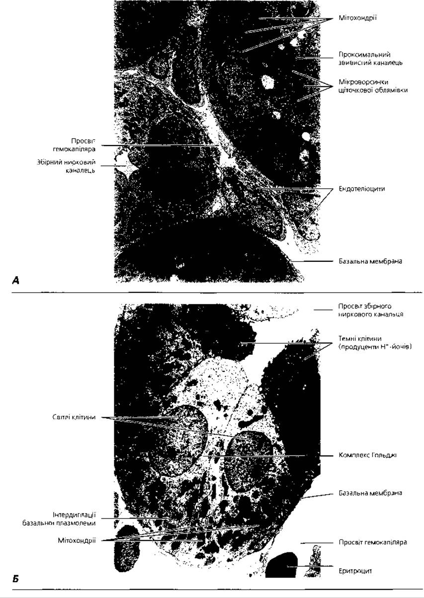
Fig. 4.87. Electron microscopy of renal tubules: A - proximal and distal tubules, x 4500; B - collecting renal tubule, x 5000
The endocrine complex of the kidney includes the juxtaglomerular apparatus (JGA), which is present in approximately 10% of cortical nephrons, as well as interstitial cells and the epithelium of the collecting tubules. The juxtaglomerular apparatus consists of the following components:
1) juxtaglomerular cells;
2) macula densa cells;
3) Goormaghtigh juxtavascular cells;
4) mesangial cells (Fig. 4.83, 4.88).
Juxtaglomerular cells are located beneath the endothelium in the wall of the afferent and, to a lesser extent, efferent arteriole. They are oval-shaped and contain renin granules in their cytoplasm, which they secrete into the blood. Renin helps to increase blood pressure by catalyzing The formation of angiotensin, which has a vasoconstrictive effect. In addition, renin stimulates The production of the hormone aldosterone in the Adrenal Glands.
The macula densa is a region of the wall of the distal nephron segment lying near the renal corpuscle between the afferent and efferent arterioles. The epithelial cells of the macula densa are taller compared to other epithelial cells of the distal segment and lack basal folds; the basement membrane here has a specific structure. Under the Electron microscope, it is revealed to form splittings containing the processes of Goormaghtigh juxtavascular cells; the surface of the basement membrane is uneven, containing folds, tunnels, etc. It is possible that this structure facilitates contact between the macula densa and juxtaglomerular cells, which is necessary for regulating renin synthesis, and also prevents desquamation of the macula densa cells. The macula densa cells act as a sodium receptor, responding to changes in sodium concentration in the urine and influencing the renin-producing juxtaglomerular cells.
Juxtavascular cells (Goormaghtigh cells) are located in the region of the vascular pole of the renal corpuscle, in the triangular space between the afferent and efferent arterioles and the macula densa. They have an oval or irregular shape and long processes through which they contact mesangial cells; their cytoplasm contains fibrillar structures. Some authors consider Goormaghtigh cells to be a type of mesangial cells, the so-called extraglomerular mesangium. Mesangial cells possess contractile microfilaments and receptors for vasoconstrictive substances. It is believed that juxtavascular cells and mesangial cells begin to produce renin in the event of depletion of juxtaglomerular cells. In addition to renin, the juxtaglomerular apparatus of the kidney also produces Erythropoietin, a factor that stimulates erythropoiesis.
Interstitial cells are of mesenchymal origin and are located in the stroma of the renal pyramids. From their transversely elongated cell bodies, processes extend, some of which wrap around the tubules of the nephron loop, while others wrap around blood capillaries. The cytoplasm of interstitial cells contains well-developed organelles and lipid granules. These cells produce a type of prostaglandin that has an antihypertensive effect, thereby lowering blood pressure. In addition to interstitial cells, Prostaglandins are also produced by the light cells of the collecting renal tubules. Thus, the endocrine complex of the kidneys participates in The regulation of systemic and Renal Circulation, thereby influencing urine formation.
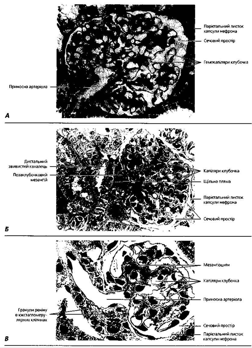
Fig. 4.88. Light microscopy of the renal corpuscle and elements of the renal endocrine complex: A - selective staining of the glomerular capillaries, x 350; B — renal corpuscle with adjacent macula densa, x 300; C - juxtaglomerular cells within the wall of the afferent glomerular arteriole, x 300
The renal Circulatory system (Fig. 4.89). Blood enters the kidney through the renal artery, which, entering via the renal hilum, branches into five segmental arteries; the latter, in turn, divide into interlobar arteries passing between the pyramids of the medulla. The interlobar arteries arch at the boundary of the cortex and medulla, forming arcuate arteries. From these, interlobular arteries branch off at a right angle into the cortex, giving rise to afferent arterioles. These break up into the capillaries of the glomerulus, which have already been described. This capillary network is called the rete mirabile because the capillaries here are situated between two arterioles. This capillary network is also known as the primary capillary network of the kidney, and its purpose is blood filtration and the Formation of primary urine. The glomerular capillaries merge into the efferent arteriole. After exiting the renal corpuscle, the efferent arteriole breaks up again into a capillary network that wraps around the nephron tubules; this is called the secondary, or peritubular capillary network. Its function is the nourishment of the nephron and Participation in the second phase of urine formation, which occurs via reabsorption. The filtration process in the renal corpuscle occurs due to high blood pressure in the glomerular capillaries, whereas the return of valuable substances from the primary urine back into the blood—The process of reabsorption—is, conversely, driven by low blood pressure in the capillaries of the peritubular capillary network. The venous System of the kidney begins from the latter. In the upper Regions of the cortex, it is formed by stellate veins, from which interlobular veins arise. In the middle regions of the cortex, the capillaries of the peritubular network merge directly into the interlobular veins. From the latter, arcuate veins begin, which pass into interlobar veins and then into the renal veins exiting the renal hilum. The described blood supply system is characteristic of cortical nephrons and is called the cortical circulation. Its features determine the active participation of cortical nephrons in urine formation.
In juxtamedullary nephrons, the afferent and efferent arterioles of the glomeruli have the same diameter, or the efferent arteriole is even slightly wider. Therefore, the blood pressure in the capillaries of these glomeruli is not as high as in cortical nephrons. The second difference of the juxtamedullary circulation is that the efferent arterioles descend into the medulla, where they break up into bundles of so-called vasa recta with thin walls that are wider than ordinary capillaries. In the medulla, branches arise from the efferent arterioles as well as from the vasa recta, forming the medullary peritubular capillary network. The vasa recta form loops at various levels of the medulla and turn back in the opposite direction, transitioning into veins. The descending and ascending limbs of these loops are called vascular bundles. The capillaries of the medulla collect into straight veins, which empty into the arcuate veins. Due to these described circulatory features, juxtamedullary nephrons do not form urine as actively as cortical ones; they act as shunts, allowing blood to pass easily and quickly through the kidneys under conditions of intensive circulation.
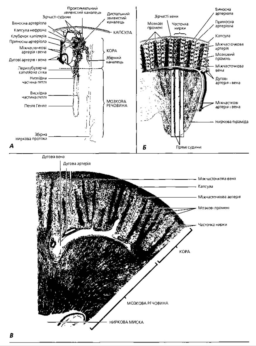
Fig. 4.89. Interaction of renal vessels with nephrons: A - diagram of nephron blood supply; B - reconstruction of the renal Vascular System; C - semi-schematic representation of a histological section of the kidney
The urinary tract (Fig. 4.90) begins in the kidneys with the renal calyces (calices renales) and pelvis (pelvis renalis), then continues into the ureters (Ureter), urinary bladder (vesica urinaria), and urethra (urethra). All of these organs, except for the urethra, have a similar structure and consist of a mucosa with a submucosa, a muscularis, and an outer coat. The mucosa is lined with transitional epithelium (Figs. 4.90, 4.91).
The thickness and number of layers of this type of Epithelial Tissue increase from the renal calyces toward the urinary bladder and decrease when the organs are distended with urine. Transitional epithelium is impermeable to water and salts and is capable of changing its shape. Its superficial cells are large, polyploid, or binucleated, changing shape from pear-shaped to flat depending on the degree of organ distension. Specific structures of these cells include invaginations of the plasmolemma and spindle-shaped vesicles in the apical part, which serve as reservoirs of the plasmolemma that insert into the latter during distension. The barrier function of the epithelium is ensured by tight junctions between superficial cells, the significant thickness of the plasmolemma, and its unique chemical composition.
The lamina propria is composed of loose connective tissue, which in its deeper regions is referred to as the submucosa. Due to the presence of the submucosa, the mucosa of the ureters and urinary bladder forms deep folds that provide The ability to stretch and expand the lumen of the ureter, which is important during the passage of kidney stones. In cross-section, the lumen of the ureter has a stellate appearance. In the region of the Cytology/practical/108.html">Fundus of the urinary bladder (the so-called trigone), where the ureters enter and the urethra exits, the mucosa has no folds; the submucosa is absent here, and the lamina propria contains small alveolar-tubular glands similar to the Glands of the prostate. Similar glands are found in the submucosa of the lower part of the ureters.
The muscular coat of the renal pelvis and calyces consists of two thin layers of smooth myocytes—an inner longitudinal and an outer circular layer—but around the papillae of the renal pyramids, only a single circular layer of Muscle cells is preserved. Its contraction compresses the papilla and facilitates urine excretion. The muscular coat of the ureters in the upper two-thirds is structured the same as in the renal pelvis, while in the lower third, it has three layers—inner and outer longitudinal, and a middle circular layer. At the site where the ureter passes through the wall of the urinary bladder, the smooth myocytes of the muscular coat run only longitudinally, which ensures the opening of the ureteral orifice regardless of the state of the bladder Muscles. The muscular coat of the urinary bladder is composed of three layers of smooth myocytes; their orientation is the same as in the lower part of the ureter, but they are much thicker and poorly demarcated. The middle circular layer is the most developed.
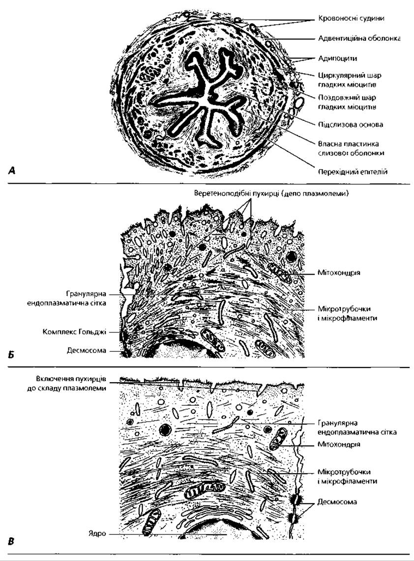
Fig. 4.90. Urinary tract: A - semi-schematic representation of a ureter specimen, x 18; B - part of a superficial epithelial cell of the urinary bladder mucosa in a contracted state; C - schematic diagram of the epithelial Cell Structure from panel B during stretching (filling) of the urinary bladder wall
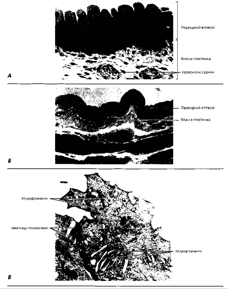
Fig. 4.91. Urinary bladder epithelium: A - light micrograph of the urinary bladder wall in a contracted state, x 1200; B - urinary bladder wall in a stretched state, x 1200; C - transmission electron micrograph of the apical portion of a superficial urinary bladder epithelial cell in a contracted state, x 21 000
In the region of the urinary bladder trigone, There is a single muscular layer in which two parts are distinguished: the muscle connecting the muscular structures of both ureters, and the sphincter muscle of the trigone, which surrounds the internal urethral orifice. The sphincter Muscle consists of Striated Muscle tissue in its outer part, and Smooth muscle tissue in its inner part closer to the urethra.
The outer coat of all the described organs is adventitial and is composed of fibrous connective tissue. Only on the posterosuperior and, partially, lateral surfaces of the urinary bladder is the outer coat serous.
The urethra has a different structure in males and females. Since the male urethra also serves as a channel for semen passage, it will be described in the chapter "Male Reproductive System". The female urethra is 2 to 6 cm long. Its wall is composed of a mucosa with a submucosa, a muscularis, and an adventitia. In cross-section, the lumen of the canal is crescent-shaped. The mucosa forms longitudinal folds. The epithelium near the urinary bladder is transitional, over most of its length it is stratified or pseudostratified columnar, and in the region of the external urethral orifice, it is stratified squamous. The epithelium invaginates into the lamina propria, forming small, gland-like pockets. True glands are also present here, being particularly numerous in the upper part of the urethra.
Development of the urinary system. During the Embryonic period, three paired excretory organs develop sequentially: the pronephros, the mesonephros, and the permanent or metanephros. The pronephros is formed from the anterior eight to ten nephrotomes (segmental stalks) of the intermediate mesoderm. In the human embryo, it is non-functional and soon regresses.
The mesonephros develops from 25 nephrotomes located in the trunk region of the embryo. The nephrotomes separate from the somites and splanchnotome, transforming into the tubules of the mesonephros - metanephridia. The tubules grow toward the mesonephric duct, which is formed during pronephros development, and fuse with it. Vessels branch off from the aorta toward the tubules, breaking up into capillary glomeruli. The blind ends of the tubules invaginate around these glomeruli, forming their capsules, and together they constitute renal corpuscles. The mesonephric duct opens into the hindgut. The mesonephros serves as the primary excretory organ During the first half of the embryonic period.
The metanephros (permanent kidney) is initiated in the embryo during the second month of Embryogenesis, but its development is completed only after birth. This kidney develops from two sources: the outgrowth of the mesonephric duct and the nephrogenic tissue, which consists of unsegmented mesoderm in the caudal region of the embryo. The outgrowth of the mesonephric duct gives rise to the ureters, renal pelvis, calyces, papillary ducts, and collecting tubules. The nephrogenic tissue gives rise to the renal tubules, which form Bowman's capsules at one end to enclose the glomeruli, and connect with the collecting tubules at the other end. Once formed, the permanent kidney begins to grow rapidly. Starting from the third month, it is positioned superior to the mesonephros, which undergoes regression during the second half of gestation.
After birth, The Development of the excretory system continues, concluding with the onset of sexual maturity. The increase in renal tissue mass is due not to the formation of new nephrons, but to the growth and differentiation of existing ones.
Terms to remember
1. Kidney. 2. Renal cortex. 3. Renal medulla. 4. Renal Column of Bertin. 5. Renal pyramid. 6. Renal papilla. 7. Renal lobe. 8. Renal lobule. 9. Medullary ray of Ferrein. 10. Nephron. 11. Cortical nephron. 12. Juxtamedullary nephron. 13. Renal (Malpighian) corpuscle. 14. Bowman's capsule. 15. Podocyte. 16. Filtration barrier. 17. Mesangial cell. 18. Proximal tubule of the nephron. 19. Thin tubule of the nephron. 20. Distal tubule of the nephron. 21. Loop of Henle. 22. Collecting duct (tubule). 23. Filtration phase. 24. Reabsorption phase. 25. Secretion phase. 26. Endocrine complex of the kidney. 27. Juxtaglomerular apparatus. 28. Juxtaglomerular cell. 29. Macula densa cell. 30. Juxtavascular cell (Goormaghtigh cell). 31. Renin. 32. Renal interstitial cell. 33. Light cell. 34. Dark cell. 35. Prostaglandins. 36. Renal artery. 37. Segmental artery. 38. Interlobar artery. 39. Arcuate artery. 40. Interlobular artery. 41. Intralobular artery. 42. Afferent arteriole. 43. Glomerulus (rete mirabile) of the kidney. 44. Efferent arteriole. 45. Secondary peritubular capillary network. 46. Stellate vein. 47. Interlobular venule. 48. Arcuate vein. 49. Interlobar vein. 50. Renal vein. 51. Vasa recta. 52. Vascular bundle. 53. Renal calyx. 54. Renal pelvis. 55. Ureter. 56. Urinary bladder. 57. Urethra. 58. Pronephros. 59. Mesonephros. 60. Metanephros (permanent kidney).
Last update: 09/08/2026
Editorial and Educational Adaptation: This material has been compiled based on the primary/original source text. The project team performed an editorial review, corrected technical inaccuracies, structured sections, and adapted the content for an educational format.
What was processed:
- elimination of formatting defects (OCR errors, structural breaks, corrupted characters);
- editorial organization of content;
- standardization of terminology in accordance with academic sources;
- verification of factual statements against the original source text.
All mentions of the author, publication year, and origin of the primary text have been preserved in accordance with the source.