Human Histology - O. D. Lutsyk 2003
Systemic Histology
Respiratory System
The Respiratory system (Fig. 4.72) performs the function of external respiration in the body, which involves gas exchange between the Blood and inhaled air. During this process, oxygen from the air enters the blood, and Carbon dioxide is removed from the blood into the air. Gas exchange occurs in the pulmonary alveoli. This part of the respiratory apparatus is known as the respiratory division. However, before reaching the pulmonary alveoli, air passes through the conducting airways. They perform the following Functions: air conduction, humidification, warming (or cooling), clearing of dust and microorganisms, and Volume Regulation. In addition, the airways facilitate vocalization, Olfaction, and immune defense.
The immune function is provided by:
1) diffusely distributed elements (dendritic Cells and lymphocytes in the airway epithelium, macrophages, plasma cells, and lymphocytes in the lamina propria, and alveolar and interstitial macrophages in the respiratory division);
2) compact specialized structures—the pharyngeal and tubal Tonsils, and in the Lungs, the so-called bronchus-associated lymphoid tissue (BALT) and diffuse lymphoid aggregates.
BALT is concentrated mainly in the areas of bronchial bifurcations and is structurally similar to a Peyer's patch.
The non-respiratory Functions of the respiratory apparatus also include: thermoregulation, blood reservoir function, Participation in the Regulation of Blood coagulation (due to The production of thromboplastin and heparin), endocrine function (synthesis of certain Hormones), and participation in Water-salt and Lipid METABOLISM.
The conducting airways. This division of the respiratory system includes the Nasal cavity, nasopharynx, Larynx, Trachea, and Bronchi with their branches, including the terminal bronchioles (Table 30). Virtually all conducting airways are lined with pseudostratified ciliated epithelium, also known as the respiratory epithelium. In humans, it contains seven Cell types: ciliated, goblet, basal (short) and intermediate (tall) cells, brush cells, bronchiolar exocrinocytes (Clara cells), endocrine cells, and dendritic (Langerhans) cells.
Ciliated cells are the most numerous; their basal end is narrowed and contacts the basement membrane, while the apical end is widened and contains long cilia (their number ranges from 15–20 per cell in the nasal cavity to 100–250 in the trachea). The cilia "beat" at a frequency of approximately 25 beats per second, moving mucus with adsorbed foreign particles toward the Pharynx.
Goblet cells are unicellular endoepithelial glands that produce mucus with antimicrobial properties. When filled with secretion, they assume a goblet shape. The number of these cells decreases distally, and they are absent in the terminal bronchioles.
Class="center">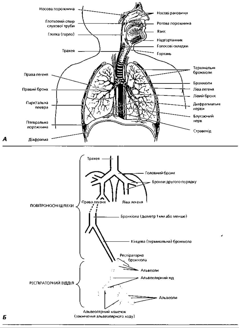
Fig. 4.72. General structural plan of the respiratory system: A - organ components; B - diagram of the conducting airways and respiratory Divisions of the lungs
Table 30. Structural Changes in the conducting airways
|
Nasal cavity |
Nasopharynx |
Larynx |
Trachea |
Bronchi |
Terminal bronchioles |
||
|
Large |
Small |
||||||
|
Epithelium |
Pseudostratified ciliated columnar |
Simple ciliated cuboidal |
|||||
|
Goblet cells |
Numerous |
Moderate amount |
Solitary |
Absent |
|||
|
Glands |
Numerous |
Moderate amount |
Absent |
Absent |
|||
|
Alar cartilages |
Absent |
Hyaline and elastic |
Incomplete rings |
Plates |
Absent |
||
|
Smooth myocytes |
Absent |
Connect the ends of cartilage |
Numerous bundles of myocytes |
||||
|
Elastic fibers |
Absent |
Moderate amount |
Numerous |
||||
1 In the nasal vestibule, the epithelium transitions from Cytology/practical/35.html">Stratified squamous keratinized to pseudostratified ciliated columnar; in the larynx, the true vocal cords and epiglottis are covered by stratified squamous non-keratinized epithelium
Basal (short) cells have a wide base and a narrow apex. These are stem (cambial) cells. It is also suggested that The primary function of these cells is to anchor the epithelium to the basement membrane. They contain numerous keratin filaments and are connected by desmosomes to neighboring cells and by hemidesmosomes to the basement membrane.
Intermediate (tall) cells are columnar in shape, and their apical pole does not reach the epithelial surface. They function as progenitor (cambial) cells.
Brush cells are columnar in shape, reaching the epithelial surface with their apical pole, which is covered with microvilli. At their basal pole, they form synapses with sensory nerve fibers, which is why a receptor function is attributed to them.
Bronchiolar exocrinocytes (Clara cells) are found only in the distal Regions of the conducting airways, as well as in the respiratory bronchioles. They have a dome-shaped apex that bulges above the epithelial surface. Clara cells produce surfactant components (see below) and function at the bronchiolar level. They possess a well-developed smooth Endoplasmic reticulum containing Enzymes involved in the detoxification of chemical compounds.
Endocrine cells are pyramidal in shape and contain secretory granules in their basal portion. These cells produce Peptide Hormones and biogenic amines. They belong to the diffuse neuroendocrine system (DNES / APUD system) of the body. The function of tracheal endocrine cells is the local regulation of smooth Muscle contraction in the airways.
Dendritic cells (Langerhans cells) are specialized antigen-presenting cells of Bone Marrow origin (sharing a common progenitor with macrophages). They form long, branching processes that lie between epithelial cells. They stimulate lymphocyte proliferation 10 to 100 times more effectively than macrophages.
Clearing of the airway mucosa from dust and microorganisms is carried out by the so-called mucociliary mechanism, which includes:
1) adhesion of particles to the mucus covering the epithelium;
2) their removal from the respiratory system through the continuous movement of mucus by the ciliated epithelium toward the pharynx, where it is swallowed or expelled into the external environment.
The mucus covering the airway epithelium consists of two layers. The outer layer is a viscous, elastic gel, 2 μm thick, which facilitates the adhesion of particles (microbes) and traps them on the mucus surface, preventing them from sinking deeper and contacting the epithelium; it has low permeability to water, thus preventing tissue desiccation. The inner layer (sol), 5 μm thick, allows free movement of the cilia. Its excess is absorbed by the epithelium.
Impairment of the mucociliary mechanism leads to The Development of infectious diseases and can occur As a result of:
1) changes in mucus volume and properties (for example, hypersecretion in smokers or increased viscosity in cystic fibrosis):
2) loss of cilia or impairment of their motility (for example, due to smoking, anesthesia, viral infections, as well as an inherited disorder — immotile cilia syndrome [Kartagener syndrome]).
The mucociliary clearance mechanism is also impaired when the ciliated epithelium is replaced in certain areas by stratified squamous epithelium, which occurs in chronic systemic respiratory diseases.
The nasal cavity (cavum nasi) consists of the vestibule and the nasal cavity proper, which includes the respiratory and olfactory regions. The vestibule is a cavity located beneath the cartilaginous part of the Nose. It is lined with stratified squamous epithelium, which is a continuation of the integumentary epidermis. The Connective Tissue layer beneath the epithelium contains Sebaceous Glands and the roots of nasal hairs (vibrissae).
The nasal cavity proper in the respiratory region is lined with a mucous membrane consisting of pseudostratified ciliated columnar epithelium and a connective tissue lamina propria. The lamina propria of the nasal mucosa is composed of loose connective tissue rich in elastic fibers and contains the secretory portions of mucous glands, whose excretory ducts open onto the epithelial surface. The secretion of these glands, combined with that of goblet cells, moistens the mucosa and traps dust particles and microorganisms, which are subsequently removed by Ciliary movement. The lamina propria also contains lymphatic nodules. Accumulations of these nodules near the auditory tubes form the tubal tonsils, and in the nasopharynx, they form the pharyngeal tonsil. Chronic inflammation of the latter leads to mucosal hypertrophy, known as adenoids, which obstruct nasal breathing.
The nasal mucosa is highly vascularized, with Blood Vessels located superficially, directly beneath the epithelium. This helps warm inhaled air during cold seasons but makes the area prone to nosebleeds. In the region of the Inferior nasal concha, There is a plexus of wide-lumen Veins. When engorged with blood, the mucosa swells, which can obstruct breathing.
The olfactory region (Fig. 4.73) of the nasal cavity proper functions as the peripheral part of the olfactory analyzer, located in the superior and posterior PARTS OF THE nasal cavity. The mucosa here has a yellowish tint. The epithelium is composed of three cell types:
1) olfactory receptor cells;
2) supporting (sustentacular) cells;
3) basal cells.
Olfactory receptor cells are modified bipolar Neurons. Each cell has a cell body, with a dendrite extending from its superficial part toward the epithelial surface, and an axon extending from its deep part. The receptor cells and the intervening supporting cells are oriented perpendicular to the mucosal surface. Their bodies are wedged between the supporting cells so that their nuclei reside in the expanded portion of the cells in the lower half of the epithelial layer, while the dendrites ascend through the gaps between the supporting cells to the surface, ending in a bulbous Swelling called the olfactory vesicle (knob). Long olfactory cilia (or hairs) project from the olfactory vesicle, lying along the epithelial surface and forming an uneven layer that covers the microvilli of the supporting cells. This layer is moistened by the secretion of the olfactory (Bowman's) glands located in the lamina propria of the mucosa. The axons of the receptor cells pass into the lamina propria, bundling to form the Olfactory nerve, which reaches the olfactory bulbs of the Brain.
The supporting Cells of the olfactory region are tall columnar cells with light nuclei located above those of the receptor cells. The basal ends of the supporting cells taper irregularly. The Cytoplasm of these cells contains a yellow-brown pigment, which gives the olfactory mucosa its yellow color. Basal cells are conical and sit on the basement membrane at some distance from one another. These are stem (cambial) cells that can differentiate into supporting or receptor cells.
The larynx. From the nasal cavity, inhaled air passes through the pharynx into the larynx, which, In addition to conducting air, is responsible for phonation (voice production). The primary function of the larynx is to prevent any substances other than air from entering the Lower Respiratory Tract. Consequently, the larynx is often referred to as the "watchdog" of the lungs. If foreign bodies or substances do enter, a cough reflex is immediately triggered. During swallowing, the laryngeal inlet is closed by the epiglottis, which plays an auxiliary role: the larynx is pulled upward and forward during swallowing so that its upper end is pressed against the posterior surface of the epiglottis below the ROOT of the Tongue.
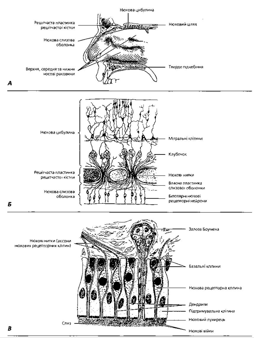
Fig. 4.73. Olfactory mucosa: A — localization of the olfactory region in the posterosuperior part of the nasal cavity; B — diagram of the Structural Organization of the olfactory pathways; C — cellular COMPOSITION OF THE olfactory region
The larynx is a tubular organ located at the level of the fourth to sixth cervical vertebrae. The laryngeal wall consists of three layers: the mucosa with its submucosa, the fibrocartilaginous layer, and the adventitia. The mucosa consists of an epithelium, a lamina propria, and a submucosa. The epithelium here is pseudostratified ciliated columnar with numerous goblet cells, except for the region of the true vocal cords and the epiglottis, which are covered by stratified squamous non-keratinized epithelium. The lamina propria and the submucosa of the larynx are composed of loose connective tissue and blend into each other without distinct boundaries. The lamina propria and submucosa contain elastic fibers that gradually merge with the perichondrium of the laryngeal cartilages and also lie between the striated Muscles of the vocal folds. Bundles of elastic fibers located at the Base of the free edges of the vocal folds are called the vocal ligaments. The space between the edges of the vocal folds forms the rima glottidis (glottis), the size of which, along with the tension of the vocal ligaments, changes depending on the contraction of the striated muscles located within the vocal folds (vocalis muscles). On the anterior surface of the larynx, the submucosa contains the secretory portions of mixed seromucous glands, as well as accumulations of lymphatic nodules that form the laryngeal tonsil.
The fibrocartilaginous layer is composed of hyaline and elastic cartilages of various shapes, surrounded by dense Fibrous connective tissue. The paired corniculate and cuneiform cartilages are elastic, while the unpaired thyroid and cricoid cartilages, as well as the paired arytenoid cartilages, are hyaline. The fibrocartilaginous layer forms the supporting framework of the larynx, which prevents its walls from collapsing and ensures a continuous flow of air into the lower respiratory tract. The adventitia of the larynx is formed by loose connective tissue.
The larynx is separated from the pharynx by the epiglottis, the core of which is formed by elastic cartilage. Here, the pharyngeal mucosa transitions into the laryngeal mucosa. On both surfaces of the epiglottis, the mucosa is covered by stratified squamous non-keratinized epithelium. The lamina propria forms papillae that interdigitate with the epithelium.
The trachea is a tube 11 cm long and 20-25 mm in diameter, extending from the level of the sixth cervical to the fifth thoracic vertebra. It is composed of the same three layers as the larynx: the mucosa with its submucosa, the fibrocartilaginous layer, and the adventitia (Figs. 4.74, 4.75). The tracheal epithelium rests on a thick basement membrane. The lamina propria of the mucosa contains elastic fibers oriented mainly in the longitudinal direction; these sometimes form a distinct layer at the boundary with the submucosa.
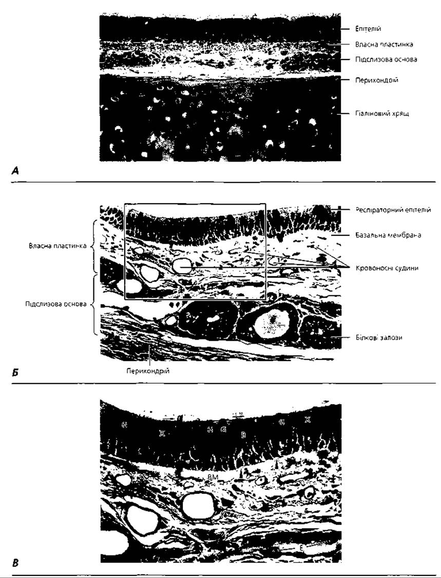
Fig. 4.74. Light Microscopy of the tracheal wall with increasing magnification. A — x 180; B — x 300; C — x 400. G — goblet cells; X — ciliated cells; B — brush (microvillous) cells; BM — basement membrane; E — endothelium of a venule; arrows indicate the direction of ciliary movement, arrowheads indicate basal cells
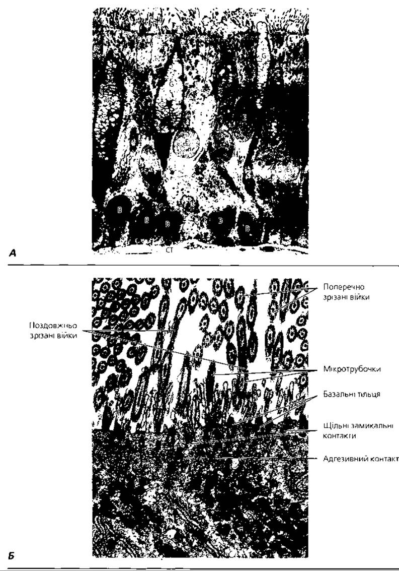
Fig. 4.75. Transmission Electron microscopy of the tracheal epithelium: A — MAIN TYPES OF tracheal epithelial cells: E — ciliated cells; B — basal cells; G — goblet cells; CT — connective tissue, x 1300; B — ULTRASTRUCTURE OF THE apical portion of the ciliated epithelium, x 6500
As in the larynx, the lamina propria of the trachea is usually not demarcated from the submucosa. Solitary lymphatic nodules may occur here. The submucosa of the trachea also contains the secretory portions of mixed (seromucous) glands, which are primarily localized in the posterior and lateral parts of the organ. The loose connective tissue of the submucosa gradually transitions into the Dense connective tissue of the perichondrium of the hyaline cartilage C-rings (semi-rings), 16-20 of which support the fibrocartilaginous layer of the trachea. On the posterior wall, these rings are open and joined by bundles of smooth muscle cells (trachealis muscle) attached to the outer surface of the cartilage. Due to the presence of this muscle, the posterior surface of the trachea is flexible and does not obstruct the passage of food through the Esophagus, which lies directly behind the trachea. The tracheal cartilages are connected to each other vertically by dense connective tissue. The adventitia consists of loose connective tissue that connects the trachea to other mediastinal Organs.
The bronchial tree (arbor bronchialis). At the level of the fifth thoracic vertebra, the trachea divides dichotomously into two main (primary) bronchi (right and left), which enter the right and left lungs, respectively, and branch into lobar (secondary) bronchi. The lobar bronchi branch into zonal bronchi (four in each lung), segmental (tertiary) bronchi (ten in each lung), subsegmental bronchi, small bronchi, and terminal bronchioles. Depending on their wall Structure and diameter, all the aforementioned bronchi are classified into main, large, medium, small bronchi, and terminal bronchioles.
The bronchi share a general structural plan similar to that of the trachea; that is, their wall is composed of three tunics: the mucosa with its submucosa, the fibrocartilaginous tunic, and the adventitia. The Specific features of these tunics depend on the caliber of the bronchus. Therefore, only the distinguishing characteristics of these tunics in Different types of bronchi will be highlighted below.
The main bronchi have a diameter of about 15 mm. Unlike the trachea, their mucosa features a muscularis mucosae that separates the mucosa from the submucosa. It is thin and consists of two layers of smooth muscle cells: an inner circular layer and an outer longitudinal layer. The mucosa of the main bronchus, like that of the trachea, does not form folds. A distinguishing feature of the main bronchi is the fibrocartilaginous tunic, which is composed of complete rings of hyaline cartilage.
Large bronchi (Fig. 4.76) have a diameter ranging from 15 to 5 mm. The muscularis mucosae is well-developed, consisting of a single layer of smooth muscle cells oriented in an oblique-circular direction. Due to their contraction, the mucosa of these bronchi forms longitudinal folds. Lymphatic nodules are frequently observed in the lamina propria of the mucosa. The lamina propria of the bronchi is rich in longitudinally arranged elastic fibers, which allow the bronchi to stretch and recoil during respiration. The submucosa contains numerous glands. Unlike the main bronchi, the fibrocartilaginous tunic is formed not by a continuous ring of cartilage, but by separate cartilaginous plates, the size of which decreases as the caliber of the bronchus diminishes. The secretory portions of mixed seromucous glands are located in large groups, primarily in those areas of the bronchial wall where cartilage is absent.
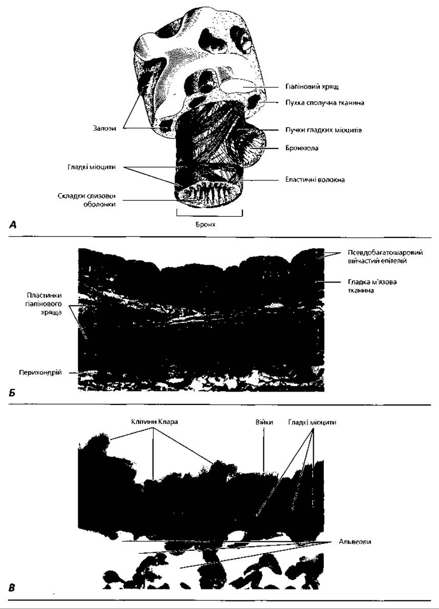
Fig. 4.76. Bronchi: A - three-dimensional reconstruction of a large bronchus; B - light microscopy of the wall of a large bronchus, x200; C - wall segment of a terminal bronchiole, x 650
Medium-sized bronchi have a diameter ranging from 5 to 2 mm. The thickness of the mucosa and the height of the epithelial layer decrease. The muscularis mucosae and its folds are well-developed. The fibrocartilaginous tunic contains only isolated islands of hyaline cartilage, with elastic cartilage appearing in some places. Glands are still present in the submucosa.
Small bronchi have a diameter of 2 to 0.5 mm. The epithelium becomes pseudostratified (two-layered). The muscularis mucosae is well-developed in small bronchi. These bronchi lack glands and cartilage. Thus, small bronchi possess only a mucosa and an outer adventitia, or four layers: epithelial, lamina propria, muscularis, and adventitia.
Terminal bronchioles have a diameter of about 0.5 mm and a length of up to 1200 µm. The general structural plan of their walls is similar to that of small bronchi, but the wall thickness is significantly smaller. The epithelium becomes simple ciliated cuboidal. The muscularis mucosae in terminal bronchioles has a net-like arrangement of smooth muscle cells, which prevents The formation of mucosal folds.
Lung (pulmo). Respiratory Portion. The lung is covered by a serous membrane—the visceral Pleura, which is composed of a simple squamous epithelium (mesothelium) facing the pleural cavity, and a lamina propria of connective tissue fused with the lung parenchyma. The lung contains part of the conducting airways (bronchial tree) as well as the respiratory portion (alveolar tree). The lung consists of lobes, segments, lobules, and acini. The right lung has three lobes, and the left has two. Each lung contains ten segments and about 800 lobules. A lobule consists of 12-18 acini, with approximately 15,000 acini in each lung.
The pulmonary lobule is the branching territory of a small bronchus (Fig. 4.77). It is pyramidal in shape, with a height of 51-27 mm and a width of 9-21 mm. A small bronchus enters the lobule through its apex and, branching dichotomously, forms terminal bronchioles at the boundary between its upper and middle thirds. The branching of terminal bronchioles gives rise to the respiratory portion of the lungs. The branching territory of a single terminal bronchiole is the Structural and functional unit of the respiratory portion of the lungs, known as the pulmonary acinus.
The acinus is composed of three parts:
1) respiratory bronchioles (I, II, III orders);
2) alveolar ducts;
3) alveolar sacs.
First-order respiratory bronchioles are formed by the dichotomous division of terminal bronchioles. They have the same length, diameter, and wall structure as terminal bronchioles, but their epithelium lacks ciliated cells. The main distinguishing feature of these bronchioles is the presence of small air sacs—alveoli—in their walls. Second-order respiratory bronchioles are shorter (up to 800 µm), and the number of alveoli in their walls increases. Third-order respiratory bronchioles are even shorter—up to 500 µm—and contain even more alveoli. Alveolar ducts have a diameter two to three times larger than respiratory bronchioles and A large number of alveoli, with only small remnants of the alveolar duct's own wall remaining between them. Alveolar sacs are composed of several adjacent alveoli.
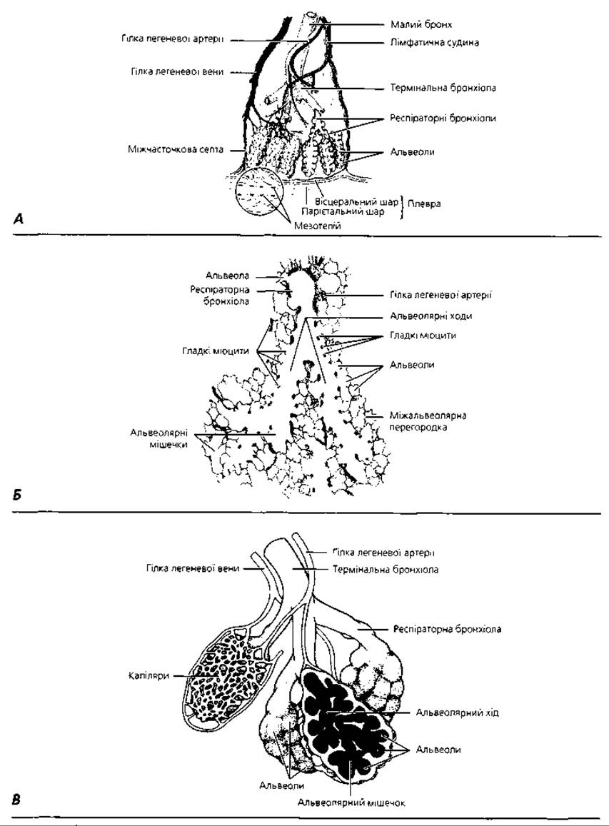
Fig. 4.77. Respiratory portions of the lungs: A - three-dimensional rendering of a pulmonary lobule; B - semi-schematic representation of a section of a pulmonary acinus; C - diagram of a three-dimensional reconstruction of a pulmonary acinus
A pulmonary alveolus is an open vesicle, an air-filled sac through whose thin wall gas exchange occurs (Figs. 4.77, 4.78, 4.79). The total number of alveoli in a single adult lung is 300-400 million. The maximum surface area of all alveoli in an adult during inhalation reaches 100 m2. In an adult, the size of the alveolar entrance is 0.15-0.25 mm, and its depth is 0.06-0.3 mm. The alveolar wall contains openings—the so-called pores of Kohn—with a diameter of 9-19 µm, which connect adjacent alveoli.
The average number of pores per alveolus is 13-21, half of which are located on the alveolar wall opposite the entrance. The average area occupied by the pores of Kohn is 1-5% of the alveolar surface area. Internally, the alveolus is lined by a continuous layer of epithelium resting on a basement membrane. Among the alveolar epithelial cells, a distinction is made between small respiratory epithelial cells (type I alveolar cells) and large secretory, granular epithelial cells (type II alveolar cells).
Type I alveolar cells (Figs. 4.78, 4.79) cover 97% of the alveolar surface, measuring 5-6 µm in the nuclear region and 0.2-0.3 µm in the cytoplasmic region. These are squamous cells. Their broad cytoplasmic extensions, called squames, are up to 10 µm long. It is these regions of the respiratory cell processes that are adapted for gas exchange, which is the primary function of these cells. Type II alveolar cells, or secretory alveolar cells (Figs. 4.78, 4.79, 4.80), cover 3-4% of the alveolar surface, being located near or at the margins of the pores. They measure up to 10 µm, have a rounded shape, bulge into the alveolar lumen, and form tight junctional complexes with respiratory cells. The cytoplasm of these cells contains osmiophilic bodies, a well-developed Golgi complex, and endoplasmic reticulum. Type II alveolar cells produce phospholipoprotein complexes that form surfactant membranes. With the involvement of the smooth Endoplasmic reticulum and the Golgi complex, Phospholipids are synthesized and accumulate as lamellar bodies. Lipoprotein molecules synthesized in the granular endoplasmic reticulum and the Golgi complex accumulate as multivesicular bodies. Before secretion from The Cell, lamellar and multivesicular bodies fuse, after which the newly formed phospholipoprotein complex is released onto the cell surface via exocytosis. In addition to type I and type II alveolar cells, alveolar macrophages are found in the alveolar wall. These cells belong to the mononuclear phagocyte system, originate from the bone marrow, and perform a protective function. When a significant amount of phagocytosed dust particles accumulates in their cytoplasm, they are referred to as dust cells.
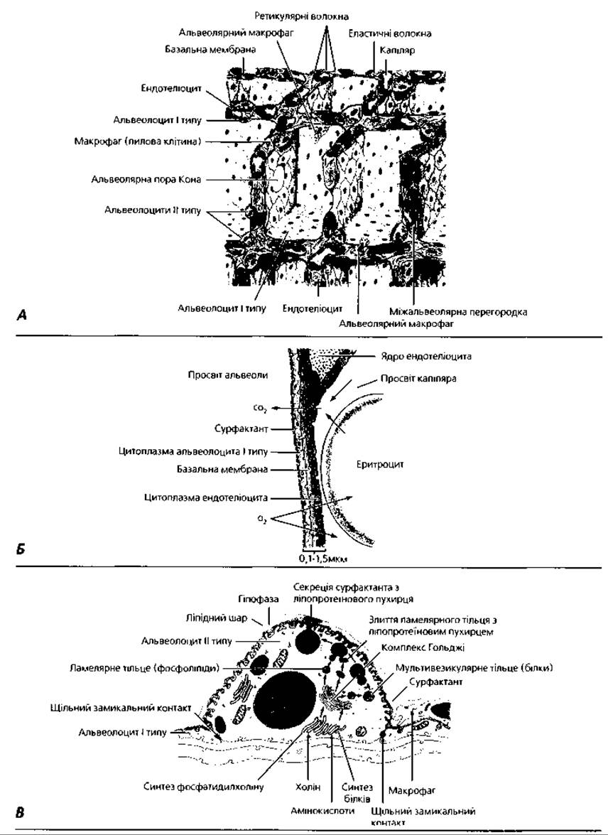
Fig. 4.78. Structure of pulmonary alveoli: A - diagram of the ultrastructural organization of alveoli; B - STRUCTURE OF THE alveolocapillary (blood-air) barrier; C - ultrastructure of a type II alveolar cell
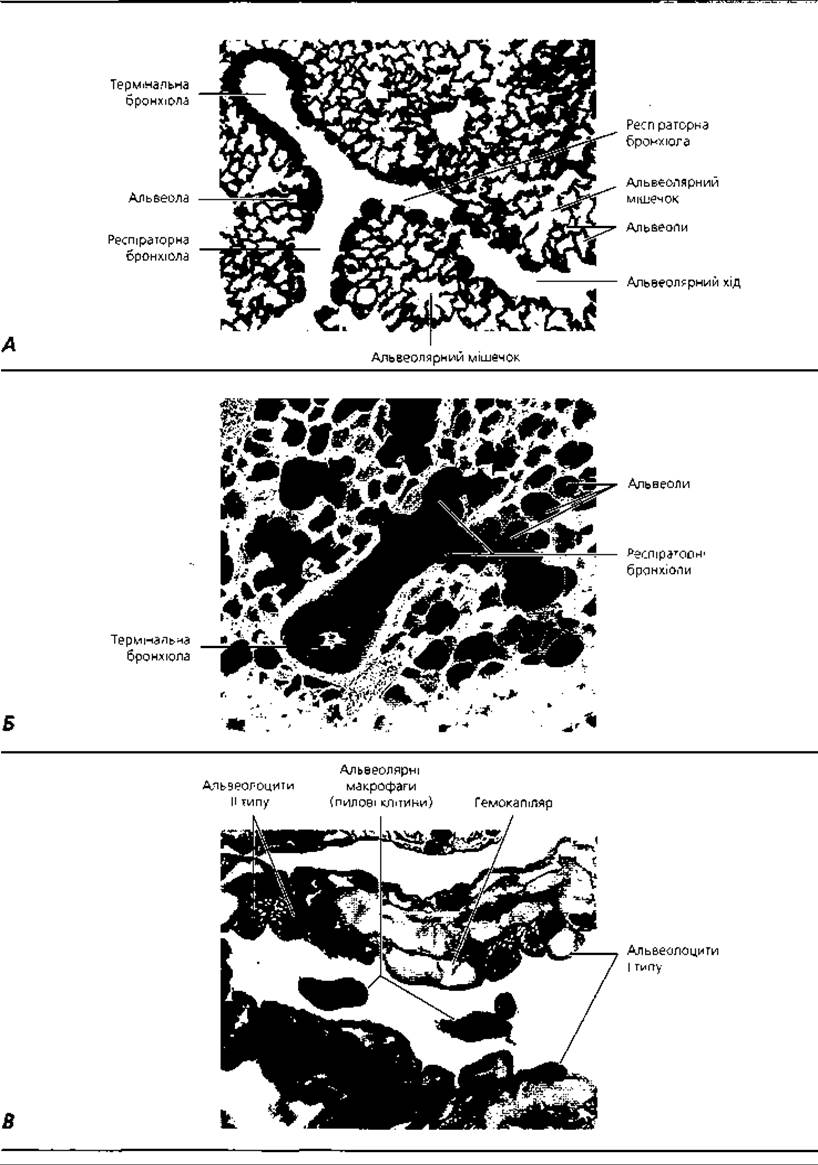
Fig. 4.79. Respiratory portions of the lungs: A - light microscopy of the branching region of a terminal bronchiole, x 65; B - scanning electron micrograph of a terminal bronchiole, respiratory bronchioles, and alveoli, x 130; C - light microscopy of the pulmonary alveolar wall, x 1100
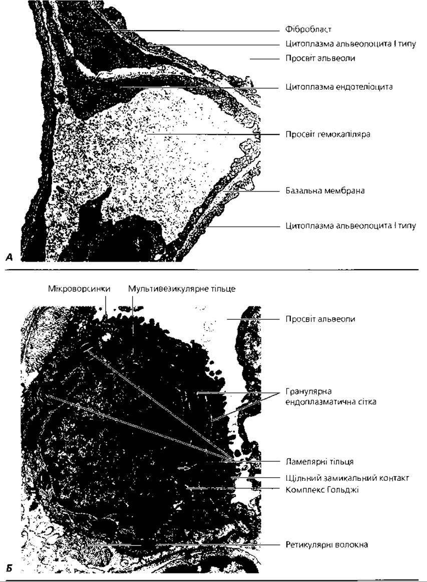
Fig. 4.80. Transmission electron microscopy of pulmonary alveoli: A - ultrastructure of the blood-air (alveolo-capillary) barrier, x 32 000; B - type II pneumocyte, x 12 000
The pulmonary surfactant system (surfactant) is a thin film that lines the inner surface of the alveoli and is in contact with air. Surfactant consists of two phases: a membranous phase and a liquid phase (hypophase). The superficial membranous component is composed of phospholipids and Proteins, while the underlying liquid hypophase consists of water-Soluble Glycoproteins. The primary function of surfactant is to reduce surface tension, thereby preventing alveolar collapse during expiration. In addition, it exerts a bactericidal effect and prevents airborne microorganisms from penetrating the alveolar wall. Surfactant also prevents the transudation of fluid from capillaries into the alveoli and facilitates the migration of alveolar macrophages and lymphocytes.
The differentiation of type II pneumocytes and their synthesis of surfactant begin at THE START OF the seventh month of human Embryogenesis. It is the presence of this surfactant film in the alveoli of a newborn that allows them to expand During the first breath and remain open thereafter. Certain developmental anomalies prevent the formation of surfactant in the lungs, rendering the newborn non-viable.
Externally, blood capillaries and a network of elastic fibers that wrap around the alveoli lie adjacent to the alveolar basement membrane. Because the alveoli are closely packed, each capillary borders several alveoli simultaneously. This provides optimal conditions for gas exchange between the blood flowing in the capillaries and the air filling the alveolar cavities. The structures through which gas exchange occurs form the so-called blood-air barrier. It comprises the alveolar wall and the Capillary Wall (Fig. 4.78, B, 4.80, A), with an average thickness of 0.5 µm. The Components of the blood-air barrier are as follows: surfactant, the anucleate regions of respiratory pneumocytes, the alveolar basement membrane, the capillary basement membrane, and the anucleate regions of the capillary endothelium. Often, the basement membranes of the alveolus and the blood capillary fuse into a single shared alveolar-capillary membrane, creating optimal conditions for respiratory function.
Development of the respiratory system. The larynx, trachea, and lungs develop from the laryngotracheal diverticulum (laryngo-tracheo-pulmonary primordium), which appears during the third to fourth week as an outgrowth of the ventral wall of the foregut. The upper part of this primordium gives rise to the larynx and trachea. In its lower part, it divides into two lung buds, which are the precursors of the lungs. During development, numerous outgrowths arise from these buds, surrounded by mesenchyme. Each terminal branch of the outgrowth ends in an expansion—the future alveolar sac. This forms the bronchial tree, which branches like a compound alveolar gland. From the sixth month until birth, alveolar development occurs in the lungs. Throughout the prenatal period, the alveoli have a narrow lumen and thick walls. With the newborn's first breath, the alveoli expand, their lumens enlarge, and their walls thin, facilitating gas exchange. Surfactant production begins at the start of the seventh month of gestation; therefore, premature infants born before this term are considered unable to breathe on their own. To support pulmonary Gas Exchange in these infants, exogenous surfactant is administered into their respiratory tracts.
Key terms
1. Conducting airways. 2. Respiratory portion. 3. Nasal cavity. 4. Nasal vestibule. 5. Nasal cavity proper. 6. Respiratory and olfactory regions. 7. Mucous membrane. 8. Brush cells. 9. Tubal tonsil. 10. Pharyngeal tonsil. 11. Olfactory receptor cells. 12. Supporting cells. 13. Olfactory vesicle. 14. Olfactory cilia. 15. Olfactory (Bowman's) glands. 16. Laryngeal tonsil. 17. Seromucous glands. 18. Tracheal endocrine cells. 19. Main bronchi. 20. Large bronchi. 21. Medium bronchi. 22. Small bronchi. 23. Terminal bronchioles. 24. Lobar bronchi. 25. Zonal bronchi. 26. Segmental bronchi. 27. Subsegmental bronchi. 28. Bronchiolar exocrine cells (Clara cells). 29. Non-ciliated cells. 30. Brush cells. 31. Pleura. 32. Pulmonary lobe. 33. Pulmonary segment. 34. Pulmonary lobule. 35. Pulmonary acinus. 36. Respiratory bronchioles. 37. Alveolar ducts. 38. Alveolar sacs. 39. Alveoli. 40. Type I pneumocytes (respiratory alveolar cells). 41. Type II pneumocytes (great alveolar cells). 42. Surfactant complex. 43. Alveolar macrophages (dust cells). 44. Blood-air barrier. 45. Alveolar pores of Kohn. 46. Alveolar-capillary membrane. 47. Laryngo-tracheo-pulmonary primordium.
Last update: 09/08/2026
Editorial and Educational Adaptation: This material has been compiled based on the primary/original source text. The project team performed an editorial review, corrected technical inaccuracies, structured sections, and adapted the content for an educational format.
What was processed:
- elimination of formatting defects (OCR errors, structural breaks, corrupted characters);
- editorial organization of content;
- standardization of terminology in accordance with academic sources;
- verification of factual statements against the original source text.
All mentions of the author, publication year, and origin of the primary text have been preserved in accordance with the source.