Special Histology and Embryology: Practical Course - V. K. Napkhanyuk 2001
Respiratory System
Respiratory Portion
The Structural and functional unit of the respiratory division of the lung is the pulmonary acinus (acinus pulmonaris). It is a system of alveoli located in the walls of the respiratory bronchioles, alveolar ducts, and alveolar sacs, which facilitate gas exchange between the Blood and alveolar air. The acini are separated from one another by thin Connective Tissue septa; 12–18 acini make up a pulmonary lobule. Each lung consists of lobes, segments, lobules, and acini.
An acinus begins with a 1st-order respiratory bronchiole, into the lumen of which alveoli open. Each 1st-order bronchiole branches dichotomously into 2nd-order bronchioles, and subsequently into 3rd-order bronchioles.
The 3rd-order respiratory bronchioles subdivide into alveolar ducts, which terminate in a pair of alveolar sacs.
Respiratory bronchioles are lined with a simple cuboidal epithelium. Ciliated Cells are rarely observed. The muscular layer thins out and breaks down into separate circularly arranged bundles of smooth myocytes. The connective tissue fibers of the adventitia merge with the interstitial connective tissue.
The walls of the alveolar ducts and alveolar sacs feature several dozen alveoli separated by thin connective tissue septa that contain blood capillaries. The alveoli communicate with one another via alveolar pores with a diameter of 10-15 µm.
Alveoli resemble open vesicles whose lumen is lined by three Cell types:
1) respiratory epithelial cells (type I pneumocytes), which cover approximately 95% of the alveolar surface. These are irregular, flattened, and elongated cells with a thickness of 5-6 µm in the nuclear region and 0.2-0.3 µm in other areas. The free surface of their Cytoplasm bears cytoplasmic projections. The cytoplasm contains small Mitochondria and pinocytotic vesicles. The anuclear regions of type I cells lie adjacent to the anuclear regions of capillary endothelial cells. Due to this structural relationship between the walls of the alveoli and capillaries, the blood-air barrier (air-blood barrier) is extremely thin—0.5 µm (micrographs 37, 38);
2) large epithelial cells (type II pneumocytes), whose number is approximately equal to that of type I cells, yet they occupy only 5% of the alveolar surface. These are cuboidal or polygonal cells whose cytoplasm contains osmiophilic lamellar bodies containing surfactant—the surface-active, amorphous lining material of the alveoli, which is released through secretion;
3) alveolar macrophages, whose cytoplasm contains a significant number of lipid droplets and Lysosomes.
Slide 2. Section of the lung (Fig. 62).
Low magnification. Examine and draw the slide. Locate the medium and small intrapulmonary Bronchi.
The cartilaginous framework of a medium bronchus on the slide is represented by isolated plates of hyaline Cartilage. Medium bronchi are lined with a pseudostratified ciliated epithelium, beneath which lies a thin lamina propria. There is also a continuous circular layer of smooth Muscle separating the submucosa, where occasional mucous gland acini are still found. Most small bronchi appear stellate in cross-section. It is evident that a small bronchus is lined with cuboidal epithelium, followed by a circular layer of smooth muscle beneath the lamina propria; a cartilaginous framework is absent, though individual small gland acini can still be detected in the submucosa. The bronchi are accompanied by bronchial Arteries, the cross-sections of which are consistently found near medium and small bronchi.
In a respiratory bronchiole, the wall is lined with a simple cuboidal epithelium, and smooth muscle Cells of the muscular layer are visible, which are intermittently interrupted by alveoli. In the wall of the alveolar duct, the alveoli are closely apposed. Bundles of smooth muscle cells and connective tissue fibers are encountered only at their openings. Alveolar sacs are blind outpouchings consisting of three to four alveoli.
Identify the following on the figure: 1) medium-sized bronchus: a) tunica mucosa of the bronchus; b) submucosa with bronchial glands and Blood Vessels; c) cartilage plate of the fibrocartilaginous tunic; d) adventitia; 4) respiratory bronchiole; 5) alveolar duct; 6) alveolar sac; 7) alveolus; 8) blood vessels.
Class="center">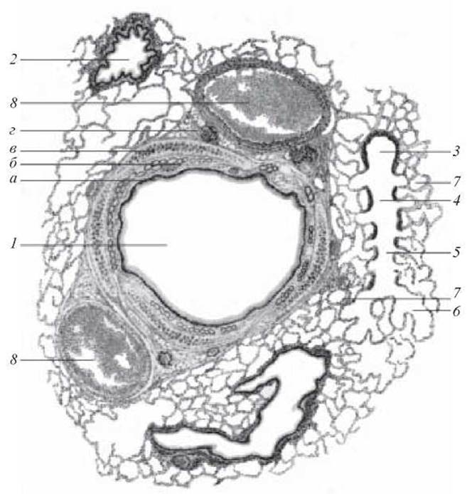
Fig. 62. Lung section. Hematoxylin and eosin staining. x 56:
1 — medium-sized bronchus (a — tunica mucosa of the bronchus; b — submucosa; c — cartilage plate of the fibrocartilaginous tunic; d — adventitia); 2 — small bronchus; 3 — terminal bronchiole; 4 — respiratory bronchiole; 5 — alveolar duct; 6 — alveolar sac; 7 — alveolus; 8 — blood vessels
1. Sources of Development of the Respiratory system.
2. What two divisions comprise the respiratory system?
3. Structure and histophysiology of the Nasal cavity.
4. STRUCTURE OF THE Larynx and its histophysiology.
5. The Trachea, its structure, and histophysiology.
6. Lungs, General structural layout.
7. Structural Features of the bronchial tree.
8. Structure of the respiratory bronchioles.
9. The acinus and its structure.
10. Structure of the alveoli.
11. Structural components of the air-blood barrier.
12. Micro- and ultrastructure and function of respiratory and large epithelial cells (alveolocytes).
13. Which structures are involved in clearing dust particles inhaled from the air through the airways down to the alveoli?
14. What is the surfactant complex and what is its significance?
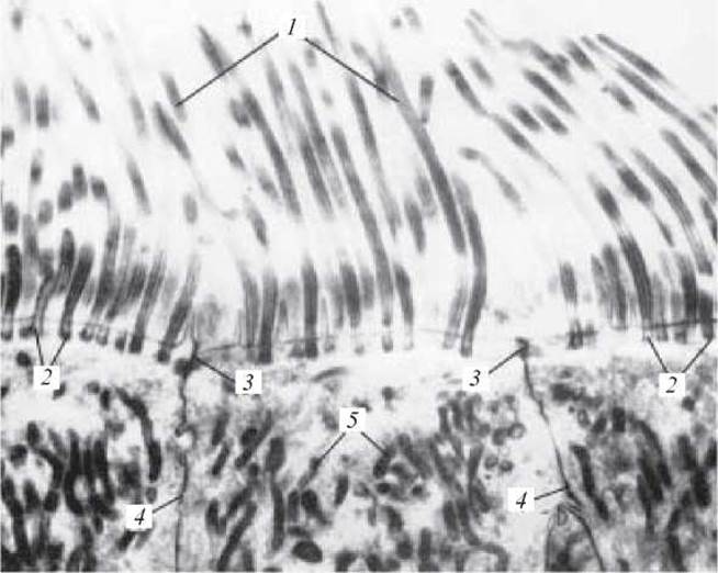
Electron micrograph 36. Ciliated epithelial cells of the trachea. x 8000:
1 — cell cilia; 2 — basal bodies; 3 — junctional complexes between cells; 4 — cell boundaries; 5 — mitochondria (from Rodin's atlas)
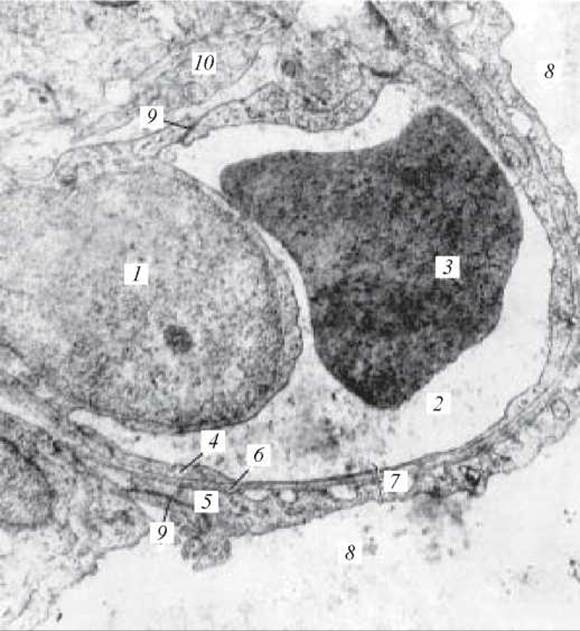
Electron micrograph 37. Alveolar wall and pulmonary blood capillary. x 25,000:
1 — Nucleus of a blood capillary endothelial cell; 2 — lumen of the blood capillary; 3 — erythrocyte in the lumen of the blood capillary; 4 — cytoplasm of the blood capillary endothelial cell; 5 — cytoplasm of the alveolar epithelial cell; 6 — basal laminae of the endothelium and epithelium; 7 — air-blood barrier; 8 — alveolar lumen; 9 — desmosomes; 10 — portion of a CONNECTIVE TISSUE CELL of the interalveolar septum (after V. A. Shakhlamov)
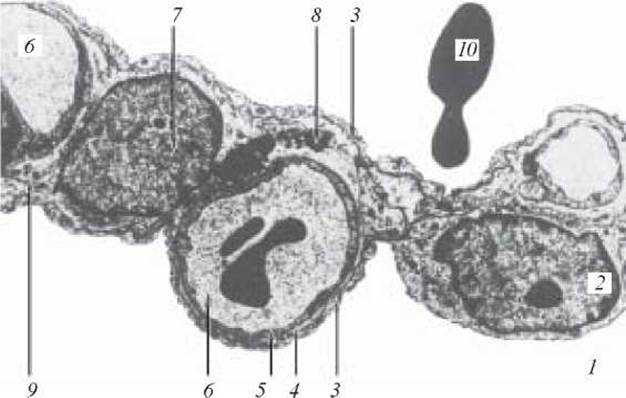
Electron micrograph 38. Air-blood barrier of the human lung. x 9000:
1 — alveolar space; 2 — epithelial cells; 3 — long cytoplasmic processes; 4 — basement membrane; 5 — endothelium; 6 — capillaries; 7 — septal cells; 8 — elastic fibers; 9 — Collagen fibers; 10 — erythrocytes (after V. V. Serov and V. S. Paukov)
Situational problems
1. In which bronchus are all tunics developed, and the fibrocartilaginous framework contains two or three large plates of hyaline cartilage tissue?
2. In Bronchial Asthma, suffocating attacks occur due to spasms of smooth muscle cells in intrapulmonary bronchi. What caliber of bronchi is primarily involved? Explain this based on their morphological features.
3. Microscopic examination of the trachea revealed excretory ducts and secretory end-pieces of glands. The cytoplasm of the glandular cells exhibited different tinctorial properties: some cells showed distinct basophilia, while the cytoplasm of other cells was pale and weakly stained. To which type, based on The chemical composition of their secretion, do these glands belong?
4. An electron micrograph of an alveolus shows an alveolocyte containing numerous electron-dense osmiophilic bodies in its cytoplasm. What cell is this, and what role does it play in protecting the alveolar cells?
5. In the regional Lymph Nodes of smokers and individuals who constantly breathe dust-laden air, A large accumulation of particles and dust is deposited. Which cells transport them here?
6. During moderate physical exertion, modifications are observed in the structures of the respiratory bronchioles, associated with enhanced plastic processes in myocytes, nuclear hypertrophy, and an increasing mitotic index. What structural changes will be observed in the wall, and what consequences will this lead to?
Sample exam questions
1. Nasal cavity. Embryonic development. Structure and histophysiology of the nasal cavity wall.
2. The respiratory system. General morphofunctional CHARACTERISTICS OF THE respiratory tract. Structure of the Trachea and Bronchi of various calibers.
3. The lungs. Sources of development. General morphofunctional characteristics. The pulmonary acinus, structure of the alveolar wall. The blood-air barrier. The pulmonary lobule. Features of the Blood supply to the lungs. Age-related changes.
Last update: 10/08/2026
Editorial and Educational Adaptation: This material has been compiled based on the primary/original source text. The project team performed an editorial review, corrected technical inaccuracies, structured sections, and adapted the content for an educational format.
What was processed:
- elimination of formatting defects (OCR errors, structural breaks, corrupted characters);
- editorial organization of content;
- standardization of terminology in accordance with academic sources;
- verification of factual statements against the original source text.
All mentions of the author, publication year, and origin of the primary text have been preserved in accordance with the source.