BOTANY VOLUME 2 - PLANT PHYSIOLOGY - 2007
6. PHYSIOLOGY OF METABOLISM
6.4. Photosynthesis. Light Reactions
The ability to synthesize Organic compounds from inorganic ones using solar energy is a hallmark of photoautotrophic organisms (see Table 6.1); this overall process is called Photosynthesis. On the one hand, CARBOHYDRATES are formed from atmospheric carbon dioxide during photosynthesis (carbon assimilation, see 6.5). On the other hand, solar energy is used to form ammonium nitrogen from absorbed nitrate (nitrate assimilation, see 6.6) and to convert sulfate into sulfide (sulfate assimilation, see 6.7). In this process, carbon, nitrogen, and sulfur are reduced; in the case of green plants, cyanobacteria, and prochlorophytes1, the necessary electrons are extracted from Water, whereas in some photosynthetically active Bacteria, they are derived from other sources (see Table 6.1). In the light phase of photosynthesis, after the absorption of a light quantum in membrane-bound photosynthetic reaction centers, electrons are released from the chlorophyll pigment and then transferred along the Electron Transport Chain to ferredoxin. Reduced ferredoxin serves as an electron donor during nitrogen and sulfur assimilation or for the reduction of oxidized pyridine NUCLEOTIDES (NADP+ in cyanobacteria, prochlorophytes, and green plants; NAD+ in other photosynthetically active bacteria), forming the reducing equivalent NADPH + H+ (or NADH+ + H+). Photosynthetic Electron transport is coupled with the directional transport of hydrogen ions across the membrane, which is utilized in ATP synthesis.
1 Prochlorophytes (Prochlorales) are close to the METABOLISM/14.html">Chloroplasts of green plants in terms of pigment composition and structural Organization: they contain chlorophyll b, have grana, etc. Sometimes they are grouped with cyanobacteria, but here, to emphasize The Role of prochlorophytes as "ancestral" chloroplasts, they are considered separately. — Editor's note.
The positive charge of the reaction centers in higher plants, cyanobacteria, and prochlorophytes is replenished by electrons from water. ATP formed during the light phase and reduced NADPH (or NADH) are used for carbon assimilation. The synthesis of carbohydrates from CO2 (see 6.5) is often called the Dark Phase of photosynthesis because it does not directly depend on light and, in the presence of ATP and NAD(P)H, could proceed in the dark. The light reactions of photosynthesis in green plants and cyanobacteria occur on thylakoid membranes. These are located in the stroma of green plant chloroplasts (see 2.2.9.1; Fig. 2.83). The thylakoid membranes of cyanobacteria consist of folds of Cell/30.html">The Plasma Membrane and lie in the Cytoplasm (see Fig. 2.89). In other photosynthetic bacteria, the light phase takes place on the plasma membrane.
Among autotrophs, the most crucial role is played by photoautotrophs, which meet their energy needs through light radiation; chemoautotrophs (see Table 6.1 and Section 6.9), which obtain their necessary energy from The oxidation of Inorganic Compounds, lag behind in quantitative terms. Thus, photosynthesis forms the basis for life on Earth. This is also supported by calculations: despite a significantly smaller number of species (approximately 400,000 plant species compared to more than 2 million animal species), the total plant biomass produced (phytomass) is almost 1,000 times greater than the animal biomass (zoomass, including humans). Phytomass, for its part, is more than 99% composed of terrestrial plants (Table 6.16). Global CO2 fixation by plants on Earth is about 275 billion tons per year.
Class="center">Table 6.16. Earth's biomass and its distribution on land and in the oceans (dry weight in 109 t)
Biomass |
Continents |
World Ocean |
Phytomass |
1837 |
3.9 |
Zoomass |
1.005 |
0.997 |
Humans |
0.052 |
|
Total biomass |
1,838.057 |
4.897 |
The basis of all photosynthetic processes is the absorption of light quantum radiation energy by pigments. Consequently, in nature, photosynthesis depends on sunlight. The electromagnetic radiation of the Sun is generated by The conversion of hydrogen atoms into helium atoms:
4H —> 42He + 2β+ + ∆E, (6.40)
where β+ are positively charged particles — positrons.
The mass defect arising during nuclear fusion (a helium atom is 0.029 mass units lighter than four hydrogen atoms) leads to the release of energy in the form of electromagnetic radiation ∆E.
Every day, the Sun radiates about 3 · 1031 kJ of energy, of which approximately 1.5 · 1019 kJ reaches the Earth's surface. According to Einstein's formula E = m · c2, every 9 · 1013 kJ corresponds to 1 kg of solar matter converted into energy. Thus, The amount of energy reaching the Earth during a day corresponds to about 165 t of matter (about 60,000 t per year). Half of this energy reaches the Earth's surface, and only a small fraction (about 0.01%) is required by plants for photosynthesis, totaling 3.6 · 1038 kJ per year (corresponding to 40 t of matter). Using this energy, plants synthesize about 2 · 1011 t of biomass annually.
Electromagnetic radiation has a dual nature: it can be represented both as waves and as a stream of particles consisting of quanta. The energy of a single quantum (∆Eq) can be derived from the formula

where h is Planck's constant, 6.626 · 10-34 J · s; c is the speed of light, 3 · 108 m · s-1; is the wavelength, nm; v is the frequency, s-1.
Fig. 6.41. Electromagnetic radiation spectrum. Biologically important processes occur in the wavelength range of 100 — 1,000 nm: A — bacterial death (maximum), B — sunburn of the Skin (maximum), C — photosynthetic region, D — light visible to the human eye, E — region of Bacterial photosynthesis. The ultraviolet light region in the range of 250 — 280 nm is called UV-C; 280 — 320 nm is UV-B, 320 — 390 nm is UV-A. Quantum energy decreases with increasing wavelength

Due to the absorption properties of atmospheric ozone in the ultraviolet region (Fig. 6.42), and of atmospheric carbon dioxide and water in the infrared, THE SPECTRUM OF radiation reaching the surface narrows to 340 — 1,100 nm. In water, the infrared region decreases particularly rapidly with depth because absorption occurs in the red, orange, yellow, and green PARTS OF THE spectrum, and the blue light range narrows so that only a narrow "window" remains in this region (Fig. 4.43). Aquatic plants must adapt to the changing quality of light with increasing depth.
According to Equation 6.41, the energy of electromagnetic radiation increases proportionally to the frequency of the radiation. It is inversely proportional to the wavelength, i.e., the amount of energy decreases as the wavelength of the radiation increases.
The region of the electromagnetic spectrum visible to the human eye is called light, and light quanta are called photons (from the Greek phos — light). Light covers the wavelength range of approximately 400 — 700 nm (Fig. 6.41); the entire solar spectrum spans 225 — 3,200 nm, thus extending from the ultraviolet to the infrared zone of the electromagnetic spectrum.
Fig. 6.42. Absorption spectrum for ozone and DNA, as well as the biological action spectrum of bacterial killing. The biological action spectrum is slightly shifted toward longer wavelengths relative to the DNA absorption spectrum, probably because bacterial Proteins, which actively absorb in the region around 280 nm, are also destroyed. The arrow indicates the radiation maximum (254 nm) of UV sterilization lamps
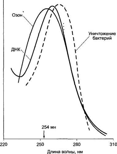
The absorption of ultraviolet radiation at wavelengths below 290 nm, which occurs in the ozone layer, is of crucial importance for life on Earth because this radiation is photochemically highly active and can destroy Nucleic Acids and Proteins. Indeed, it is frequently used to destroy harmful microorganisms (see Fig. 6.42). Therefore, the ozone layer protects the nucleic acids and Proteins of the biosphere from photochemical damage. Chlorofluorocarbons (CFCs), used in refrigeration systems as refrigerants or as propellants in aerosol cans, are responsible for disrupting the equilibrium between the formation and destruction of the ozone layer.1
1 The ozone shield is also depleted by other factors, such as gases released during volcanic activity. The leading role of CFCs (chlorofluorocarbons) has not been rigorously proven and remains one of several hypotheses explaining the occurrence of "ozone holes" in the atmosphere.
Fig. 6.43. Changes in the solar radiation spectrum as light passes through the atmosphere and water. Solid line: maximum radiation intensity; dashed line: short-wave and long-wave BOUNDARIES OF THE spectrum (these boundaries can be considered approximate average values). Green, brown, and red Algae show peak distribution in the sea at different depths.
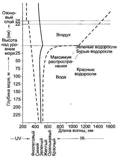
Table 6.17. Dependence of energy (or free enthalpy of reaction) and Electrochemical Potential per Einstein of photons on different wavelengths
Wavelength, nm |
Color |
∆G, kJ/Einstein |
∆E, V |
400 |
Violet |
297,5 |
-3,08 |
500 |
Blue-green |
238,0 |
-2,47 |
600 |
Yellow |
198,3 |
-2,05 |
650 |
Red |
183,1 |
-1,90 |
700 |
Purple-red |
170,0 |
-1,76 |
800 |
Infrared |
148,7 |
-1,54 |
900 |
Infrared |
132,2 |
-1,37 |
To characterize biochemical processes, moles are often used as Units of Measurement (1 mole = 6,023 1023 molecules — Avogadro's number, NA). Photochemical processes are also frequently calculated on a molecular basis. The energy of one mole of quanta (1 Einstein) and the maximum free enthalpy of a photochemical reaction required upon absorption of 1 Einstein of photon energy are expressed by the following equation:

Using the constants for , we obtain

Applying Equation 6.23, ∆G = -zF∆E (z=1) for the oxidation potential equivalent to 1 Einstein of absorbed photons ( in nm), we have

Some calculated values are presented in Table 6.17.
6.4.2. Photosynthetic Pigments
The process of photosynthesis begins with the absorption of photons by photosynthetic pigments, which thereby transition into an excited state. Chlorophyll is of primary importance for photoautotrophic organisms. Chlorophyll a plays the leading role in all organisms where photosynthesis proceeds with the evolution of oxygen arising from the photooxidation of water (Fig. 6.44). In Higher Plants and certain groups of algae (see Table 11.3), chlorophyll b is also found. The ratio of chlorophyll a to chlorophyll b is 3:1'. Instead of chlorophyll b, chlorophyll c can be found in some algae. Cyanobacteria and red algae possess exclusively chlorophyll a. Instead of chlorophyll a, photoautotrophic bacteria contain so-called bacteriochlorophyll (Fig. 6.45).
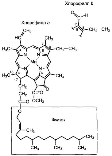
Fig. 6.44. Structure of chlorophylls a and b (bottom: delocalized π-electron system)
The special significance of chlorophyll a for photosynthetically active organisms lies in its presence within the reaction centers, i.e., the sites of the primary reactions of photosynthesis. Most chlorophyll molecules are part of Light-Harvesting Complexes, which act as antennas for the Reaction Centers and are responsible for more efficient Light absorption. Other antenna pigments include carotenoids. Collectively, antenna pigments are also referred to as accessory photosynthetic pigments. Light-absorbing photosynthetic pigments do not exist in a free state; they are bound to proteins (see Fig. 6.53). In the case of chlorophylls and carotenoids, these bonds are non-covalent. The accessory pigments of cyanobacteria and red algae also include phycobilins, which are Chromoproteins with covalently bound chromophore groups.
1 The value shown should not be considered a constant: the chlorophyll ratio varies depending on the species, light conditions, developmental stage, and plant tissue. — Editor's note.
Chlorophyll is a system of four pyrrole rings—porphyrin—with magnesium at the center and characteristic substituents on the rings (see Figs. 6.44, 6.45). The four pyrrole rings are linked by methine bridges. Magnesium is covalently bound to two nitrogen atoms and forms a coordinate bond with the other pair of nitrogen atoms.1Chlorophylls a and b differ in the substituent group at the seventh carbon atom: chlorophyll a carries a methyl group, while chlorophyll b carries a formyl group. At C17, all chlorophylls have a propionic acid residue esterified to a lipophilic alcohol, which is phytol in the case of chlorophylls a and b. It serves to anchor the chlorophyll molecules in the lipophilic region of chlorophyll-containing antenna proteins or reaction centers. Phytol is a diterpene containing 20 carbon atoms (for terpene Biosynthesis, see 6.16.2).
1 Due to the presence of a conjugated double bond system in the porphyrin ring, the electrons of all four nitrogen atoms are delocalized, and all four bonds with magnesium can be considered equivalent. — Editor's note.
Fig. 6.45. Structural relationships between different chlorophylls and bacteriochlorophyll
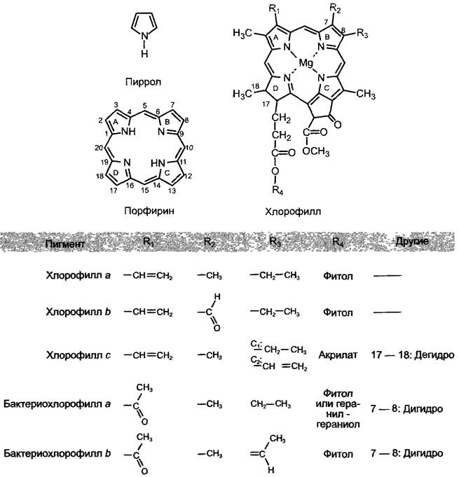
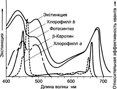
Fig. 6.46. Absorption spectrum and action spectrum (gray line) of Chlorella photosynthesis compared to the absorption spectra of the major photosynthetic pigments (in organic Solvents). The action spectrum is obtained by irradiating Cells with monochromatic light of different wavelengths but equal intensity (mol photons m-2), using a universal method to determine the necessary photosynthetic parameters (e.g., oxygen evolution). Often, the maximum observed effect is set to 100%, and the action spectrum is presented as relative quantum efficiency.
Chlorophyll stripped of its phytol tail is chlorophyllide, while chlorophyllide lacking the central atom is called pheophorbide. If the central magnesium atom is removed from chlorophyll (by mild acid Treatment), pheophytin is obtained. It acts as an electron carrier and is also a component of reaction centers (Photosystem II, see 6 4 5; reaction center in purple bacteria, see 6 4 10). Porphyrin biosynthesis is discussed in Ch 6 15.
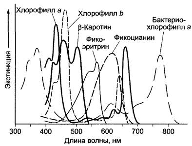
Most chlorophylls absorb light in the 400–480 nm (blue) and 550–700 nm (yellow to red) regions (Fig. 6 46). Bacteriochlorophyll a of purple bacteria absorbs light around 400 nm in the UV region and in the far-red and infrared parts of the spectrum between 700 and 850 nm. The absorption maximum of bacteriochlorophyll b of green sulfur bacteria is around 1000 nm. Between 480 and 550 nm, in the green light region, the absorption capacity of chlorophyll is very low (creating the so-called "green gap"). Therefore, chlorophyll solutions and chlorophyll-containing plant parts appear green to the human eye. The "green gap" of chlorophyll a is partially filled by the absorption of accessory pigments, chlorophyll b and carotenoids. Cyanobacteria and red algae fill this spectral region, left open by the light absorption of green algae, thanks to their accessory pigments, the phycobilins, which include phycoerythrin and phycocyanin. Consequently, cyanobacteria and red algae can find light for photosynthesis even in deep-water zones, below the canopy of green algae. Bacteria containing bacteriochlorophyll a or b are able to utilize wave energy from the part of the spectrum that other photosynthetic bacteria cannot absorb (see Figs. 6.46, 6.47).
Fig. 6.47. Absorption spectra of the major photosynthetic pigments (chlorophylls and β-carotene in organic solvents, phycobiliproteins in aqueous solution)
Although chlorophyll does not utilize sunlight optimally (due to the "green gap"), it evolved at an early stage of evolution (bacteriochlorophyll a of purple bacteria is already over 3 billion years old) and has changed little since then. The evolutionary dominance of chlorophylls as the primary photosynthetic pigments is due to The properties of these molecules: the porphyrin ring and its various substituents (see Fig. 6.45) form a system of conjugated double bonds. The participating π-electrons form a shared molecular orbital in which the electrons not only oscillate but can also circulate within the ring system. This phenomenon is one of the reasons for the high stability of this class of compounds. In fact, Porphyrins are among the most stable chemical compounds and are found, for example, in petroleum and coal (which are 400 million years old) in an almost chemically unaltered form.
The highly delocalized π-electrons of the porphyrin ring system can be raised to a higher energy level only by a specific quantum of energy, for example, through the absorption of photons with a relatively long wavelength (Fig. 6.48). This promotes the molecule to an excited state, in which it can characteristically participate in subsequent reactions.
Fig. 6.48. Excited states of chlorophylls (exemplified by chlorophyll). The ground energy level gives rise to several sublevels As a result of intramolecular vibrational movements; furthermore, the rotation of certain atomic groups within the molecules causes slight deviations in the energy state (so-called vibrational and rotational sublevels). In organic molecules, such processes lead to the appearance of more or less broad absorption peaks instead of the narrow-band spectrum characteristic of atoms. The diagram shows electron spins in the excited singlet and Triplet States compared to the ground state, as well as the part of the absorption spectrum of chlorophyll a resulting from the S0 —> S1 transition.
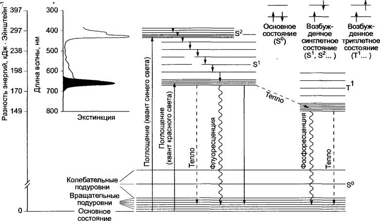
In molecules with an even number of electrons, all orbitals are occupied by pairs (singlet state, S0). Upon absorbing a photon, an electron occupies a higher energy level while maintaining its spin direction (excited singlet state S1, S2, etc., depending on the absorbed energy). After a short period, electrons in such an excited state return to their ground state, releasing the excitation energy, or alternatively, a spin flip of the excited electron occurs (triplet state). Thus, the unpaired electrons will have parallel spin directions (see Fig. 6.48). A spin flip can occur when an electron remains in an excited singlet state for a longer period than required for the spin inversion process (about 10-9 s).
The excited states important for chlorophyll are the first singlet state (corresponding to the absorption of a red light quantum), the second singlet state (absorption of a blue light quantum), and the first triplet state, which is reached only from the S1 level because its lifetime is sufficiently long (about 15 • 10-6 s). The excited S2 state proves to be too short-lived (10-12 s) for a spin flip.
As shown in Figure 6.48, the release of absorbed energy from excited chlorophyll occurs in various ways. Only some of these processes can be utilized to perform chemical work. This is associated with the S1 —> S0 transition. In this case, a radiationless energy transfer (exciton transfer) can occur from one chlorophyll molecule to another, provided they are sufficiently close to each other (distance less than 10 nm) and the absorption maximum of the second pigment molecule is lower (longer-wavelength form) than the energy-releasing capacity of the first excited molecule (shorter-wavelength form). This mechanism is particularly important for the directed transport of absorbed light quantum energy into the antenna complex and for the energy transfer to chlorophyll a of the reaction center. The absorption spectrum of a pigment molecule depends on its environment (in the case of chlorophylls, this is the protein environment); therefore, in an antenna complex containing chlorophylls with different absorption properties, excitons are directed toward pigment molecules that can absorb longer wavelengths of light, i.e., transfer occurs from chlorophyll b molecules to chlorophyll a molecules, and within this group, to forms of chlorophyll a that absorb light in the longer-wavelength region. The ultimate destination of exciton transfer is chlorophyll a of the reaction center, which is surrounded by specific proteins. Its structure can be represented as a dimer (special pair, Chl a2), and it possesses the lowest excitation energy in the entire complex. Chlorophyll in the reaction center is extremely rarely excited by direct photon absorption; most often, this occurs due to exciton transfer from the adjacent antennas.
Unlike antenna chlorophylls, the chlorophyll a dimer of reaction centers does not transfer its excitation energy immediately; first, in the excited state, it loses an electron, forming a positively charged radical (Chl a1+2). Upon receiving an electron, it returns to the ground state (Fig. 6.49). Under optimal illumination, this process occurs approximately 100 to 200 times per second.
Fig. 6.49. Charge Separation after excitation of dimeric chlorophyll a (Chl a2) in the reaction centers of Photosystems and transition to the ground state upon electron attachment. Under optimal illumination, this cycle occurs 100–200 times per 1 s
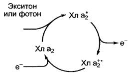
The process of primary charge separation

is a crucial step in photosynthesis. The short-lived excitation energy of photons is converted into a much longer-lived electrical potential,
which can be used for chemical work. The absorbed energy is partially lost as heat. This is true for the S2 —» S1 transition, because the lifetime of the S2 state is too short for exciton transfer. Therefore, in experiments studying photosynthesis, it is sufficient to apply red light to induce the S0 —> S1 transition. The energy of the excited S1 state can also be completely lost as heat or emitted as fluorescence. Chlorophyll in the triplet state does not play a major role. Only one chlorophyll molecule out of 10 million is in the triplet state at any given time when a plant is illuminated.1 Upon transition to the ground state, which occurs very slowly due to the spin flip required at this moment (the lifetime of the T1 state is 10-4 — 10-2 s), light can also be emitted (phosphorescence). However, chlorophyll in the triplet state can stimulate the transition of oxygen to the singlet state. Oxygen in this form is chemically highly reactive and can damage The Cell; therefore, various protective mechanisms have evolved (see carotenoids below, and also 6.48).
1 The formation of triplet states depends on light intensity, CO2 availability, and (indirectly) on water supply. Under extreme conditions, there will be more triplet-excited chlorophyll molecules than under normal physiological conditions. Excessive formation of chlorophyll triplets can lead to photodestruction (see below). — Ed. note.
Carotenoids are considered accessory photosynthetic pigments that absorb light in the blue and blue-green Regions of the spectrum, thereby narrowing the "green gap" in the absorption spectrum of chlorophylls (see Figs. 6.46; 6.47). There are two groups of carotenoids: carotenes (pure Hydrocarbons, the most famous being β-carotene2) and oxygenated carotenes (xanthophylls), the main representative of which in higher plants and green algae is lutein. The characteristic coloration of brown algae and diatoms is due to the xanthophyll fucoxanthin; lycopene1 can be found in purple bacteria (Fig. 6.50). Carotenoids transfer energy relatively poorly; their efficiency corresponds to approximately 20–50% of chlorophyll efficiency (this value reaches 80% only for fucoxanthin in brown algae). For this reason, in the absorption region characteristic of carotenoids (approximately 460–500 nm), there is also a clear discrepancy between Light Absorption and the action spectrum of photosynthesis (see Fig. 6.46). Xanthophylls of green algae and higher plants do not transfer excitation energy to chlorophyll a. Their main function in light-harvesting complexes (antennas) is to prevent the Formation of the triplet state of chlorophyll and thereby prevent the formation of highly dangerous singlet oxygen. Carotenoids are terpenoids, like phytol, but they contain 40 carbon atoms and therefore belong to the group of tetraterpenes (see 6.12.2). Light absorption in the visible region is due to A large number of conjugated double bonds, whose π-electrons form a single molecular orbital where the electrons are highly delocalized and easily transition to an excited state.
1 First isolated from tomato fruits (Lycopersicon). — Ed. note.
2 First isolated from carrot roots (Daucus carota). — Ed. note.
Fig. 6.50. Structure of carotenoid photosynthetic pigments. Carotenes do not contain oxygen; xanthophylls are oxygen-containing molecules. The system of delocalized π-electrons is highlighted in the formulas

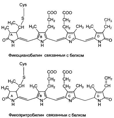
Phycobiliproteins are accessory photosynthetic pigments of cyanobacteria, red algae, and Cryptophyta. Phycocyanins (blue pigments) and phycoerythrins (red pigments) are present in these groups in various quantitative ratios and predominate over chlorophyll. The light-absorbing structures (chromophores) of phycobilins are open-chain tetrapyrroles (Fig. 6.51), similar to Bile pigments that arise during The breakdown of Hemoglobin (hence the name from Greek bilis — bile). Via a vinyl group at ring A of the tetrapyrrole, the chromophores are covalently linked to a Cysteine residue of the carrier proteins (thioether bond). Phycocyanobilin is a component of phycocyanin and allophycocyanin, while phycoerythrobilin is a component of phycoerythrin. Phycobiliproteins are part of highly organized light-harvesting structures — phycobilisomes (see Figs. 2.89; 6.53). Phycobilisomes lie on the side of the thylakoid membranes that contacts the cytoplasm and, acting as antennas, absorb light very efficiently. The absorbed excitation Energy is transferred to chlorophyll a of the reaction centers with nearly 95% efficiency via an exciton mechanism. Due to this feature and the absorption properties of phycobiliproteins (see Fig. 6.47), blue-green and red algae are capable of photosynthesis deep in the water Column. This phenomenon has its "price": up to 40% of the total weight of cellular proteins and up to 30% of the dry weight of cells are accounted for by phycobilisomes.
Fig. 6.51. Structure of phycocyanobilin and phycoerythrobilin. The system of delocalized pi-electrons is highlighted in the formulas. Chromophores are covalently bound to the cysteine residue of the apoprotein
When cyanobacteria and red algae are grown under light of various spectral compositions, phycobilins adapt to these conditions—a phenomenon known as chromatic adaptation. This is most likely due to the differing transcriptional activity (see 7 2 2) of the genes encoding the protein moiety of various phycobilins. Although such adaptation seems rational, from a physiological standpoint, it remains unclear whether it occurs under natural conditions and how widespread it is.
The efficiency of light utilization in different spectral regions, and consequently THE CONTRIBUTION OF various pigments to photosynthesis, can be determined by comparing the absorption spectrum of photosynthetically active organisms or Organs with the action spectrum of photosynthesis. In green plants (see Fig. 6.46), There is a significant discrepancy between the absorption and action spectra in the region of carotenoid absorption maxima, because carotenoids, as previously mentioned, have a limited capacity to transfer energy to chlorophyll. The fact that the absorption spectrum of intact cells or Tissues exhibits absorption bands as broad as those of isolated pigments (see Fig. 6.46) is explained by alterations in the absorption Properties of the pigments resulting from their association with light-harvesting complex proteins. This association also narrows the so-called "green gap" in the absorption spectrum. Subtle differences in the absorption spectra of photosynthetic pigments, arising from neighboring proteins, form the basis for directed exciton transfer within the antenna complexes.
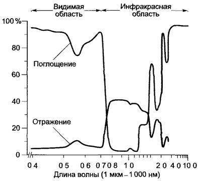
Comparing the absorption and reflection spectra of leaf blades (Fig. 6.52) reveals that absorption not only decreases in the green region, but also drops sharply in the infrared between 700 and 2 000 nm, while reflection here is at its maximum. Since leaf blades reflect more strongly in the infrared region than conifer needles, deciduous forests can be easily distinguished from coniferous ones using infrared aerial photography. Since infrared radiation does not contain sufficient energy to drive photosynthesis, and on the other hand, nearly half of the solar energy reaching the Earth's surface is dissipated, utilizing these wavelengths would only lead to the heating of the leaf blades; thus, from a biological perspective, it is disadvantageous. Intense absorption is also unnecessary at very long wavelengths (>3 000 nm), as they barely reach the Earth's surface. However, because the regions of best absorption are also the zones of optimal emission, the leaf can rapidly dissipate the heat absorbed along with sunlight.
Fig. 6.52. Absorption and reflection spectra of poplar leaves (Populus deltoides). Absorption still occurs. Note the high level of reflection in the infrared region ("the cool shade of the forest")
Leaf pubescence can significantly increase reflection in the visible spectrum and consequently reduce absorption. For example, heavily pubescent leaves of the desert plant Encelia farinosa absorb only 30% of radiation between 400 and 700 nm, whereas glabrous leaves of an Encelia species with the same chlorophyll content absorb 84%.
6.4.3. Structure of light-harvesting antennas
In all photosynthetic organisms, light energy is absorbed by highly organized antennas in which photosynthetic pigments are bound to proteins by covalent or non-covalent bonds. The precise orientation of pigment molecules ensures radiationless energy transfer within the antenna (transport via an exciton mechanism). The structural coupling of the antennas to the photosynthetic reaction center also enables The transfer of excitation energy from the antennas to the reaction center in the form of excitons. As a result, light-harvesting antennas increase the efficiency of the reaction center, because its constituent pigment molecule would only rarely reach an excited state through direct photon absorption.
The structure of antennas varies among different groups of photosynthetically active organisms and is not yet fully understood (Fig. 6.53). Antennas can be located within photosynthetically active membranes, as in purple bacteria and green plants, consisting of small integral proteins embedded in the membrane, or they can form large complexes located on the cytoplasmic side of the membrane (as in cyanobacteria and green sulfur bacteria). These large antennas are likely adaptations that allow cyanobacteria and sulfur bacteria to carry out photosynthesis using very low-intensity light, such as that which penetrates deep into the sea.
Fig. 6.53. Cytology/cytology/92.html">SCHEMATIC STRUCTURE OF various light-harvesting antennas of photosynthetic organisms: A — chlorosome of green sulfur bacteria (Chlorobium), proposed structure; B — phycobilisome of cyanobacteria and red algae (Rhodophyta). Phycobilisomes are associated with a photosystem II dimer. The pigment COMPOSITION OF THE antenna can vary among different species; C — structure of purple bacterial antennas as viewed from above The cell membrane; 9–11 LH2 complexes are associated with a single LH1 complex; the STRUCTURE OF THE purple bacterial reaction center is shown in more detail in Fig. 6.65; D — structure of the green plant antenna; surface view of the thylakoid membranes; CP43 and CP47 form the core antennas, while CP26, CP29, and LHCII form the peripheral antennas, with LHCII serving as the major antenna. There are likely 4 (only 2 shown) trimeric LHCII antenna complexes per dimeric photosystem II. AP — allophycocyanin; CP — chloroplast protein (the number indicates the molecular mass in kDa); PC — phycocyanin; PE — phycoerythrin; LH1 — core antenna; LH2 — peripheral antenna; LHC — light-harvesting complex
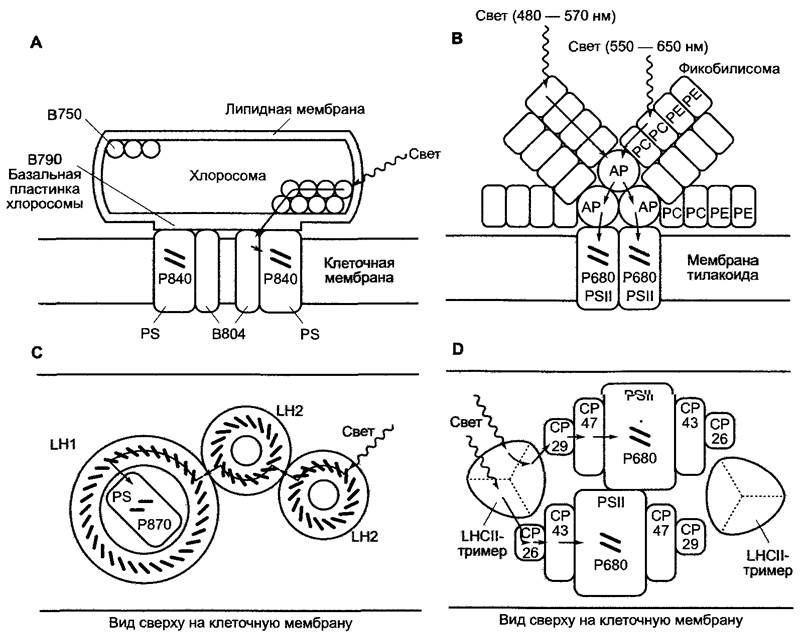
In the chlorosomes of green sulfur bacteria (Fig. 6.53, A), the light-harvesting complex on the cytoplasmic side of the membrane consists of 10,000 protein-bound bacteriochlorophyll molecules (predominantly bacteriochlorophyll c). They are surrounded by lipid envelopes and contact at their base (which contains bacteriochlorophyll a) with the membrane-integrated light-harvesting complex surrounding the reaction center. Exciton transfer occurs from bacteriochlorophyll c, which absorbs at a wavelength of around 750 nm (B750), via the baseplate bacteriochlorophyll a molecules (B790) to the bacteriochlorophyll a of the membrane-integrated light-harvesting complex (B804), and finally to the bacteriochlorophyll a of the reaction center (P840).
Phycobilisomes (see Fig. 6.53, B) lie on the thylakoid membrane, which branches off from the plasma membrane. They form a dense layer (approximately 400 phycobilisomes per 1 µm2) on the cytoplasmic side (see Fig. 2.89) and are protein-linked to the reaction centers embedded in the thylakoid membranes. Thus, exciton transfer can occur from phycoerythrins (absorbing at 480–570 nm) via phycocyanin (absorbing at 550–650 nm) to allophycocyanin (absorbing at 600–680 nm), and then to the chlorophyll a dimer of the photosystem II reaction center (see 6.4.5).
The antennas of purple bacteria (Fig. 6.53, C) are integral complexes of the plasma membrane. The core antenna (LH1, from light-harvesting) probably surrounds the reaction center in a ring, which, along with the pigment-binding protein complex, contains carotenoids and 32 symmetrically arranged bacteriochlorophyll a molecules. In some species, each reaction center is accompanied by an additional 8–10 peripheral antennas (LH2), which are also ring-shaped and carry 27 bacteriochlorophyll a molecules per ring. They are arranged in two stacked layers containing 18 and 9 molecules, respectively. Thus, a total of 250–300 pigment molecules are associated with each reaction center. Exciton transfer occurs from LH2 via LH1 to the bacteriochlorophyll a dimer of the reaction center (P870).
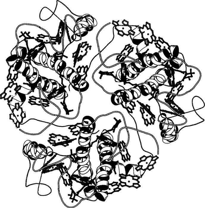
In green plants, antennas are closely associated with the Two Types of reaction centers (photosystem II and Photosystem I, see 6.4.4); these are called core antennas (hereinafter referred to as "integrated antennas"). In photosystem I, they consist of approximately 100 chlorophyll a molecules per photosystem and serve a single reaction center (see Fig. 6.61). The integrated antennas of photosystem II are two subunits of the protein complex (CP43 and CP47) with 15 associated chlorophyll a molecules (see Fig. 6.59). These antennas are in contact with peripheral, mobile light-harvesting complexes integrated into the membranes, with the pigment-Protein Complexes CP26, CP29, and LHCII being in contact with each other. The main antenna is formed by LHCII (LHC stands for light-harvesting complex) (see Fig. 6.53, D). Structural studies of LHCII have shown that each protein is bound to 7 chlorophyll a molecules, 5 chlorophyll b molecules, and 2 lutein molecules. Chlorophyll b is located in the peripheral region, while chlorophyll a is in the center of the protein (Fig. 6.54). The chlorophyll molecules are only 0.5–3 nm apart, which ensures efficient exciton transfer. LHCII exists as a trimer in the thylakoid membrane. It is believed that LHCII can interact with the structurally similar core antennas of photosystem I, thereby regulating energy distribution between both photosystems (see 6.4.4). Each reaction center of photosystem II is served by approximately 300 pigment molecules within the antennas. Exciton transfer occurs from the peripheral chlorophyll b via the chlorophyll a molecules of the complex to the chlorophyll a of the integrated and core antennas, and finally to the reaction center of photosystem II (chlorophyll a dimer, P680). The light-harvesting complex of photosystem I may function in a similar manner, but its structure has not yet been studied in detail. A single "photosynthetic unit", i.e., one complete electron transport chain from photosystem II to photosystem I (see 6.4.4, Fig. 6.55), can be served by approximately 500 pigment molecules (chlorophylls a and b, carotenoids).
Fig. 6.54. Structural model of the chlorophyll a/b-binding protein and the spatial arrangement of photosynthetic pigments in the trimeric light-harvesting complex of photosystem II (LHCII, see Fig. 6.53, D)
6.4.4. Electron and Proton Transport in Photosynthesis
For a better understanding of the subject, we will not consider antennas further, but will focus only on the exit of the exciton from the antennas. In this section, we will examine the processes occurring in green plants (the processes taking place in cyanobacteria and prochlorophytes are analogous). Other light reactions characteristic of bacteria are discussed in Section 6.4.10.
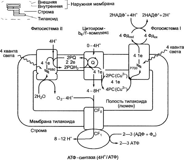
Fig. 6.55. General scheme of photosynthetic electron and hydrogen ion transport, as well as Photophosphorylation in green plants. For further comments, see the text; the localization of the photosynthetic complex and ATP synthase in the grana or stromal regions of the thylakoid membrane is shown in Fig. 6.57. In cyanobacteria, the systems and reaction pathways are analogous. However, in these organisms, plastocyanin is replaced by cytochrome. Fd — ferredoxin, PC — plastocyanin, PQ — plastoquinone, Q — Q-cycle (see Fig. 6.60)
As early as 1937, Hill observed that an illuminated leaf extract (isolated thylakoid membranes) in the presence of artificial electron acceptors (A), such as Fе3+, or reduced pigments, releases О2. In the Hill reaction, the presence of Н2О as an electron donor is mandatory; СО2 is not involved in it:

This means that the oxygen released during photosynthesis originates from water molecules, and that illuminated thylakoid membranes, by extracting electrons from water, reduce dissolved electron acceptors rather than CO2. Consequently, the process of CO2 reduction to form carbohydrates is separated from the light reactions. It is referred to as the dark phase (see 6.5.1–6.5.3).
Under natural conditions, the electron acceptor in the Hill reaction occurring in chloroplasts is NADP+:
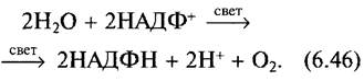
The Gibbs Free energy (at pH 7; see 6.1, equation 6.7) for this redox reaction is ∆G0' = +218 kJ • mol-1. In this reaction, two moles of electrons are transferred from a system with a high positive redox potential (H2O/1/2O2 E0' = +0.82 V) to a system with a strongly negative potential (NADPH + H+/ NADP+ : E0' = -0.32 V). (∆E0' = -0.32 V - 0.82 V = -1.14 V per mole of electrons; according to equation 6.27, z = 2, ∆G0' = 218 kJ per mole of NADPH + H+ formed.)
To reduce NADP+ with electrons from water, a series of two light reactions occurring in photosystems I and II is required (the numbering of the photosystems corresponds to the order of their discovery), each involving specific chlorophyll a dimers of both reaction centers. These chlorophyll a dimers of the two reaction centers can differ in their absorption properties: P680 is the reaction center with an absorption maximum at 680 nm, which is part of photosystem II (PSII); P700 is the reaction center within photosystem I (PSI), with its absorption maximum at 700 nm.
The existence of Two Photosystems was first revealed by determining the quantum yield (moles of O2 produced per mole of absorbed quanta) as a function of wavelength. A sharp decline in quantum yield was observed in the far-red region, at wavelengths longer than 680 nm (the "red drop" effect), as the absorption and photosynthetic spectra differ significantly here (see Fig. 6.46). Conversely, exposure to shorter wavelengths of the red spectrum (650 nm) resulted in a synergistic increase in quantum yield (the quantum yield under simultaneous illumination with 650 and 700 nm light is significantly higher than the sum of the quantum yields under illumination with either 650 nm or 700 nm light alone). This phenomenon was named the Emerson enhancement effect after the scientist who discovered it. Emerson was the first to demonstrate the interaction between two photosystems with different light-absorption properties.
The overall process of the light reaction, starting from water and ending with the formation of NADPH + H+ is shown in Fig. 6.55. Clearly, light energy is used not only for electron transport but also for the coupled, directional transport of hydrogen ions into the thylakoid lumen. The chemical potential of the hydrogen ion drives ATP synthesis. Finally, the complex sequence of reactions occurring in The electron transport chain is presented as a simplified scheme. The structures of the individual Components of the redox systems involved in the light phase of photosynthesis are shown in Fig. 6.56.
Fig. 6.56. Structure of the key carriers involved in photosynthetic electron transport in green plants

P680, having entered an excited state after exciton transfer (P680*), donates an electron, which is transferred via the internal electron transport chain of photosystem II (see 6.4.5; Fig. 6.59) to mobile plastoquinone molecules (PQ). The addition of a second electron occurs after a subsequent excitation of P680, accompanied by the uptake of two hydrogen ions from the stroma, yielding plastohydroquinone (PQH2, Fig. 6.56).
Oxidized P680 is reduced by extracting electrons from water, thereby returning to its ground state. Photolysis of water1 is carried out by the oxygen-evolving complex, which is an integral part of photosystem II. Plastohydroquinone leaves photosystem II and diffuses within the thylakoid membrane. The membrane contains a pool of dissolved plastoquinone molecules (approximately 7 per photosystem II, with at least 4 molecules existing as PQH2 in the light). Plastohydroquinone moves to the second membrane-bound complex, the cytochrome b6/f complex (see Fig. 6.60), where PQH2 is oxidized to PQ.
1 The term "photolysis" does not entirely accurately reflect The Mechanism of the ongoing processes. It is more correct to speak of the photooxidation of water. Hereafter, the term has been corrected in the text. — Editor's note.
The two electrons released in this process are sequentially transferred along the electron transport chain components bound within the protein complex (see 6.4.6; Fig. 6.60) to plastocyanin molecules. This forms reduced Cu+-plastocyanin. Plastocyanin is a soluble protein localized in the intrathylakoid space (lumen). Hydrogen ions released during plastoquinone oxidation are transferred through the cytochrome b6/f complex into the thylakoid lumen. It is assumed that an internal plastoquinone-plastohydroquinone redox cycle (Q-cycle) occurs at the cytochrome b6/f complex, in which electrons separated from a plastohydroquinone molecule are transferred back to plastoquinone. During this process, hydrogen ions are repeatedly taken up from the stroma and, upon subsequent oxidation of plastohydroquinone, are translocated into the thylakoid lumen. The cytochrome b6/f complex acts as a proton pump in the Q-cycle, enhancing the hydrogen ion concentration gradient between the stroma and the thylakoid lumen. Due to the Q-cycle, two hydrogen ions are transferred into the thylakoid lumen per electron, whereas without the Q-cycle, the theoretical ratio would be 1:1.
The excited chlorophyll a dimer of photosystem I (P700*) transfers an electron via the bound components of the electron transport chain (see 6.4.7; Fig. 6.61) to ferredoxin (Fd) (a soluble iron-sulfur protein) located in the stroma, which serves as an electron donor for NADP+. NADP+ sequentially accepts two electrons to form NADPH + H+ (see Fig. 6.56). The electron deficit of oxidized P700 is replenished by reduced plastocyanin (Cu+ form).
Thus, the electron transport chain contains carriers that transfer either one or two electrons. To reduce a total of two molecules of NADPH + H+, 4 electrons are transported, producing one molecule of O2; to drive this process, 8 light quanta are required. The functioning of mobile soluble carriers in the redox system (soluble plastoquinone/hydroquinone in the thylakoid membranes between PSII and the cytochrome b6/f complex, and soluble plastocyanin in the thylakoid lumen between the cytochrome b6/f complex and PSI) is advantageous for several reasons.
It creates local conditions for the operation of each photosystem when they are in an excited state, since PSI and PSII do not need to operate synchronously.1 This would be extremely difficult due to the very high rate of the primary photosynthetic processes.
1 The argument presented is not sufficiently justified. The operation of the two photosystems is usually described as a sequence of reactions, each of which must follow the previous one. This description creates the illusion that synchronous operation of the photosystems is prohibited. In reality, there is no such logical restriction. — Editor's note.
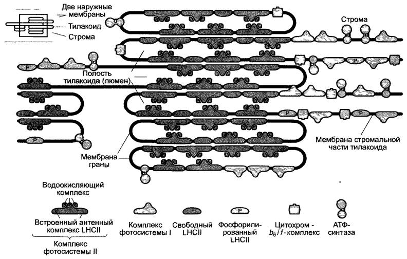
The spatial distance between the three transmembrane complexes is overcome. These complexes are unevenly distributed within the thylakoid membrane. Photosystem II, along with its antenna complexes, is located in the grana thylakoid membranes. In turn, photosystem I and the cytochrome b6/f complex reside predominantly in the stromal thylakoids (Fig. 6.57). The light-harvesting complex LHCII mediates the stacking of thylakoids into grana (see Fig. 2.83).
Fig. 6.57. Lateral heterogeneity in the distribution of photosynthetic complexes in the thylakoid membrane. The diagram is drawn to scale. The side view is presented as seen under an Electron microscope (for example, see Fig. 6.63 for the ATP synthase image)
The directional (vectorial) transport of electrons during the light phase of photosynthesis is coupled with The transport of hydrogen ions in the cytochrome b6/f complex, which proceeds from the stroma into the thylakoid lumen. Additionally, during the photooxidation of water, H+ ions are released into the thylakoid lumen. In this case, there are 8 H+ ions per O2 molecule formed (4 from the photooxidation of water, and 4 hydrogen ions supplied from the stroma via PQH2).
If the Q-cycle operates at maximum efficiency, an additional 4 H+ enter, so that 8 to 12 H+ accumulate in the thylakoid lumen per O2 molecule formed. The hydrogen ion concentration gradient, which rapidly develops upon illumination, can be calculated from the pH value: in the stroma of illuminated chloroplasts, it is about 8, and in the thylakoid lumen, it is 4.5–5. A ∆pH of about 3–3.5 units corresponds to a difference in hydrogen ion concentration between the stroma and the lumen of approximately 1:1,000 to 1:3,000. Since chloride ions are transported into the thylakoids simultaneously with hydrogen ions to balance the charge (presumably via a chloride channel), the potential difference across the thylakoid membranes is small.
The energy of the hydrogen ion gradient (proton-motive force, equation 6.19) drives the photosynthetic synthesis of ATP (Mitchell's chemiosmotic model of photophosphorylation). ATP synthase is localized in the stroma thylakoids (see 6.4.9; Fig. 6.63) and, coupled with the formation of one ATP molecule, translocates 4 H+ ions. Depending on the contribution of the Q-cycle, 8 to 12 H+ ions accumulate in the thylakoid lumen per O2 molecule formed. This is sufficient for the synthesis of 2 (without the Q-cycle) or a maximum of 3 (with a fully operating Q-cycle) ATP molecules. The NADP:ATP ratio in the light reaction is 1:1 to 1:1.5. The dark reaction (CO2 fixation in The Calvin Cycle, see 6.5) requires NADP and ATP in a ratio of 1:1.5.
What is the energy yield of the light phase of photosynthesis? The standard molar free enthalpy of NADPH + H+ formation is ∆G0 = +218 kJ mol1, and for the formation of ATP from ADP and inorganic phosphate, this value is ∆G0 = +30.5 kJ mol-1. Consequently, the yield of the endergonic light reaction is at least 2 mol × 218 kJ mol-1 + 2 mol × 30.5 kJ mol-1 = 497 kJ per 1 mol of O2 formed. To achieve this, 8 mol of excitation energy excitons must be used, and at least 8 mol of photons at a wavelength of 700 nm must be absorbed.1 This corresponds to an energy of 8 mol × 170 kJ mol-1 = 1360 kJ of absorbed light energy. Thus, the energy yield (= efficiency of the light phase) is 497 : 1,360 = 0.36 (36%). The remaining energy is lost as heat. Such losses are inevitable, as they are a consequence of charge separation. This ensures the irreversibility of the process: recombination of the electrons donated by the excited chlorophyll a dimer and the oxidized dimer (Chl a2+) does not occur. Because of these inevitable energy losses as heat, photosynthesis requires two sequentially operating photosystems to utilize electrons from water for the reduction of NADP+. Photosynthetic bacteria, which extract electrons from a substrate with a relatively low negative standard redox potential (e.g., H2S, E0' = -1.24 V), require only a single photosystem to reduce NAD+, and can therefore utilize low-energy light from longer wavelength regions.
1 The energy of quanta with  = 700 nm is insufficient for the entire light phase to proceed. Half of the absorbed quanta must have a wavelength of
  (see 6.4.5). Thus, the energy of the absorbed quanta in this case is underestimated, and the efficiency is overestimated. — Editor's note.
Table 6.18. Carriers of the photosynthetic electron transport chain in plants; arranged in order of increasing standard redox potential
Redox couple |
E0′, V |
Redox couple |
E0′, V |
P700* |
Below -1.10 |
P680* |
Below -0.6 |
A0 |
-1.10 |
Pheophytin |
From -0.66 to -0.45 |
A1 |
-0.88 |
Plastoquinone, bound |
From -0.25 to -0.05 |
FeSx |
-0.70 |
Plastoquinone, free |
+0.11 |
FeSB |
-0.59 |
FeSR (Rieske protein) |
+0.29 |
FeSA |
-0.53 |
Cytochrome f |
+0.35 |
Ferredoxin |
-0.43 |
Plastocyanin (PC) |
+0.37 |
Fd-NADP+ reductase (FNR) |
-0.35 |
P700+ + e- ⇄ P700 |
+0.45 |
NADP+ + 2H+ + 2e- ⇄ NADPH + H+ |
-0.32 |
O2 + 4H+ + 4e- ⇄ 2H2O |
+0.82 |
Cytochrome b6 |
-0.02 |
P680+ + e- ⇄ P680 |
Above +0.82 |
If a redox reaction is not listed in the table, the E0′ values for the reduced form in equilibrium with the oxidized form apply. The excited state is marked with an asterisk (*).
In addition to the non-Cyclic electron transport from water to NADPH shown in Fig. 6.55, under certain conditions, cyclic electron transport also occurs, in which light energy via photosystem I is used solely for ATP synthesis (see Fig. 6.61, B), as well as pseudocyclic electron transport, where electrons from photosystem I are transferred to oxygen (Mehler reaction, see 6.47).
If the characterization of the redox system carriers involved in non-cyclic electron transport is based on their standard redox potential (Table 6.18) and their association with a complex, the electron transport chain can be represented as the so-called Z-scheme (Fig. 6.58). We will consider the individual steps later, focusing in more detail on the structure of the photosystems. Excited-state P700*, which has a standard redox potential below -1.1 V, is currently the most active reducing agent known in living cells.
Fig. 6.58. Sequence of electron transport chain carriers involved in the light reaction of photosynthesis, arranged according to their standard redox potentials (Z-scheme). The Z-scheme reflects changes in the free enthalpy of redox reactions at individual stages under standard conditions; however, under natural conditions (influenced by changes in concentration, Temperature, and pH), deviations from standard values are observed in chloroplasts. Furthermore, the Z-scheme shows that the carriers are associated with membrane complexes, but their precise spatial localization is not depicted. P680*, P700*: chlorophyll a dimer in the excited state in the reaction centers of photosystem II or I

6.4.5. Photosystem II
Photosystem II (Fig. 6.59) consists of at least 16 different proteins, of which two (D1 and D2 proteins) form the actual reaction center and two others (CP43 and CP47) form the core antennas (see Fig. 6.53, D). The D1 and D2 proteins are homologous to each other and to the reaction center of purple bacteria (see 6.4.10), meaning they arose from a common ancestor during evolution. Bound to the heterodimer are 4–5 molecules of chlorophyll a, 2 pheophytins, 2 plastoquinones, and 1–2 carotenoids. At some distance, the protein D1/D2 complex holds a cluster consisting, most likely, of 4 manganese ions (manganese cluster). The manganese ions face the thylakoid lumen and are held by proteins that stabilize the cluster (MSP331). The chlorophyll a dimer (reaction center) is bound to both the D1 and D2 proteins.
Fig. 6.59. Schematic representation of the structure and electron flow in photosystem II. The structure of the integrated (core) antennas actually looks somewhat different from the figure (see Fig. 6.53, D). MSP33—manganese-stabilizing protein (33 kDa), standard abbreviation, see 6.4.5
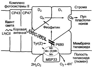
Following excitation of P680, electrons are transported via the pheophytin of the D1 protein to plastoquinone QA, which is tightly bound to the D2 protein. It is converted to a semiquinone radical (see Fig. 6.58) and transfers an electron to QB of a second plastoquinone molecule, which is weakly bound to the D1 protein. In the second step, QB receives another electron from the reduced QA, which has again become a semiquinone radical, as well as 2H+ ions, dissociates as PQH2 from the binding site, and enters the plastoquinone pool dissolved in the thylakoid membrane. The nonpolar terpenoid side chain (prenyl residue) of plastoquinone promotes solubility in the lipid phase.
1 From the English name of the protein: Mn-stabilizing protein with a mass of 33 kDa. — Ed. note.
Oxidized P680+ with a standard potential above +1.1 V is a highly active oxidizing agent. It compensates for the electron deficit by oxidizing a specific Tyrosine residue (Z) on the D1 protein; the resulting tyrosine radical abstracts 1 electron from the manganese cluster mentioned earlier. This cluster acts as an accumulator of 4 electrons, in which the 4 manganese atoms can exist in oxidation states of Mn2+, Mn3+, or Mn4+. Upon the sequential transfer of 4 electrons, the manganese group (also called the S-system) is sequentially oxidized:
S0 —> S1 (+1) —> (+2) —> S3 (+3) —> S4 (+4).
The S4 state returns to the ground state upon the oxidation of 2 water molecules and the simultaneous acceptance of 4 electrons1:
S4 (+4) + 2H2O —> O2+ 4H++ S0.
1 The presented information on the operation of the water-oxidizing system is outdated. According to current data, sequential oxidation of water occurs, rather than just the Mn cluster. Proton release and oxygen release from the complex are separated in time (i.e., they are not simultaneous events). The equations shown reflect only the overall process, but not the reaction mechanism. — Ed. note.
Thus, during water splitting, the harmful effect of highly reactive oxygen radicals on cells is prevented.
With the help of certain inhibitors, electron transport in PSII can be stopped. Triazines (e.g., atrazine) displace QB from its niche on the D1 protein. Dichlorophenyldimethylurea (diuron) acts in a similar manner. This substance is used as a herbicide. Even a single point mutation in the D1 protein can prevent the action of the herbicide (without disrupting QB binding) and thus confer herbicide resistance.
6.4.6. Cytochrome b6/f complex
The cytochrome b6/f complex Functions in photosynthetic electron transport as a plastohydroquinone-plastoquinone oxidoreductase and simultaneously as a proton pump. This transmembrane complex, homologous to the cytochrome b/c1 complex of the Mitochondrial Electron Transport chain (see 6.10.3.3), consists of numerous components, among which cytochrome f, cytochrome b6, and the Rieske protein function as redox systems (Fig. 6.60).
Fig. 6.60. Hypothetical model of the structure of the Main Components of the cytochrome b6/f complex in the thylakoid membrane. The movement of electrons and hydrogen ions during the functioning Q-cycle is depicted step-by-step. For every electron transferred to plastocyanin, 2 hydrogen ions are transported into the thylakoid lumen via the Q-cycle; without the Q-cycle, this ratio would be 1:1. PC — plastocyanin
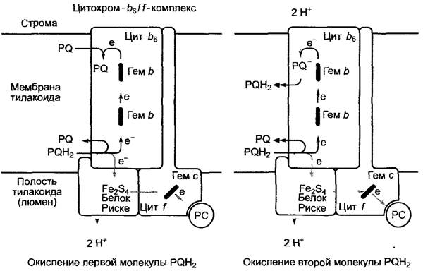
Cytochromes, like chlorophyll, are based on a porphyrin ring system, but instead of magnesium, they contain iron as the central atom of the tetrapyrrole ring. The porphyrin ring (see Fig. 6.45) with an iron atom at its center is called a heme, and the central iron atom is heme iron (see Fig. 6.56). During electron transfer, the central atom changes its valence state (Fe3+/Fe2+). Based on the structure of the bound Hemes, cytochromes are divided into three main groups: a, b, and c (corresponding to heme a, b, and c, respectively). They also differ depending on THE POSITION OF specific absorption bands of their reduced forms (α-bands) and are often characterized by their respective absorption maxima (e.g., cytochrome c555). Cytochromes of the b- and c-types play an important role in photosynthesis. In c-type cytochromes, the heme is covalently bound to the protein (the SH groups of cysteine are attached to the two vinyl groups of the heme). A distinguishing feature of cytochrome f (from Latin frons — foliage) is that it is present in chloroplasts; from a chemical standpoint, it belongs to the c-group (cytochrome c555). Cytochrome f is predominantly located closer to the thylakoid lumen.
Heme c is also located here. The protein is anchored in the membrane by hydrophobic Amino acid sequences. Cytochrome b6 is an integral membrane protein. It contains two heme b molecules, arranged one above the other and perpendicular to the membrane.
The Rieske protein is a peripheral protein that is not tightly bound to the membrane; its redox component is a Fe2S2 cluster consisting of two iron atoms and two sulfur atoms coordinated with them (see Fig. 6.56). Sulfur can be easily leached from the structure (for example, by a weak acid), which is why it is referred to as acid-labile sulfur (as opposed to the acid-stable sulfur of cysteine, which cannot be released by acid treatment). The iron within the Fe2S2 cluster is called non-heme iron. Like cytochromes, this cluster acts as a single-electron carrier.
Electrons delivered by plastohydroquinone, which is associated with the cytochrome b6/f complex, are transferred via the Fe2S2 cluster of the Rieske protein and cytochrome f to oxidized plastocyanin in the thylakoid lumen. Plastocyanin is a small protein with a molecular mass of about 10.5 kDa. Its central Cu atom is coordinated by cysteine and Methionine residues and two Histidine residues (see Fig. 6.56). During the reversible valence change Cu2+ —> Cu+, plastocyanin either accepts one electron or donates it again.
The protons released during the oxidation of PQH2 are directed into the thylakoid lumen by the cytochrome b6/f complex. It is possible that a closed Q-cycle, which has not yet been studied in detail, facilitates the translocation of additional H+ ions from the stroma into the thylakoid lumen with the participation of cytochrome b6 (see Fig. 6.60).1
1 According to current data, the Q-cycle begins with PQH2, which donates two electrons: one to the Rieske protein and the other to cytochrome b6. In this process, the Rieske protein changes the conformation of the entire complex, and the stored energy allows the second electron to move against the electrochemical potential. Two b-type hemes are used to transfer the electron from the lumenal to the stromal side of the membrane. Subsequently, the electron is transferred to PQ, which is fully reduced over two turns of the cycle, taking up 2H+ from the stroma. PQH2 leaves the complex and diffuses to the lumenal side of the membrane, where it re-enters the Q-cycle. — Ed. note.
6.4.7. Photosystem I
The third transmembrane complex involved in the light reactions of photosynthesis, photosystem I, receives electrons from reduced plastocyanin (PC) and transfers them to ferredoxin (Fd). From there, via the enzyme ferredoxin-NADP+ reductase (FNR), they are transferred to NADP+ to form NADPH + H+. Photosystem I is homologous to the reaction center of green sulfur bacteria and consists of 12 or more different subunits. PSI (Fig. 6.61) is a heterodimer of proteins A and B, which, along with electron carriers and the P700 chlorophyll a dimer, contain integrated light-harvesting antennas. Protein A is homologous to proteins D1 + CP43 of photosystem II, and B to D2 + CP47. Subunit F interacts with plastocyanin, subunit D with ferredoxin, and C is responsible for Electron transfer from the reaction center to ferredoxin.
Fig. 6.61. Schematic representation (A, B) of the structure and electron flow through the photosystem I complex During cyclic electron transport (after H.W. Heldt).
During cyclic electron transport, the cytochrome b6/f complex and photosystem I operate, involving the plastoquinone pool as a light-driven 'proton pump' to produce ATP without the formation of NADPH + H (cf. Fig. 6.55). Explanations in the text

Charge separation after excitation of P700 occurs similarly to this process in PSII. The electron lost by the excited P700 is transferred via two monomeric chlorophyll a molecules (A, A0) to phylloquinone (see Fig. 6.56), which, like chlorophyll, possesses a phytol residue. Phylloquinone (Q, also designated as A1, Fig. 6.61, A) is attached to subunit B of the reaction center and corresponds to QA of photosystem II. Like QA, it accepts an electron, forming a semiquinone radical. The next step of electron transfer differs from the processes in PSII. The semiquinone radical transfers the energized electron to ferredoxin via three Fe4S4 clusters (see Fig. 6.56) (carriers designated as FeSX, FeSB, and FeSA). Ferredoxin binds to PSI from the stromal side via subunit D. Ferredoxin is a small protein (molecular mass 11 kDa) that contains a Fe2S2 cluster (see Fig. 6.56) and acts as a single-electron redox system.
From reduced ferredoxin, electron transfer can occur via the cytochrome b6/f complex and plastocyanin back to P700+, rather than to NADP+. This cyclic transport (Fig. 6.61, B), involving the cytochrome b6/f complex, leads to the translocation of hydrogen ions from the stroma into the thylakoid lumen, driving ATP synthesis ('cyclic photophosphorylation'), although NADPH is not formed in the process. Cyclic electron transport occurs when the ratio of the reduced to oxidized form, NADPH + H+/NADP+, is sufficiently high, meaning there is insufficient substrate for the operation of ferredoxin-NADP+ reductase.
Under highly active reduction of the ferredoxin pool, electron transfer from ferredoxin to O2 occurs, forming Н2O (Mehler reaction, Fig. 6.62, A).
This process is called pseudocyclic electron transport because it resembles cyclic transport in that only ATP, rather than NADPH + H+, is generated. However, under Mehler reaction conditions, the ATP/ADP ratio is often high, so the available amount of ADP is insufficient for ATP synthesis. Thus, the Mehler reaction generates a very high hydrogen ion gradient across the thylakoid membrane.
Fig. 6.62. Conversion of O2- in the ascorbate-Glutathione system of chloroplasts (A). The xanthophyll (violaxanthin) cycle of photosystem II for dissipation of excitation energy as heat (B). The xanthophyll cycle is also coupled to the ascorbate-glutathione system. GSH — reduced glutathione; GSSG — oxidized glutathione, consisting of two glutathione molecules linked by a disulfide bridge (2GSH ⇄ GSSG + 2Н+ + 2е-)

Oxidation of ferredoxin in the Mehler reaction ultimately generates the superoxide anion radical (O-2). Thanks to the enzyme superoxide dismutase, it is converted into O2 and Н2O2. Subsequently, Н2O2 is reduced to water under The Influence of several Enzymes (Fig. 6.62, A), thereby preventing the formation of the highly reactive hydroxyl radical (OH). In the presence of Metal Ions and O-2, this radical forms spontaneously from Н2O2 and damages Lipids, proteins, and nucleic acids.
Superoxide dismutases (SODs) are metalloenzymes. Chloroplasts contain Fe-SOD, Mn-SOD, and CuZn-SOD. These enzymes are also found in the cytoplasm (CuZn-SOD), Mitochondria (CuZn-SOD, Mn-SOD), and Peroxisomes (Mn-SOD).
6.4.8. Mechanisms of Regulation and Protection of the Light Reaction
Spatial separation of PSII, PSI, and their antennas (see Fig. 6.57) reduces the uncontrolled spillover of excitons from PSII to PSI and simultaneously promotes dynamic distribution of excitation energy between both photosystems. If PSI receives too little excitation energy, this leads to the accumulation of PQH2 in the plastoquinone pool (over-reduction)1. As a result, a protein kinase is activated, which phosphorylates the light-harvesting complex LHCII. Recall that the light-harvesting complex LHCII establishes an electrostatic connection between adjacent thylakoid membranes. Grana are formed from stacked thylakoids. To associate with PSI, LHCII diffuses from the stacked thylakoid region to the stromal region.2 Thus, the energy of absorbed quanta is redirected from PSII to PSI 3 (see Fig. 6.57; 7.6). Dephosphorylation of LHCII leads to dissociation from PSI and re-association of the LHCII complex with PSII.
1 Such conditions can only be created experimentally by irradiating plants with monochromatic light of  = 680 nm. Under natural conditions, over-reduction of the plastoquinone pool occurs under excessively high light intensity and/or closed Stomata (= CO2 starvation). The reason is not that PSI receives insufficient light quanta, but that the excited electron cannot be transferred to NADP+, because it is also in a reduced state under these conditions. — Editor's note.
2 Phosphorylation and migration of LHCII lead to the disassembly of grana. Thus, the presence of grana correlates with the active operation of PSII, while their absence correlates with low activity of this photosystem, and decreased production of O2 and NADPH + H+. — Editor's note.
3 The described redistribution of light quantum energy is necessary to switch the operation of the electron transport system from non-cyclic (PSII is active, NADPH + H+ must be formed) to cyclic mode (only PSI is active, no new electrons from water enter the plastoquinone pool). — Editor's note.
High light intensity combined with a low demand for ATP and NADPH (for example, when stomata close to reduce water loss at high temperatures, resulting in limited CO2 for assimilation) causes excessive activation of pigment systems without electron outflow. Consequently, there is a risk of intensive formation of triplet states of excited chlorophyll, and thus singlet oxygen (see 6.4.2). Both carotenoids and α-tocopherol, which are abundant in the thylakoid membrane, return the triplet excited state of chlorophyll and singlet oxygen back to the ground unexcited state. Apparently, this protection is not always sufficient under high light intensity. Damage to photosystem II (photoinhibition) often occurs, likely due to the intensive degradation of the already short-lived D1 protein. Thus, the bleaching of needles during spring damage to forest trees is explained by photooxidative processes caused by a sharp increase in light intensity.
To reduce harmful light effects, the excess energy of absorbed quanta is converted into heat. The xanthophyll zeaxanthin is involved in this process (see Fig. 6.50). It is formed from the xanthophyll violaxanthin present in the antennas by de-epoxidation (reduction) of the latter, when a high hydrogen ion gradient exists between the thylakoid lumen (acidic reaction) and the stroma (alkaline reaction), which indicates a highly reduced state of all components of the electron transport chain during the light phase (see Fig. 6.62, B).
Since the epoxidizing enzyme catalyzing the reverse reaction has a pH optimum in the weakly alkaline range (pH 7.6), and the de-epoxidases in the acidic range (pH
5.0), a decrease in the hydrogen ion gradient across the thylakoid membrane causes zeaxanthin to be converted back into violaxanthin (the xanthophyll or violaxanthin cycle).
The hydrogen ion gradient of 3 pH units formed across the thylakoid membrane upon illumination represents a proton-motive force (see Equation
4.19), which is used for ATP synthesis. This process is called photophosphorylation. The change in standard molar free enthalpy for the synthesis of ATP from ADP and inorganic phosphate is ∆G0' - 30.5 kJ • mol-1. However, taking into account the actual concentration ratios of the substances involved in the reaction within the cell, it can be ∆G = 45 — 50 kJ • mol-1. From Equation 6.19, it follows that the free enthalpy of the hydrogen ion gradient (∆pH = 3 at 25 °C) is ∆G = -17 kJ • mol-1. Therefore, for the synthesis of one ATP molecule, at least 3 H+ ions would have to move down their electrochemical potential gradient.
ATP Synthesis in isolated thylakoid membrane preparations also occurs in the dark in the presence of ADP and inorganic phosphate, provided that a suit-
able buffer system is used to establish a hydrogen ion concentration gradient across the thylakoid membrane. This experiment Supports Mitchell's chemiosmotic hypothesis.
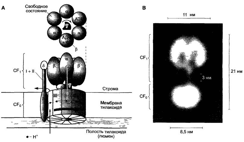
ATP synthase is localized in the stromal region of the thylakoids; its structure is similar to that of bacterial and mitochondrial enzymes (see 6.10.3.3). ATP synthase consists of a hetero-oligomeric headpiece projecting into the stroma, designated CF1 (CF from coupling factor), and another hetero-oligomeric, transmembrane part. This part is designated CF0 (the "O" is used to reflect a specific property and indicates the inhibition of the F0 part of mitochondrial ATP synthase by oligomycin. This terminology is also used for the chloroplast enzyme, although in this case CF0 is not sensitive to oligomycin. Often, instead of the letter "O", a zero is used as a subscript, as is the case here). CF0 forms a channel through which hydrogen ions are conducted, while CF1 catalyzes ATP synthesis.
ATP synthase is a rotary mechanism that transports H+ ions. It is the smallest known motor of its kind, with a size of about 20 nm. The asymmetric γ-subunit of the CF1 headpiece, together with the 12 subunits III of the CFo part, which form a ring-like structure, rotates at speeds of up to 100 rps. During this time, H+ ions flow through the CF0 channel. During one revolution, 12 H+ ions pass through the channel (one for each subunit III). The headpiece consists of three alternating α and β subunits. Contact with the rotating γ-subunit induces Conformational Changes in the α and β subunits, so that during each revolution, each of the three catalytic centers localized on the β subunits passes through three states (Fig. 6.63, A): 1) nucleotide-free; 2) ADP and inorganic phosphate bound; 3) the reaction ADP + Pi —> ATP takes place, and finally, ATP is released from the catalytic center of the enzyme. It is believed that the third state occurs upon the removal of water from the catalytic center, preventing the reverse reaction (Hydrolysis!) from competing with the Transfer of phosphate to ADP (cf. Fig. 6.8). Accordingly, 3 ATP molecules are synthesized per one revolution of the motor, which implies that the H+ : ATP ratio must be 4:1. Many details of the ATP synthesis reaction mechanism remain to be elucidated.
Fig. 6.63. ATP synthase. Side view. Electron micrograph, highly magnified: A — structural model of the rotary Mechanism of ATP synthase. Subunits III of the CFo part form a 12-part chamber, and together with the γ and ε subunits of the CF1 part, they form the rotor; the α3β3 headpiece, together with the δ subunit of the CF1 part and parts I, II, IV of CFo, form the stator. The rotor turns within the stationary stator, with 12 H+ ions (one per subunit III) being conducted through the proton channel, which is probably formed between subunits III and IV. The interaction of the rotating asymmetric γ-subunit with the α and β subunits of the ATP synthase headpiece promotes conformational changes in the enzyme's catalytic center located on the β-subunit, which passes through the following three stages: free center —> ADP + Pi bound —> ATPbound; B — reconstruction of ATP synthase
Due to disulfide-dithiol transitions on the γ-subunit (see Fig. 6.71), the rotary mechanism is switched on in the light and switched off in the dark. In the light, ferredoxin reduces the disulfide bridge in the protein molecule to a dithiol via thioredoxin, whereas in the dark, the disulfide form is regenerated, changing the conformation of the γ-subunit. This process prevents the reverse reaction catalyzed by ATP synthase in the dark, which could otherwise pump hydrogen ions into the thylakoid lumen by hydrolyzing ATP. Thioredoxin is a low-molecular-weight protein (about 10 kDa) found in all prokaryotic and Eukaryotic cells. Several of its isoforms are known. They all share a specific Amino Acid Sequence in their catalytic center: Cys-Gly-Pro-Cys. Both cysteines form disulfide bridges in oxidized thioredoxin.
Certain substances uncouple photosynthetic electron transport from ATP synthesis: the Hill reaction proceeds, but photophosphorylation does not occur. Such uncouplers include, for example, NH4+ ions and carbonyl cyanide para-trifluoromethoxyphenylhydrazone, which can penetrate the membrane in both protonated and deprotonated forms, thereby reducing the hydrogen ion gradient across the thylakoid membrane.
6.4.10. Light Reactions of Bacterial Photosynthesis
The light phase occurring in organisms capable of oxygenic photosynthesis was discussed in the previous chapters. This highly organized sequence of reactions involves two sequentially functioning photosystems that use electrons from water to reduce NADP+. These organisms evolved from ancestors that possessed only a single photosystem and used electron sources with a significantly lower standard redox potential. Such relatively simple light reactions occur in currently existing photolithotrophic and photoorganotrophic prokaryotes (see Table 6.1). Photolithotrophs include purple sulfur bacteria (Chromatiaceae) and green sulfur bacteria (Chlorobiaceae), while photoorganotrophs include purple bacteria (Rhodospirillaceae) and green non-sulfur bacteria (Chloroflexaceae).
All representatives of these groups possess light-driven cyclic electron transport, which leads to the formation of a hydrogen ion concentration gradient across the thylakoid membrane. In all groups, it is used for ATP synthesis, and also—with the exception of Chlorobiaceae—for NADH synthesis. In Chlorobiaceae, NADH is formed during non-cyclic electron transport via ferredoxin and ferredoxin-NAD+ reductase (Fig. 6.64). In this case, electrons do not return to the reaction center, so their deficit must be replenished by an electron donor, in most cases H2S:
H2S —> S + 2H+ + 2e- (E0'= -0.24 V).
Fig. 6.64. Light reactions of photosynthesis in green sulfur and purple bacteria. Ared, for example, is succinate, Aox is fumarate
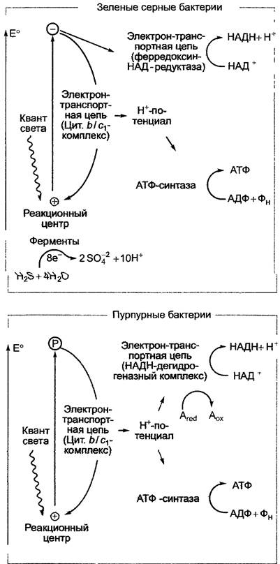
The difference in standard redox potentials between H2S/S and NAD+/NADH+H+ (E0' = -0.32 V) is very small. Therefore, a single photosystem and very low-energy light are sufficient for electron transfer from H2S to NAD+ (for example, the photosystem reaction center in Chlorobium absorbs quanta with a wavelength of  = 840 nm, which corresponds to approximately 142 kJ per Einstein). The resulting molecular sulfur can be oxidized to sulfite and then to sulfate, so that up to 8 electrons are available for photosynthetic electron transport:
H2S + 4H2O —> SO-24 + 10H+ + 8e-.
NADH formation is characteristic of purple bacteria, which possess only cyclic Electron Transport and an NADH dehydrogenase complex functioning at the expense of the hydrogen ion gradient energy. This energy is also used for ATP synthesis. The electrons required by the NADH dehydrogenase complex are derived from organic compounds: for example, in Rhodospirillum from succinate, which is oxidized to fumarate (E0' = -0.03 V). In this case, too, a single photosystem (P870) absorbing long-wavelength light is sufficient to ensure electron transfer from the organic substrate to NAD+.1
The reaction centers of photolithotrophic and photoorganotrophic bacteria are very similar in structure and homologous to each other. They evolved from a common ancestor. At the same time, the photosystems exhibit characteristic features inherent to organisms with oxygenic photosynthesis: the structure of the reaction center of purple bacteria corresponds to that of photosystem II, and the reaction center of green sulfur bacteria corresponds to that of photosystem I. The cytochrome b/c1 complex (see Fig. 6.64), which participates in cyclic electron transport and the coupled generation of a hydrogen ion gradient, is homologous to the cytochrome b6/f complex of organisms with oxygenic photosynthesis. The ferredoxin-NAD+ reductase system and ferredoxin, which are involved in the non-cyclic electron transport of green sulfur bacteria, are very similar to those in homologues of green plants, cyanobacteria, and prochlorophytes. Apparently, these are functional modules that arose at a very early stage of evolution. They can be combined in various ways, and some, in addition to photosynthesis, have assumed other functions (for example, functioning in the mitochondrial Respiratory Chain and in bacterial Respiration).
1 Flaw in logic: in this case, light energy is only required to generate a proton gradient; it cannot be used directly to reduce NAD+. — Ed. note.
It is believed that in the course of evolution, heterotrophic prokaryotes eventually gave rise to prokaryotes capable of cyclic photophosphorylation, thanks to The Emergence of photosystem I (or its ancestor). Later, the ability for non-cyclic electron transport from substances with a relatively low redox potential to NAD+ appeared, thereby enabling photoautotrophy in relatively small areas (where such reductants, e.g., H2S, are present). Only with the acquisition of photosystem II did The Use of water, which is ubiquitous, as a reductant become possible. Thus, photosynthesis became a quantitatively dominant process. Oxygen, which arises as a by-product of photosynthesis, gradually enriched the atmosphere from this point onward, creating the Prerequisites for the development of aerobic organisms.
Fig. 6.65. Structure of the pigment system in the photosynthetic reaction center of Rhodopseudomonas viridis (Rhodospirillaceae). The polypeptide backbones of the M, L, and H subunits are shown as grey lines, as is the reaction center embedded in The Lipid Bilayer of the bacterial plasma membrane. The reaction center carries the following pigments: 4 molecules of bacteriochlorophyll b, 2 of which form a dimer that is ionized upon excitation and donates one electron; the other two are located on either side of the dimer (BChlA and BChlB); 2 molecules of bacteriopheophytin (BPheoA, BPheoB); and 2 Quinones (QA — menaquinone, QB — ubiquinone), with an iron atom (Fe) located between them. A carotenoid lies adjacent to BChlB. The electron flow proceeds predominantly According to the scheme D —> BPheoA —> QA —> QB. According to the latest data, electrons flow from QA to QB via an iron ion with A change in valence (Fe3+ —> Fe2+). To avoid cluttering the diagram, the nonpolar side chains of the pigment molecules are only partially shown.
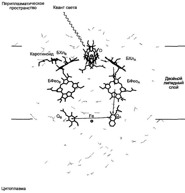
With the greening of chloroplasts during photomorphogenesis (see 7.7.2.1), photosystem I is formed first, and only later photosystem II.
Scientists succeeded in crystallizing the reaction centers of the purple bacteria Rhodopseudomonas viridis and Rhodobacter sphaeroides and resolving their structures at high resolution using X-ray crystal diffraction (X-ray crystallography). By comparing them with homologous structures of photosystem II, researchers were also able to reconstruct their structure (until then, the atomic composition had not been determined). X-ray crystallographic data have also been obtained for photosystem I, though at a lower resolution. The mode of photosystem operation is best understood in purple bacteria.
The reaction center of Rhodopseudomonas viridis (Fig. 6.65) consists of three proteins: the L, M, and H subunits. The L subunit is homologous to the D1 protein, and the M subunit to the D2 protein of photosystem II. Electron carriers are bound to the L and M proteins: 4 molecules of bacteriochlorophyll a (2 of which form the P870 reaction center dimer), 2 molecules of bacteriopheophytin a, 2 quinone molecules (1 ubiquinone, QB, 1 menaquinone, QA), 1 iron atom, and 1 carotenoid molecule. According to the latest data, the iron atom is involved in electron transfer, while the carotenoid serves to return chlorophyll from the triplet state to the ground state. In addition, however, a structural function is also under Discussion. The direction of electron flow in the reaction center is shown in diagram 6.65. It has been found that while the reaction center is structurally symmetrical, the electron flow is asymmetrical, proceeding mainly along the A-branch (this is also true for photosystem II — see Fig. 6.59). The extremely rapid electron transfer from the bacteriochlorophyll a dimer to bacteriopheophytin primarily prevents the recombination e- + P870+ —> P870, and thus the conversion of excitation energy into heat. The kinetics of the electron transfer step in photosystem II are analogous. From an electrostatic standpoint, the direction of electron flow along the A-branch is favored because it has a larger positive net charge compared to the flow direction along the B-branch.
A light-driven proton pump does not always operate using chlorophyll. In the non-photosynthetic1 Halobacterium halobium, under certain environmental conditions (low O2 concentration), Bacteriorhodopsin—a transmembrane chromoprotein resembling the pigment rhodopsin2—is found in the outer cell membrane; bacteriorhodopsin molecules are localized in specific regions of the membrane (purple membrane).
1 In Russian scientific literature, the prevailing view is that halobacteria are photosynthetic organisms. — Editor's note.
2 In the light-sensitive Cells of the animal retina. — Editor's note.
Using the light energy absorbed by bacteriorhodopsin, a hydrogen ion gradient is ultimately established between the cell interior and the medium. The dissipation of this gradient drives ATP synthesis, amino acid uptake, and salt exchange (Na+/K+) (Fig. 6.66). Thus, this Organism is capable of performing photophosphorylation without chlorophyll and maintaining its Energy balance. In the presence of oxygen, a proton gradient is generated via the electron transport of the respiratory chain, with energy derived from the oxidation of organic compounds. In this case, ATP synthesis occurs via Oxidative Phosphorylation (see 6.10.3.3). A second light-driven ion pump, halorhodopsin, transports chloride ions (Cl) into the cell. The system serves for osmotic regulation in biotopes with very high salt concentrations (up to 4M NaCl) in archaea.
Fig. 6.66. Photophosphorylation in Halobacterium halobium. Upon absorption of a light quantum, the bacteriorhodopsin of the purple bacteria is converted from the P560 form to P412. This conversion is reversible. During this process, an H+ ion is transported from the cell into the external medium ('proton pump'). The membrane-bound ATP synthase can utilize the resulting hydrogen ion concentration gradient to synthesize ATP.

Last update: 07/08/2026
Editorial and Educational Adaptation: This material has been compiled based on the primary/original source text. The project team performed an editorial review, corrected technical inaccuracies, structured sections, and adapted the content for an educational format.
What was processed:
- elimination of formatting defects (OCR errors, structural breaks, corrupted characters);
- editorial organization of content;
- standardization of terminology in accordance with academic sources;
- verification of factual statements against the original source text.
All mentions of the author, publication year, and origin of the primary text have been preserved in accordance with the source.