BOTANY VOLUME 2 - PLANT PHYSIOLOGY - 2007
6. PHYSIOLOGY OF METABOLISM
6.5. Photosynthesis: Carbon Assimilation Pathways
During carbon assimilation, CO2 is converted into CARBOHYDRATES (CH2O)n. The formation of a single hexose molecule can be represented by the photosynthetic equation:
6СO2 + 12Н2O —> С6Н12O6 + 6H2O + 6O2.
This reaction is highly endergonic (∆G0 = 2,862 kJ mol-1, which corresponds to 477 kJ mol-1 for the fixation of one CO2 molecule). Oxygen is derived from Water (see 6.4.4: Photooxidation of water). Formally, carbon is reduced from CO2 in the +IV oxidation state to (CH2O)n in the 0 oxidation state. Consequently, the reduction of one C atom requires 4 electrons supplied by NADPH + H+ produced during the light reactions of Photosynthesis (or NADH + H+ in some Bacteria — see 6.4.10). Additionally, carbon assimilation reactions utilize energy in the form of ATP, which is also generated in the light reactions of photosynthesis (see 6.4.4). The synthesis of one hexose molecule from 6 CO2 can be represented by the following reaction:
6СO2 + 12 NADPH + 12 H+ + 18 ATP —> С6Н12O6 + 12 NADP+ + 18 ADP + 18 Pi + 6H2O.
It takes place in the stroma of METABOLISM/14.html">Chloroplasts (and in the Cytoplasm of photosynthetic prokaryotes) and is often called the Dark Phase of photosynthesis, as it does not
depend directly on light. In principle, in the presence of NADPH and ATP, these reactions could occur in the dark. Nevertheless, in The Cell, CO2 assimilation occurs exclusively in the light, because only under these conditions are NADPH and ATP generated. Furthermore, the Reactions of the dark phase of photosynthesis are activated by light and inactivated in the dark (see 6.5.5). The sequence of dark-phase reactions is complex, involving numerous enzyme-catalyzed intermediates that form a cyclic process known as the Calvin cycle in honor of its discoverer. M. Calvin exposed Algae (and later, isolated chloroplasts) to radioactive 14CO2 in the light for a few seconds, then extracted the reaction products with hot ethanol and separated them using two-dimensional paper Chromatography. The separated radiolabeled products could be visualized on an autoradiogram and identified by comparing the positions of the radioactive spots on the chromatogram with those of reference substances of known chemical Structure (standards).
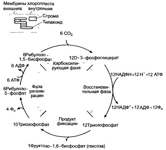
The Calvin Cycle (Fig. 6.67) can be divided into three phases: carboxylation, reduction, and regeneration.
This reaction sequence can also be called the reductive pentose phosphate cycle, as it represents the reverse of the oxidative pentose phosphate cycle (see 6.10.3.5). Consequently, many of the Enzymes involved in the Calvin cycle are not unique to photosynthesis.
Class="center">Fig. 6.67. General scheme of the three Phases of the Calvin cycle, showing the formation of one hexose molecule from 6 molecules of fixed carbon dioxide (CO2)
6.5.1. Carboxylation Phase of the Calvin Cycle
The first reaction product of the Calvin cycle is D-3-phosphoglycerate. It is formed by the carboxylation of the CO2 acceptor ribulose-1,5-bisphosphate (RuBP). Fig. 6.68 shows the reaction catalyzed by ribulose-1,5-bisphosphate carboxylase/oxygenase (RubisCO). The oxygenase function of the enzyme will be discussed later (see 6.5.6: Photorespiration).
The reaction is highly exergonic (∆G0' = -35 kJ mol-1) and therefore proceeds spontaneously. Ribulose-1,5-bisphosphate loses its enediol form upon addition of dissolved CO2. The immediate reaction product, 2-carboxy-3-keto-D-arabinitol-1,5-bisphosphate, is highly unstable and is rapidly cleaved by water into two molecules of D-3-phosphoglycerate. The product of dephosphorylation at the C-5 position and Hydration of the CO2 adduct — 2-carboxy-D-arabinitol-1-phosphate — is a potent inhibitor of the carboxylation reaction. It is thought to participate in the in vivo regulation of RubisCO activity. The newly fixed carbon appears in the carboxyl group of one of the two D-3-phosphoglycerate molecules (see Fig. 6.68).
RubisCO binds CO2 rather than the HCO3- ion, which is predominant in aqueous solution. In the alkaline stroma of illuminated chloroplasts (pH 8), the equilibrium is shifted even further toward bicarbonate. The establishment of equilibrium in the reaction CO2 + H2O ⇄ HCO3- + H+ is catalyzed by the enzyme Carbonic anhydrase; however, there is no direct evidence that The activity of this enzyme determines The rate of the dark reactions of photosynthesis.
Chloroplast ribulose-1,5-bisphosphate carboxylase/oxygenase is one of the key specific enzymes of photosynthesis. It is a hexadecamer composed of 8 large and 8 small subunits and is active only in this form. The large subunit (51–58 kDa) is encoded by plastid DNA (see 7.2.1.2) and translated on 70S plastid Ribosomes using their mRNA. The small subunit (12–18 kDa), encoded by nuclear DNA, is synthesized on 80S ribosomes in the cytoplasm. It initially possesses an N-terminal signal sequence that facilitates The transport of the small RubisCO subunit into the chloroplast (see 7.3.1.4). Assembly of the subunits into the holoenzyme proceeds with the assistance of chaperones (see 7.3.1.2).
Fig. 6.68. CO2 fixation process in the Calvin cycle. The reaction is catalyzed by the enzyme ribulose-1,5-bisphosphate carboxylase (RubisCO). A potent inhibitor of the enzyme is 2-carboxy-D-arabinitol-1-phosphate, an analog of the hydrated form of 2-carboxy-3-keto-D-arabinitol-1,5-bisphosphate

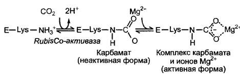
Fig. 6.69. Activation of ribulose-1,5-bisphosphate carboxylase (RubisCO) by Mg2+ ions and CO2
The catalytic site is located on the large subunit. In some purple bacteria, the enzyme has a different structure, consisting of a dimer of two large subunits.
Although the Km value (see 6.1.6.3) of RubisCO for CO2 is about 10–15 µM and thus roughly corresponds to the concentration of dissolved CO2 (an atmospheric CO2 concentration of 350–360 ppm1 corresponds to an equilibrium concentration of 10 µM CO2 in aqueous solution), catalysis is relatively slow: the turnover number of the enzyme is only 3.3 s-1 per catalytic subunit (by comparison, carbonic anhydrase achieves about 105 catalytic events per second). Therefore, efficient catalysis requires a very large amount of the enzyme: RubisCO can constitute up to 50% of the total leaf protein. RubisCO is the most abundant enzyme in the biosphere.
1 ppm — part per million; 350 ppm corresponds to 0.035%. — Ed. note.
CO2 is not only a substrate. It also acts as an allosteric activator of RubisCO: together with a specific Lysine of the large subunit, CO2 forms a carbamate complex; upon the subsequent binding of an Mg2+ ion, the enzyme becomes active (Fig. 6.69). Lysine carbamylation is catalyzed by the enzyme RubisCO activase, requiring ATP energy. Since the concentration of Mg2+ in the stroma increases in the light, ATP-dependent carbamylation and Mg2+ binding serve as effective mechanisms ensuring that CO2 fixation occurs only when all necessary components are present.
6.5.2. The reduction phase of the Calvin cycle
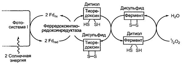
The primary product of CO2 fixation, D-3-phosphoglycerate, is reduced to D-glyceraldehyde 3-phosphate In the second stage of the Calvin cycle. This highly endergonic reaction is driven by ATP. The reducing agent in this reaction is NADPH + H+. Both ATP and NADPH are products of the light reactions of photosynthesis (Fig. 6.70). During the reaction, D-3-phosphoglycerate is converted by phosphoglycerate kinase into 1,3-bisphosphoglycerate, which is then reduced to D-glyceraldehyde 3-phosphate with the release of a phosphate group by glyceraldehyde 3-phosphate dehydrogenase (GAPDH). Isoforms of both aforementioned enzymes are also present in the cytoplasm (Glycolysis/Gluconeogenesis); however, the plastid GAPDH is NADPH/NADP+-specific, whereas the cytoplasmic isoenzyme uses NADH/NAD+. NADPH-glyceraldehyde 3-phosphate dehydrogenase is converted to its active dithiol form by the ferredoxin-thioredoxin system (Fig. 6.71) and is therefore activated only in the light. We have already encountered a similar light activation in the case of ATP synthase (see 6.4.9). The activities of many other Calvin cycle enzymes are regulated by the same principle (see 6.5.3).
D-glyceraldehyde 3-phosphate is in equilibrium with dihydroxyacetone phosphate. This equilibrium is maintained by the enzyme triose phosphate isomerase. D-glyceraldehyde 3-phosphate and dihydroxyacetone phosphate are also referred to as triose phosphates, and they are already carbohydrates (trioses). A series of Condensation reactions serves to:
✵ synthesize further carbohydrates (e.g., hexoses) from triose phosphates as the final products of photosynthesis;
✵ regenerate the CO2 acceptor ribulose 1,5-bisphosphate.
6.5.3. The regeneration phase of the Calvin cycle
To ensure continuous CO2 fixation and reduction, the CO2 acceptor—ribulose 1,5-bisphosphate (RuBP)—must be constantly regenerated. From 6 molecules of RuBP and 6 molecules of CO2, 12 molecules of triose phosphate are formed (see Fig. 6.67). Two of these molecules are used to synthesize one hexose molecule (yielding fructose 1,6-bisphosphate first), while the remaining 10 triose phosphate molecules are used to regenerate six RuBP molecules. This constitutes a cyclic process (see Fig. 6.67). Some reactions of this process are shown in Fig. 6.70. The regeneration of the CO2 acceptor directly from ribulose 5-phosphate requires ATP. Thus, the fixation of one CO2 molecule in the Calvin cycle overall requires two NADPH + H+ molecules and three ATP molecules (two in the phosphoglycerate kinase reaction, one in the ribulose-5-phosphate kinase reaction).
The enzymes catalyzing the irreversible reactions of the reduction and regeneration phases—ribulose-5-phosphate kinase, fructose-1,6-bisphosphatase, and sedoheptulose-1,7-bisphosphatase—are activated in the light by the ferredoxin-thioredoxin system (see Fig. 6.71), just like NADPH-glyceraldehyde-3-phosphate dehydrogenase (see 6.5.2). Furthermore, like RubisCO, both Phosphatases are activated by Mg2+ and have a pH optimum of around 8.0. Since light-driven proton transport into the thylakoid lumen increases the pH in the chloroplast stroma from 7.2 to 8.0, and the concentrations of Mg2+ ions and reduced thioredoxin also rise, the dependence of these key Calvin cycle enzymes on pH, Mg2+, and thioredoxin represents a highly efficient system for light activation (and dark inhibition) of all processes in the light-independent phase.
6.5.4. Processing of the primary products of carbon assimilation
Triose phosphate is the end product of the CO2 fixation and reduction reactions (see Fig. 6.70; Fig. 6.72). A portion of it is exported from the chloroplasts and used for hexose synthesis in the cytoplasm. From hexose, the most important transport sugar—sucrose (cane sugar)—is synthesized. The "surplus" triose phosphate not consumed for sucrose synthesis or RuBP regeneration (up to 30% of total photosynthetic yield) is used within the chloroplasts for starch synthesis. In this way, reduced carbon is stored in an osmotically inactive form. Assimilatory starch (also called transit starch) is converted in the dark into glucose and triose phosphates, which are transported into the cytoplasm and used for sucrose synthesis. Along with sucrose, other Organic compounds, especially certain additional carbohydrates and Amino Acids, are intensively formed during photosynthesis. These assimilates leave the cell and are transported via the phloem to the sink Organs of the plant.
Fig. 6.70. Overview of the Calvin cycle. For clarity, only the sequence of reactions leading to the formation of one triose phosphate molecule from three CO2 molecules is shown. Irreversible reactions are indicated by gray arrows. These are the primary sites of regulation (also shown in gray). The multiple arrows indicate how many molecules react in each step to yield one triose phosphate molecule as the fixation product from three CO2 molecules. To form one hexose molecule from 6 CO2 molecules (see Fig. 6.67), the process shown here must occur twice
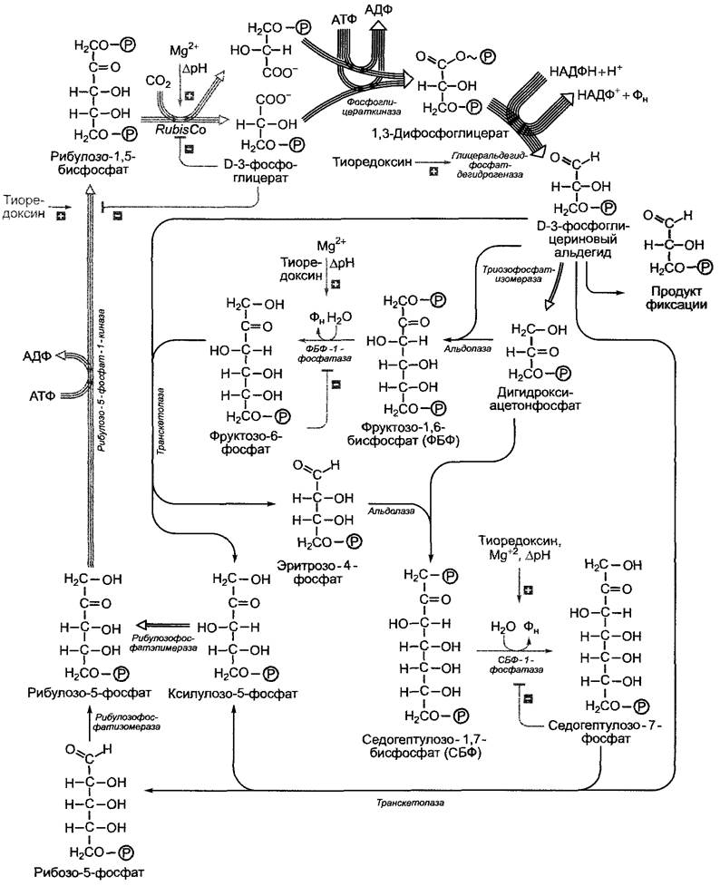
Fig. 6.71. Light-dependent REGULATION OF ENZYMATIC Activity via disulfide-dithiol conversion by thioredoxin. In the dark, thiol groups are oxidized by molecular oxygen to form disulfide bridges
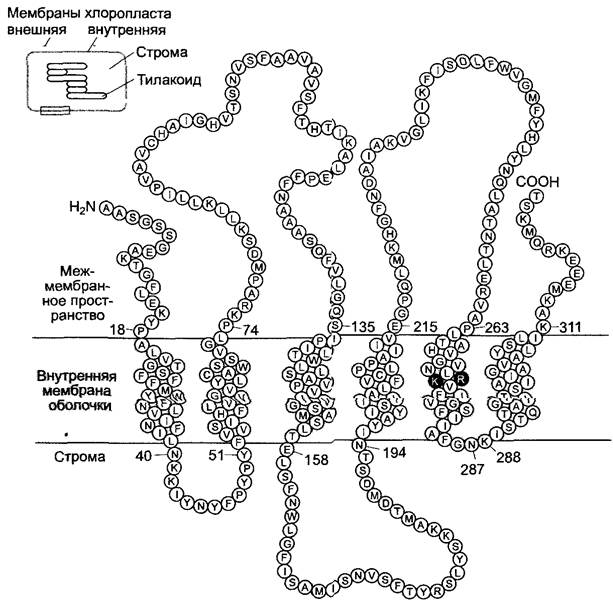
The exchange of triose phosphates between the cytoplasm and the chloroplast is mediated by a passive carrier, the triose phosphate translocator, which performs a counter-exchange with phosphate ions, i.e., antiport. This prevents phosphate depletion in the chloroplast due to triose phosphate export and ensures continuous ATP synthesis. The triose phosphate translocator is likely a homodimer whose monomers have a molecular mass of about 30 kDa, representing the most abundant protein of the inner chloroplast envelope membrane (15% of the total protein content of this membrane). It is encoded in the Cell Nucleus, synthesized as a precursor with an N-terminal transit peptide, and imported into the chloroplast, where it acquires its final conformation (see 7.3.1.4). The monomer is thought to span the inner envelope membrane 6 times via hydrophobic α-helices. Two positively charged amino acids in the fifth α-helical domain—Arginine (R) and lysine (K)—serve as the binding sites for the negatively charged substrate (Fig. 6.73).
Sucrose synthesis takes place in the cytoplasm. It begins with fructose 6-phosphate and uridine diphosphate glucose (UDPG). First, sucrose phosphate is formed (sucrose-phosphate synthase reaction), which is then dephosphorylated by sucrose-phosphate phosphatase to yield sucrose. This final step is irreversible and drives efficient sucrose synthesis. Synthesized sucrose is transported to sinks and storage sites via the phloem (see 6.8: Transport of Assimilates). As a non-reducing sugar of the trehalose type, sucrose lacks a free reducing end and is chemically inert. It is therefore an ideal transport metabolite, unlike free hexoses, which are highly reactive due to their carbonyl groups; their hemiacetal forms isomerize in aqueous solution (mutarotation, see 1.4.1; Fig. 1.18). UDPG is formed from UTP and glucose 1-phosphate (UDP-glucose pyrophosphorylase reaction). Glucose 1-phosphate is in equilibrium with fructose 6-phosphate, which in turn is produced by the irreversible dephosphorylation of fructose 1,6-bisphosphate (fructose-1,6-bisphosphatase reaction). This is preceded by the condensation of triose phosphates, as fructose 1,6-bisphosphate and triose phosphates are in equilibrium (aldolase reaction) (see Figs. 6.70; 6.72).
Fig. 6.72. Metabolism and transport of carbohydrates in photosynthetic and storage Tissues
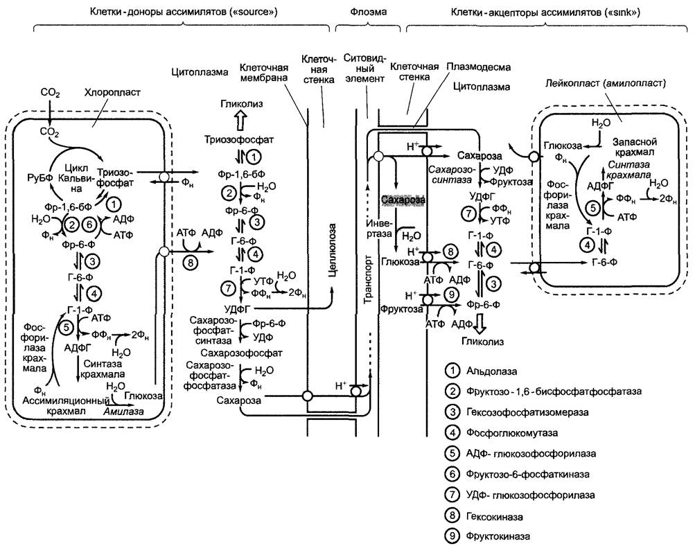
Fig. 6.73. Model of the polypeptide chain arrangement of the monomeric triose phosphate translocator in the inner chloroplast envelope membrane.
The amino acids shown in dark gray, lysine (K)-273 and arginine (R)-274, bind to the substrate (one-letter code, see Fig. 1.11). The native translocator likely exists in the membrane as a dimer
Plastid isoforms of cytoplasmic enzymes catalyze the synthesis of hexose phosphates from triose phosphates in the chloroplast stroma. Furthermore, starch synthesis, which is described in more detail in Section 6.17.1.2, utilizes activated glucose, specifically adenosine diphosphate glucose (ADPG), which is produced from ATP and glucose 1-phosphate in a reaction catalyzed by the enzyme ADP-glucose pyrophosphorylase. In the dark, assimilatory starch is converted either to glucose by amylase and maltase (Hydrolysis) or to glucose 1-phosphate by phosphorylation (starch phosphorylase) (see 6.17.1.2).
Both glucose and glucose 1-phosphate are used in sucrose synthesis. For this purpose, glucose is transported into the cytoplasm via a translocator, while glucose 1-phosphate is converted in the stroma to triose phosphate, which is exported to the cytoplasm by the triose phosphate translocator (see Fig. 6.72). The Formation of fructose 1,6-bisphosphate from fructose 6-phosphate is catalyzed by its own enzyme, fructose-6-phosphate 1-kinase; because the reverse reaction catalyzed by the aforementioned fructose-1,6-bisphosphatase is highly exergonic, it cannot easily run in reverse. The subsequent fate of photosynthetic products will be discussed later (see 6.8; 6.10–6.17).
6.5.5. Regulatory mechanisms of carbohydrate synthesis and partitioning
We have already mentioned regulatory processes in connection with the Light regulation of the Calvin cycle. Enzymes that catalyze irreversible reactions of the Calvin cycle are additionally inhibited by the end product (see Fig. 6.70). Thanks to such precise regulation, the accumulation of currently unnecessary metabolic intermediates is avoided. Nevertheless, coordinating metabolic activity in chloroplasts and cytoplasm during the light and dark Phases of Photosynthesis requires additional control, which primarily serves to partition triose phosphates according to cellular demands. For instance, an excessively high consumption of triose phosphate from the Calvin cycle for starch or sucrose synthesis can impair the regeneration of the CO2 acceptor, ribulose-1,5-bisphosphate, and ultimately lead to the interruption of the cycle itself. The regulatory checkpoints here are primarily those enzymes that catalyze irreversible reactions: in sucrose synthesis, cytoplasmic fructose-1,6-bisphosphatase, and in starch synthesis, plastid ADP-glucose phosphorylase. Regulatory mechanisms in the cytoplasm are primarily directed at sucrose-phosphate synthase, and in chloroplasts, at starch phosphorylase.
Precise control of triose phosphate consumption for sucrose synthesis is achieved by altering the concentrations of fructose-6-phosphate, phosphate, and triose phosphate (Fig. 6.74). High concentrations of phosphate and fructose-6-phosphate activate fructose-6-phosphate 2-kinase. It promotes the synthesis of fructose-2,6-bisphosphate (Fru-2,6-BP), a potent inhibitor of fructose-1,6-bisphosphatase. Fru-2,6-BP simultaneously activates pyrophosphate-dependent Fru-6-P kinase, meaning that the concentration of fructose-6-phosphate decreases while the concentration of triose phosphate increases. The latter inhibits fructose-6-phosphate 2-kinase and activates Fru-2,6-BP phosphatase, lowering the concentration of fructose-2,6-bisphosphate and increasing the utilization of fructose-1,6-bisphosphate. In this way, a highly coordinated Metabolic control of triose phosphate utilization is achieved (see Fig. 6.74). This also includes the activation of sucrose-phosphate synthase by glucose-6-phosphate (an indicator of hexose deficiency in the cytoplasm).
Fig. 6.74. Regulation of triose phosphate utilization in the cytoplasm via metabolic control of the fructose-2,6-bisphosphate system

The regulation of starch synthesis in chloroplasts is less understood. The activity of ADP-glucose pyrophosphorylase is increased by D-3-phosphoglycerate. An increase in phosphoglycerate concentration in the stroma indicates that more CO2 is being fixed than is required in the form of the reaction product D-3-phosphoglycerate for export to the cytoplasm and to sustain the Calvin cycle. Phosphate inhibits ADP-glucose pyrophosphorylase. Its concentration increases mainly during the dark phase of photosynthesis, when Photophosphorylation does not occur. At this time, the activator of ADP-glucose pyrophosphorylase, D-3-phosphoglycerate, is also absent. Phosphate activates starch phosphorylase and, along with starch, simultaneously serves as its substrate; therefore, the regulation of starch Synthesis and degradation via changes in phosphate concentration leads to the efficient mobilization of transitory starch
In a specific side reaction, RubisCO catalyzes the fixation of one molecule of O2 instead of CO2, with ribulose-1,5-bisphosphate also serving as the acceptor. In contrast to carboxylation, the oxygenase reaction produces only one molecule of D-3-phosphoglycerate and one two-carbon compound, 2-phosphoglycolate (Fig. 6.75). Under intense illumination, the oxygenase activity of RubisCO accounts for about 20–30% of all reactions, and at high temperatures, it can even reach
50%. The reason for this Temperature dependence is that the affinity of RubisCO for CO2 decreases with increasing temperature, and at the same time, the solubility of CO2 in water decreases much more rapidly than that of O2. The plant has a strong need to reclaim the carbon withdrawn from the Calvin cycle from 2-phosphoglycolate (see Fig. 6.75). This process involves oxygen consumption and CO2 release; therefore, by formal analogy with cellular Respiration, it is called photorespiration.
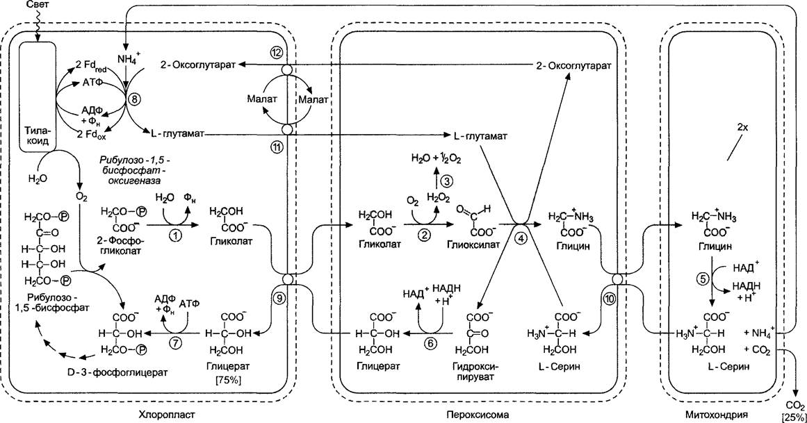
Fig. 6.75. Reaction sequence and compartmentalization of photorespiration. Two Glycine molecules are used for Serine synthesis: the ribulose-1,5-bisphosphate oxygenase reaction and the synthesis of glycine from 2-phosphoglycolate occur twice (on a gray Background): (1) — phosphoglycolate phosphatase; (2) — glycolate oxidase; (3) — catalase; (4) — serine-glyoxylate aminotransferase and glutamate-glyoxylate aminotransferase; (5) — glycine decarboxylase complex; (6) — hydroxypyruvate reductase; (7) — glycerate kinase; (8) — glutamate synthase/Glutamine Synthetase cycle (see 6.6.1); (9) — glycerate-glycolate translocator; (10) — amino acid translocator; (11) — malate-glutamate translocator; (12) — malate-2-oxoglutarate translocator
✵ Very often, when discussing the oxygenase activity of RubisCO, it is mentioned that it does not distinguish substrates with sufficient Specificity. In the Cytology/cytology/16.html">Early stages of the enzyme's evolution, The ability to distinguish between O2 and CO2 was of no consequence due to the absence of molecular oxygen, and therefore was not subject to natural Selection. Only with the advent of oxygenic photosynthesis did the atmosphere gradually become saturated with oxygen. Although nearly 1.5 billion years have passed since then, it is evident that evolutionary optimization of the catalytic center of RubisCO is constrained; thus, a complex biochemical salvage pathway (to prevent carbon loss) had to evolve across three cellular Organelles to counteract the Damage caused by the oxygenase activity of RubisCO. Alongside this, the potential role of photorespiration as an additional photoprotective mechanism against oxidative damage to the Photosystems is under Discussion. This mechanism would be engaged when, under water deficit and consequently closed Stomata, less CO2 is consumed, while high light intensity promotes the intensification of ATP and NADPH synthesis, and the partial pressure of O2 is high (photooxidation of water!). Photorespiration consumes O2, ATP, and NADPH1, and also promotes the internal release of CO2, thereby ensuring the normal operation of the Calvin cycle.
1 The reference to The Use of NADPH in the chloroplast during photorespiration is incorrect: compare with Fig. 6.75. — Editor's note.
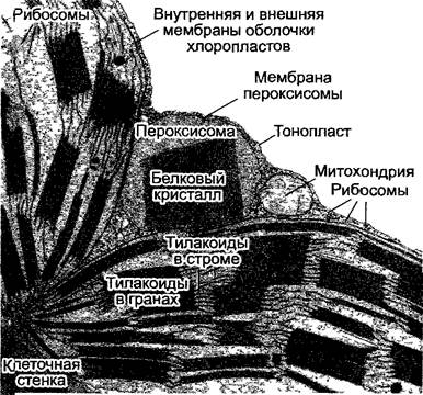
Photorespiratory reactions take place in chloroplasts, Peroxisomes, and Mitochondria. Peroxisomes, like glyoxysomes, belong (see 6.12) to Microbodies (see 2.2.6.6). In mesophyll Cells, peroxisomes, chloroplasts, and mitochondria are very often located close to each other (Fig. 6.76), which is evidence of intensive metabolism between these organelles.
Fig. 6.76. Organelles in tobacco mesophyll leaf cells (section, 17,000x)
The sequence of photorespiratory reactions is shown in Fig. 6.75. As a result, 2 molecules of phosphoglycolate (twice 2 C atoms) are converted into one molecule of D-3-phosphoglycerate, which enters the Calvin cycle. Consequently, 75% of the carbon removed from the cycle as 2-phosphoglycolate is recovered (3 out of 4 C atoms), while a quarter of the carbon is released as CO2 during the formation of L-serine from 2 glycine molecules in the mitochondria. The NH4+ ion synthesized in the glycine decarboxylase reaction is very efficiently re-fixed in the chloroplasts to form glutamate1. This reaction will be discussed in more detail in Section 6.6. Glycine decarboxylase is a multi-enzyme complex similar to mitochondrial Pyruvate dehydrogenase, and in green plant parts, it can constitute up to 30–50% of the total protein in the mitochondrial matrix, whereas in non-green plant parts, this enzyme is either completely absent or present in very small amounts. This explains the enormous expenditure of matter and energy that the plant incurs during photorespiration. In addition, peroxisomes contain large amounts of catalase, their main enzyme. Some crystalline inclusions in peroxisomes visible under Electron Microscopy consist of catalase. The enzyme catalyzes the disproportionation of hydrogen peroxide (H2O2), which arises from the glycolate oxidase reaction, into H2O + 1/2O2, thereby protecting cells from strong oxidants.
1 According to modern data, NH+4 fixation can occur in the cytoplasm and mitochondria. The transport of toxic ammonium from the mitochondrion to the chloroplast is unlikely. — Ed. note.
The exchange of metabolites between the compartments involved in photorespiration is carried out by translocators in the inner membrane of chloroplasts or mitochondria; the exchange of substances occurs through simple porins in the peroxisome membrane, which are integral pore-forming Membrane Proteins that perform direct non-selective transport of low-molecular-weight compounds.
Photorespiration requires much more energy than CO2 fixation. In the Calvin cycle, a total of 3 ATP and 2 NADPH molecules are spent per CO2 (see 6.5.3). To achieve a complete carbon balance (i.e., not to lose carbon in the oxygenase reaction), the metabolites resulting from two oxygenase cycles — two 2-phosphoglycolates and two D-3-phosphoglycerates — must be processed, and the resulting CO2 must be re-fixed by RubisCO. Since one molecule of D-3-phosphoglycerate is formed from two molecules of 2-phosphoglycolate, as a result of the Calvin cycle, three molecules of phosphoglycerate must be converted into three molecules of triose phosphate, regenerating three RuBP, i.e., 1 molecule of CO2 is fixed, and the costs of photorespiration are also compensated (1 ATP: glycerate kinase, 1 ATP and 2 Fdred corresponding to 1 NADPH: for the re-fixation of NH+4)1. In total, 10.5 ATP and 6 NADP (i.e., slightly more than 5 ATP and 3 NADP per O2 molecule) are spent per 2 molecules of O2 just to restore the carbon balance2. The ratio of carboxylation to oxygenation in the leaf is intermediate between 2:1 and 4:1, so photorespiration consumes about 50% of ATP and NADP. Thus, about a third of the light energy received from the antenna complexes is spent on this side reaction.
1 This part can be omitted from the calculations if we assume that the re-fixation of NH+4 occurs in the mitochondria using NADH formed in reaction 5 (see Fig. 6.75). — Ed. note.
2 The energy costs of photorespiration are clearly exaggerated. Often, the plant is not faced with the task of completely closing the photorespiration cycle. Thus, this process can be used to form glycine or serine, which are necessary for Protein Synthesis. Under severe drought, it is fundamentally impossible to close the photorespiration cycle: CO2 is lost as a result of diffusion, and the 're-fixation costs' are not realized. — Ed. note.
6.5.7. CO2 Uptake by the Plant
The natural concentration of CO2 in the atmosphere is currently about 0.036–0.037 vol. % (360–370 ppm). In the mid-1960s, this value was about 320 ppm. Since then, the average CO2 concentration has increased almost linearly to its current value. The concentration gradient between the ambient and intracellular air is very low, which is insufficient to drive CO2 through the diffusion barrier of the cuticle and epidermis when the stomata are closed. The situation is completely different during respiration in the case of O2 uptake: a high concentration gradient between the ambient air (about 21 vol. %, 210,000 ppm) and respiring mitochondria (close to 0%) provides a diffusion rate that meets the O2 demand of small-volume plant organs even when the stomata are closed. CO2 enters the plant only through open stomata, so the degree of stomatal opening has a decisive influence on photosynthesis. Since, due to the high affinity of RubisCO for CO2 (10–15 µmol, see 6.5.1), the natural concentration of CO2 (and the concentration of CO2 dissolved in water in equilibrium with it: at 25 °C — about 10 µmol) is below the optimal level for the enzyme, the lowest possible diffusion resistance from the stomata (the widest stomatal aperture) is required during photosynthesis to efficiently supply the chloroplasts with CO2. Because this simultaneously leads to severe water loss through Transpiration, the plant's water supply is also crucial for the normal course of photosynthesis. As already mentioned (see 6.3.4, equation 6.38), the transpiration coefficient for an average leaf surface is about 200–800 (grams of transpired water per gram of fixed CO2). This means that for every molecule of fixed CO2, 500–2,000 molecules of water evaporate. These figures show how critical optimal control of stomatal Functions is for the plant. In addition to CO2 concentration and water supply, the degree of stomatal opening is also regulated by light and temperature. Stomata (see 3.2.2.1; 6.3.4; 8.3.2.5) function as turgor-driven adjustable Valves (Fig. 6.77). The direct trigger for the opening of the stomatal pore is A change in turgor in the guard cells and the cells bordering them, which make up the stomatal complex due to their specific morphological structure (see 3.2.2.1). An increase in turgor in the guard cells relative to its normal state in the surrounding cells leads to the opening of the stomata, while a decrease in turgor leads to the closure of the stomatal pore. The regulated change in turgor in the guard cells occurs as a result of Changes in the osmotic potential in the cells, which is based on changes in the concentrations of potassium (K+) and chloride (Cl-) ions and/or malate (malate2-) ions as counterions. These are controlled by several interconnected Ion Exchange processes in which the guard cells function as regulated osmotic cells.
Fig. 6.77. Simplified diagram of the feedback system in the regulation of stomatal function:
 — water potential; ABA — Abscisic acid. Temperature regulation is not shown. Explanations in the text
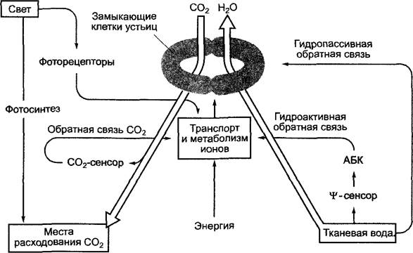
First, the osmotic potential of guard cells is regulated by the water available in the tissues. The Nature of The water potential sensor ( sensor) is unknown. When a certain threshold water potential is reached (from -0.7 to -1.8 MPa in the leaf), the phytohormone abscisic acid (ABA, see 7.6.4) is released, which induces stomatal closure within a few minutes. Along with this hydroactive feedback, stomata can also react hydropassively, i.e., without changing their osmotic potential. In this case, guard and neighboring cells lose or absorb different volumes of water.
For example, during rain, the epidermal cells of a water-stressed plant absorb water faster than the guard cells. The resulting increase in the turgor of epidermal cells relative to that of the guard cells leads to the hydropassive closure of the stomatal pore.
Guard cells react to the concentration of CO2 inside the leaf. The sensory mechanism is probably localized in the guard cells, and its nature is not yet understood. A decrease in CO2 concentration in the guard cells leads to an increase in their osmotic potential, subsequent water entry into the cells, which increase in volume, and finally, to stomatal opening. When the CO2 concentration in the guard cells increases, the osmotic potential decreases again, and the stomatal pores close.
Light, on the one hand, has a direct effect on guard cells (presumably mediated by blue light receptors)1 and leads to
an increase in their osmotic potential, and consequently, to the opening of stomatal pores. However, light also opens stomata indirectly. Upon illumination, photosynthesis is initiated, reducing the CO2 concentration in the intercellular spaces, and subsequently in the guard cells.
In general, the temperature dependence of stomatal opening corresponds to that of photosynthesis. In a well-watered plant at high temperatures, the dependence of stomatal opening on CO2 may disappear. This is ecologically advantageous, as transpirational cooling at high temperatures prevents leaf overheating and brings its temperature as close as possible to the optimum level for photosynthesis.
The degree of stomatal opening can vary across the same leaf. Guard cells respond to local conditions. This allows the plant to achieve significant optimization of gas exchange.
The Mechanism of stomatal movement and its control, as well as the features of gas exchange regulation in plants with additional photosynthetic pathways (see 6.5.8 and 6.5.9), are discussed in Section 8.3.2.5.
CO2 uptake by the plant can be described by an equation derived from Fick's first law of diffusion (see Equation 6.30):
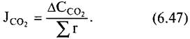
The diffusion flux of CO2 (JCO2) is directly proportional to the difference in carbon dioxide concentration (∆CCO2) and inversely proportional to the diffusion resistance r, which is equal to the sum of the individual diffusion resistances of each stomatal pore (Fig. 6.78). In air, CO2 (like O2) can diffuse approximately 105 times faster than in water (CO2 in the gas phase is 1 cm s-1, in the aqueous phase — 10-5 cm s-1). Therefore, the primary task for the plant is to deliver metabolic gases to the reaction sites in the gas phase as much as possible. The intercellular space system serves this purpose (see 3.2.1, Fig. 3.7).
1 According to recent data, in addition to cryptochrome, Phytochrome (a red light receptor) is also involved in photoreception. — Ed. note.
The barriers encountered by CO2 on its way to the photosynthetic chloroplasts of land plants (see Fig. 6.78) include the boundary layer, i.e., the air layer adjacent to the leaf, whose resistance is proportional to its thickness, or the water layer surrounding the leaves of aquatic plants. No convection processes occur in this layer. The thickness of the air layer can be several millimeters; in strong wind or current, this layer can completely disappear. The thickness and Stability of the boundary layer also depend on leaf anatomy (e.g., the presence of hairs). With high boundary layer resistance, CO2 enters the leaf from it faster than it enters the boundary layer from the outside, thus depleting the air layer adjacent to the leaf of carbon dioxide. Cuticular resistance is practically insurmountable, but CO2 penetrates through the stomata via diffusion. Stomatal diffusion resistance is regulated by the physiological state of the plant and varies widely. When stomata are open, it is 4 to 5 times lower than mesophyll resistance, which consists of diffusion resistance in the intercellular space system, surface tension resistance during transition into the liquid phase in cell walls (e.g., in palisade cells), and diffusion resistance within the cytoplasm and chloroplasts. Since the magnitude of the CO2 gradient is ultimately determined by the activity of carboxylating enzymes, there is also the so-called "carboxylation resistance," which is non-diffusional.
Fig. 6.78. CO2 concentration difference and transport resistance in the leaf of a C3 plant with stomata only on the lower side during photosynthesis
CO2 concentration in the ambient air (Ca) and intercellular air (Ci) becomes minimal at the site of carboxylation (Cc). CO2 enters the intercellular space system not only from the outside, but also as a result of mitochondrial respiration (Cm) and photorespiration in peroxisomes (Cp).1 The boundary layer resistance ra, the regulated stomatal resistance rs, the intercellular diffusion resistance ri, the resistance to dissolution and transport of CO2 in the liquid phase of The Cell wall rw and protoplasm rp, and rx — "carboxylation resistance" act as transport resistance*

1 During photorespiration, CO2 release occurs in the mitochondria rather than in the peroxisomes (cf. Fig. 6.75); thus, this part of the diagram is incorrect. — Ed. note.
Furthermore, the plant maintains the intercellular CO2 concentration at a constant level by adjusting the stomatal diffusion resistance, unless some "disturbances" interfere with this regulation (e.g., water deficit, see 8.3.2.5).
Many plants of arid and warm regions, the so-called C4 plants (see 6.5.8) and CAM plants (see 6.5.9), have developed additional mechanisms that ensure increased water-use efficiency and enable plants to have transpiration coefficients ranging from 200 (C4 plants) down to 30 (some CAM plants), respectively (see 6.3.4.1). This has been achieved through a primary CO2 fixation mechanism, which is compartmentalized in such a way that it can serve as a "CO2 pump" for the Calvin cycle. Compartmentalization in C4 plants is achieved by spatial Separation, whereas in CAM plants it is temporal. As a result, during the light period, C4 plants can keep their stomata more closed than C3 plants, thereby reducing their water requirement. CAM plants shift primary CO2 fixation to the dark period, which reduces transpiration. These processes are discussed in more detail in the next two sections.
Fig. 6.79. Reactions associated with the carboxylation of phosphoenolpyruvate during photosynthesis in C4 plants
In addition to the individual reactions of Different types of C4 photosynthesis, see Table 6.20 and text. BSC — bundle sheath cells, MC — mesophyll cells

6.5.8. Primary CO2 Fixation in C4 Plants
In contrast to C3 plants, the primary photosynthetic product in C4 plants is not the three-carbon compound D-3-phosphoglycerate, but a four-carbon compound. Oxaloacetate is formed first, which, as shown by experiments with labeled 14CO2 (Fig. 6.80), is rapidly converted into malate or aspartate (Fig. 6.79); phosphoglycerate appears later.
C4 plants are characterized by a distinctive leaf anatomy (Kranz anatomy): the vascular bundles form a wreath-like structure1, surrounded by a layer of large cells (bundle sheath cells), whose chloroplasts differ in size from the Plastids of mesophyll cells. If a C4 plant synthesizes malate, the chloroplasts of the bundle sheath cells lack grana and synthesize starch abundantly (chloroplast dimorphism, Fig. 6.81; 6.82). The mesophyll typically surrounds the bundle sheath cells and is not differentiated into spongy and palisade parenchyma.
1 The term "Kranz anatomy" originates from the German word Kranz — wreath, crown. The Anatomical Features characteristic of grasses are described here. — Ed. note.
Mesophyll cells and bundle sheath cells possess a high degree of functional specialization, which is determined by the varying Abundance of Key Enzymes in each cell type (Table 6.19). Both cell types are interconnected by numerous plasmodesmata. Frequently, though not always, apoplastic exchange is prevented by an impermeable suberin lamella in the cell wall, which separates the mesophyll cells from the bundle sheath cells.
Fig. 6.80. Accumulation of 14C radiolabel in organic compounds in C4 plants after photosynthesis of varying duration using 14CO2
A — sugarcane leaves under "steady-state" photosynthesis (i.e., with a constant supply of 14CO2), B — sorghum leaves under "pulse-chase labeling" conditions (brief exposure to labeled 14CO2). The leaves assimilated for 15 s, absorbing 14CO2, and were then supplied with non-radioactive 12CO2. In both cases, the radioactive carbon atoms remained in C4 acids for a very short time and only then appeared in D-3-phosphoglycerate and, finally, in sucrose or starch (α-glucan)
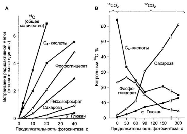
Fig. 6.81. Kranz anatomy of a C4 plant (Zea mays) (courtesy of I. Ezgg). Bundle sheath cells in a leaf cross-section surround the vascular bundle in a ring and are clearly distinct from mesophyll cells. The chloroplasts of the bundle sheath cells are noticeably larger than those in the mesophyll cells
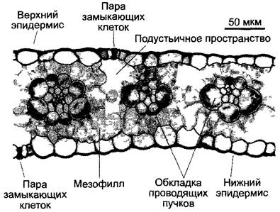
Fig. 6.82. Section through mesophyll cells and bundle sheath cells (fragments) of a maize leaf. The cell wall running obliquely in the figure contains a suberin lamella (arrows) that surrounds each bundle sheath cell and, among other things, greatly reduces the diffusion of CO2 out of the bundle sheath cells. Exchange of substances between cells occurs only via plasmodesmata (12,000x)
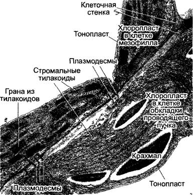
Table 6.19. Localization of certain enzymes in the Two Types of chloroplasts in C4 plants
Mesophyll chloroplasts |
Bundle sheath chloroplasts |
PEP carboxylase |
RuBP carboxylase (RubisCO) |
NADP-malate dehydrogenase* |
Malic enzyme |
Glutamate-aspartate aminotransferase* |
Aldolase |
Pyruvate-phosphate dikinase |
Starch synthase |
NADP-glyceraldehyde phosphate dehydrogenase |
RuBP kinase |
— |
NADP-glyceraldehyde phosphate dehydrogenase |
* Chloroplasts with high malate dehydrogenase content exhibit low aminotransferase activity, and vice versa.
Four-carbon compounds are formed in the mesophyll from phosphoenolpyruvate and HCO3. The bicarbonate ion is in equilibrium with diffusing CO2: CO2 + H2O ⇄ HCO3- + H+. This reaction is catalyzed by carbonic anhydrase, see 6.5.1. In the first step, oxaloacetate is formed (see Fig. 6.79). The reaction is catalyzed by the enzyme phosphoenolpyruvate carboxylase (PEP carboxylase). Its affinity for HCO3- (Km = 10 µmol/L) is not very different from the affinity of RubisCO for CO2 (Km = 10—15 µmol/L). Since RubisCO is not synthesized in the Chloroplasts of mesophyll cells, competition between Enzymes for the CO2 substrate is avoided. During malate synthesis (Fig. 6.83) in C4 plants, which include important crops such as maize, sugarcane, and millet, the resulting oxaloacetate is immediately converted into L-malate. This reaction is catalyzed by the chloroplast-localized NADP-dependent malate dehydrogenase. Via a translocator, malate is exported from the chloroplasts into the cytoplasm of mesophyll cells and moves through plasmodesmata into the bundle sheath cells. There, again with the participation of a specific translocator, it enters the chloroplasts, where it is decarboxylated to pyruvate and CO2. This reaction is catalyzed by malic enzyme1, yielding NADPH + H+ (see Fig. 6.79; 6.83). Due to the high concentration of malate in the stroma of bundle sheath cells, the concentration of free CO2 in the stroma reaches about 70 µmol/L. This ensures its efficient fixation by ribulose-1,5-bisphosphate carboxylase (RubisCO). The accumulated pyruvate is transported back to the mesophyll cells, where it is converted into phosphoenolpyruvate in the chloroplasts by pyruvate-phosphate dikinase (see Fig. 6.79; 6.83), which in turn is transported into the cytoplasm via a triose phosphate translocator in exchange for phosphate, serving there as a substrate for the next CO2 fixation reaction (see Fig. 6.83).2
1 Malic enzyme is the abbreviated name for decarboxylating malate dehydrogenase. — Ed. note.
2 This metabolic cycle is named the Hatch–Slack cycle after its discoverers. Simultaneously, the C4 pathway was investigated by Karpilov. — Ed. note.
Fig. 6.83. Metabolic reactions in mesophyll and bundle sheath cells, and the exchange of substances between these cells in the leaf of a malate-type C4 plant
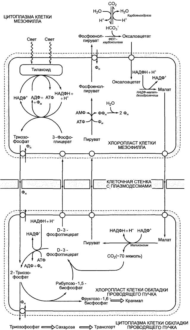
In contrast to mesophyll cells, the complete Calvin cycle takes place in the bundle sheath cells. Since these chloroplasts lack grana, Photosystem II activity is very low, and in the light, Cyclic electron transport occurs in the thylakoids involving Photosystem I and the cytochrome b6/f complex. This leads to ATP synthesis without the formation of NADPH + H+ (see 6.4.7). The Calvin cycle's requirement for NADPH is half-met by the malic enzyme. Thus, malate transports both CO2 and its reducing equivalent (one NADPH equivalent per CO2 molecule) from the mesophyll chloroplasts to the bundle sheath chloroplasts. Nevertheless, 2 NADPH + 2 H+ are required per fixed CO2 molecule (see 6.5.2). It is believed that half of the synthesized D-3-phosphoglycerate leaves the bundle sheath chloroplasts and is reduced in the mesophyll chloroplasts to triose phosphate, which, via the triose phosphate translocator, again
enters the bundle sheath chloroplasts (see Fig. 6.83).
A consequence of the insufficient photosystem II activity in the bundle sheath chloroplasts is a major reduction in the photo-oxidation of water. The low oxygen concentration in the stroma, combined with the elevated CO2 concentration, inhibits the oxygenase reaction of RubisCO. As a result, photorespiration is significantly suppressed. Therefore, C4 plants are characterized by increased photosynthetic productivity compared to C3 plants.
During photosynthesis, malate-forming C4 plants require not 3 molecules of ATP and 2 NADPH + 2 H+ per CO2 molecule, like C3 plants, but 4 ATP and 3 NADPH + 3 H+, or more precisely, 2 ATP + 2 NADPH + 2 H+ in the mesophyll chloroplasts and 2 ATP + 1 NADPH + H+ in the bundle sheath chloroplasts. Nevertheless, C4 plants eliminate the energy waste of photorespiration, so that in the end, C3 and C4 plants expend approximately the same amount of energy on photosynthesis. At low temperatures, and consequently inactive photorespiration, C3 plants have an advantage over C4 plants1, while at high temperatures (>25 °C), C4 plants benefit due to the activation of the oxygenase reaction of RubisCO. It should be added here that, thanks to the CO2-concentrating mechanism in C4 plants, RubisCO can ensure a reaction with the substrate even in the case of water scarcity, and therefore when stomata are closed to reduce transpiration, or under intense light when stomata are fully open but CO2 is limiting. The efficiency of the CO2 pre-fixation mechanism by PEP carboxylase lies not in a high affinity of the enzyme for the substrate, but in the fact that in the stroma of illuminated chloroplasts (pH 8), the HCO-3:CO2 ratio is approximately 50:1. Thus, PEP carboxylase2, unlike RubisCO, can bind the dominant form of carbonic acid in this equilibrium reaction and effectively perform CO2 fixation, even if, with partially closed stomata, the concentration of CO2 dissolved in water drops below the level acceptable for RubisCO
1 The northward distribution of C4 plants is limited by the July isotherm of +12 °C. At temperatures below +4 °C, transport through plasmodesmata is disrupted, and the mesophyll and bundle sheath cannot exchange metabolites. — Ed. note.
2 This reasoning contradicts data on the cellular localization of enzymes: PEP carboxylase operates in the Cytosol, not in the stroma. Therefore, discussing the stroma pH and reactions in this compartment in connection with PEP carboxylase activity is incorrect. — Ed. note.
From all that has been said, it follows that C4 plants have an advantage over C3 plants under water scarcity, high temperatures, and intense solar radiation. They primarily grow in warm, arid regions3 with high insolation. In California's Death Valley, 70% of all growing species are C4 plants. It is estimated that about 17% of all cultivated land is populated by C4 plants, and they carry out about 30% of global photosynthesis.
3 This refers to semi-arid conditions (water is present but not always sufficient). Under hyper-arid conditions, CAM plants predominate. — Editor's note.
The "СO2 pump" principle and its associated ecophysiological advantages also apply to C4 plants that synthesize aspartate. They differ from malate-synthesizing plants in structure and certain enzymes (Table 6.20). Based on the СO2-releasing reactions, aspartate-synthesizing plants are divided into NAD-malic enzyme and PEP-carboxykinase types. In both cases, aspartate is formed by glutamate-aspartate aminotransferase in the cytoplasm of mesophyll cells. Aspartate moves symplastically through plasmodesmata into the bundle sheath cells. In NAD-malic enzyme-type C4 plants, it is transported via an amino acid translocator into the mitochondria, where it is converted to oxaloacetate by an isoform of glutamate-aspartate aminotransferase. Oxaloacetate is then converted to malate and decarboxylated to pyruvate and СO2.
The malate dehydrogenase (malic enzyme) is NAD-specific. The released СO2 diffuses from the mitochondria into the chloroplasts and is fixed by RubisСО. Pyruvate is converted to Alanine, which is exported from the mitochondria of bundle sheath cells (via an amino acid translocator) into the cytoplasm of mesophyll cells, where it is converted back to pyruvate. Two isoforms of alanine-glutamate aminotransferase participate in the reversible conversion of pyruvate to alanine. Pyruvate, as is the case in malate-synthesizing C4-plants, is then converted back to phosphoenolpyruvate.
In the PEP-carboxykinase type of C4 metabolism, part of the СO2 released in the bundle sheath cells is provided by oxaloacetate, which is converted to phosphoenolpyruvate by PEP-carboxykinase and ATP. This reaction results in the release of СO2. In these plants, oxaloacetate is synthesized from L-aspartate (see Fig. 6.79). The reactions take place in the cytoplasm of the bundle sheath cells. A smaller fraction of СO2 is released through the action of the mitochondrial isoform of NAD-malic enzyme. Mesophyll cells also synthesize and supply malate to the mitochondria (see malate synthesis in Fig. 6.83). A dicarboxylic acid translocator is responsible for transporting malate into the mitochondrion.
C4 metabolism is activated by light. In PEP carboxylase, a serine residue is phosphorylated in response to light, thereby activating the enzyme. In this form, the enzyme is inhibited only by high concentrations of malate. In the dark, the dephosphorylated enzyme has low activity, and even very low concentrations of malate can inhibit its function. NADP-dependent malate dehydrogenase is activated in the light by thioredoxin (see Fig. 6.71), while in pyruvate-phosphate dikinase, a Threonine residue is dephosphorylated in the light, which gives it a catalytically active conformation.
Plant species with C4 photosynthesis belong to highly diverse taxonomic groups, and they are particularly abundant in certain taxa, such as Poaceae. This includes major crops like maize, sugarcane, and millet, as well as common weeds like Bermuda grass. Many C4 species are also found within the Amaranthaceae (formerly Chenopodiaceae). In this family, the genus Atriplex contains both C3 and C4 species. These C4 species are halophytes and, on saline soils, also suffer from (physiological) water scarcity.
Table 6.20 Classification of C4 plant subgroups by the primary product of СO2 fixation and the decarboxylation pathway
Primary product of СO2 fixation (synthesized in MC, transported to BSC) |
Decarboxylating enzyme |
Reducing equivalents or ATP produced during decarboxylation |
Main substance transported from BSC to MC |
Cytological features of BSC (in grasses) |
Species (Examples) |
Malate |
NADP-malic enzyme |
Generation of 1 NADPH per СO2 |
Pyruvate |
Suberin lamella present, chloroplasts with reduced grana arranged centrifugally |
Zea mays, Saccharum officinarum, Sorghum bicolor, Digitana sanguinahs |
Aspartate |
NAD-malic enzyme |
Generation of 1 NADH per СО2 |
Alanine/Pyruvate |
Suberin lamella absent, chloroplasts with grana arranged centripetally |
Amaranthusf retroflexus, Portulaca1 * * olerácea, Pamcum mihaceum |
Aspartate |
PEP-carboxykinase |
Consumption of 1 ATP per СO2 |
PEP/alanine |
Suberin lamella present, chloroplasts with grana arranged randomly or centrifugally |
Pamcum máximum, Chlons gayana |
1 The examples provided do not belong to grasses and have different anatomical features compared to those listed in the table. — Editor's note.
MC — mesophyll cells, BSC — bundle sheath cells
To identify C4 plants, several Methods are used: determining the primary products of photosynthesis (after short-term fixation of labeled 14СO2), and determining the carbon dioxide compensation point of photosynthesis (i.e., finding the ambient СO2 concentration at which no net fixation of СО2 occurs, meaning photosynthetic СО2 uptake and respiratory СО2 release balance each other out). Leaf anatomy is also examined, and the rate of photorespiration is assessed (which is low or completely absent). Finally, the carbon isotope ratio 13C/12C in the plant is determined. This last method is based on the fact that plants assimilate natural carbon isotopes in different ratios during photosynthesis (atmospheric СО2 contains 98.89% 12C and 1.11% 13C). Plants prefer 12СО2, assimilate 13СО2 to a lesser extent, and 14СО2 even less. Discrimination against 13СО2 is more pronounced during RuBP carboxylase-mediated СО2 fixation than during PEP carboxylase-mediated fixation. In C4 plants, RubisСО fixes virtually all of the СO2 previously captured by PEP carboxylase; thus, the percentage of 13C in a C4 plant reflects the product of the PEP carboxylase reaction, whereas a C3 plant is characterized by the isotope ratio typical of RubisСо. Consequently, C4 plants contain a relatively higher percentage of 13C, and carbohydrates extracted from C4 plants are heavier than sugars from C3 plants.
The 13C/12C ratio is determined by mass spectrometry and is expressed as a δ13C value.

where the standard is a reference limestone sample. The more negative the δ13C value, the lower the 13C isotope content. In C4 plants, the δ13C value is about -14 ‰, and in C3 plants it is about -28 ‰. Since sugarcane is a C4 plant and sugar beet is a C3 plant, THE ORIGIN OF sucrose can be determined by mass spectrometry based on the 13C isotope content. In this way, for example, genuine rum (made from sugarcane) can be distinguished from blended rum (with added sugar derived from sugar beet).
6.5.9. Preliminary CO2 Fixation in CAM Plants
In many succulents—plants that possess water-storing tissue—the preliminary fixation of CO2 to malate and the separate final fixation of CO2 via RubisCO constitute a sequence of reactions analogous to the cycle in malate-synthesizing C4 plants. However, these processes are separated in time rather than in space. A characteristic feature of this reaction sequence (Fig. 6.84) is the nocturnal storage of a large amount of the primary CO2 fixation product, malate, in the vacuoles (the increase in vacuolar osmotic pressure is likely associated with succulence). During the day, malate is released and metabolized. Cell acidity fluctuates in a specific rhythm with the change of day and night, which gave rise to the name of this phenomenon—diurnal (circadian) acid rhythm. This process was first discovered in Crassulaceae (stonecrop family), which is why it is called Crassulacean Acid Metabolism, or CAM (from the English crassulacean acid metabolism).
Fig. 6.84. Reactions characteristic of Crassulacean acid metabolism (CAM) during the night (black arrows) and day (gray arrows) and their compartmentalization. Characteristic enzymes: (1) — PEP carboxylase; (2) — NAD-malate dehydrogenase; (3) — malic enzyme; (4) — pyruvate, phosphate dikinase. Gray box: PEP carboxylase is active at night (phosphorylated form, PEPC-P). This form is very weakly inhibited by malate. During the day, a dephosphorylated enzyme (PEPC) highly sensitive to malate appears. Activation is carried out by a specific PEP carboxylase kinase (PEPC kinase), which is present only at night. Ki — inhibition constant (the concentration of inhibitor required for 50% inhibition of the enzyme)

In all CAM plants, PEP is synthesized at night from starch via triose phosphate through glycolysis, followed by CO2 fixation by PEP carboxylase (the substrate is HCO3-) and the formation of oxaloacetate. Malate, derived from it by cytoplasmic NAD-dependent malate dehydrogenase, is transported into the vacuole via a malate channel (see Fig. 6.5). The transport of malate against its concentration gradient occurs due to the transmembrane proton motive force generated in the tonoplast by the H+-ATPase, which simultaneously delivers counterions to the malate anion. Since the pH of the vacuolar contents drops over time, malate at night must exist primarily in the form of protonated malic acid. Compared to the malate2- anion, it penetrates the tonoplast membrane more easily, so the rising concentration of hydrogen ions limits the capacity of vacuoles to store malate. An increase in cytoplasmic malate content inhibits PEP carboxylase. This negative feedback loop ultimately limits CO2 fixation as the dark period is prolonged.
During the day, the malate stored overnight is exported from the vacuole via the malate channel through mechanisms that are not fully understood. As in C4 plants, Three types of daytime decarboxylation can be distinguished: NADP-malic enzyme type (e.g., Cactaceae, Agavaceae), NAD-malic enzyme type (Crassulaceae), and PEP carboxykinase type (Asclepiadaceae, Bromeliaceae, Liliaceae). Refixation of CO2 (released in the light by one of these three enzymes) by PEP carboxylase instead of RubisCO is prevented because PEP carboxylase is converted in the light from the active (phosphorylated) 'night form' with low sensitivity to malate (50% inhibition at approximately 3 mmol/L malate) to a highly inactive (dephosphorylated) 'day form' with high sensitivity to malate (50% inhibition at 0.3 mmol/L malate). Consequently, during the day, the malate exported from the vacuoles inhibits the already catalytically weak enzyme so strongly that it cannot perform CO2 fixation, leaving the CO2 derived from malate available for RubisCO.
As in C4 plants, phosphorylation of a serine residue of PEP carboxylase also occurs in CAM plants (see 6.5.8). The responsible enzyme, PEP carboxylase kinase, is under strict control by the biological clock and exhibits a circadian rhythmicity (see 7.7.2.3). PEP carboxylase kinase degrades rapidly, so The amount of enzyme in the cell is determined by the rate of Gene Transcription (see 7.2.2.3 — Transcriptional Control). This rate is high at night and negligibly low during the day. Under constant light conditions (and likewise in constant darkness), this rhythmicity persists, indicating its endogenous nature (cf. 7.7.2.3).
The ecological advantage of CAM plants is that CO2 uptake through stomata open at night leads to a significant reduction in water loss compared to the daytime. This is made possible by the very low night temperatures in the habitats of these plants and the correspondingly high relative humidity. Under well-watered conditions, CAM plants not only fix CO2 released during malate decomposition, but also open their stomata to fix ambient CO2 via RubisCO once malate reserves are depleted. During drought, to which these plants are actually adapted, they conversely close their stomata, thereby greatly reducing the fixation of ambient CO2 during the day compared to the night. The transpiration ratio in this case ranges from 30 to 150 (CO2 fixation occurs predominantly at night, see 6.3.4.1), meaning that the water requirement of CAM plants is only a small fraction of that of C3 plants. However, due to the limited capacity of vacuoles to store malate, when CO2 can be fixed exclusively in the dark, the daily biomass accumulation is very small. Consequently, CAM plants are competitive primarily in arid habitats, where cool nights favor malate synthesis and storage, and occasional, very rare, yet heavy rainfall allows them to replenish their water reserves. Some CAM plants, such as species of the genus Mesembryanthemum, switch to standard C3 photosynthesis when water is abundant. Water deficit or salinity induces the synthesis of CAM enzymes. Under extreme conditions, desert plants (such as cacti) experiencing severe water shortage keep their stomata closed even at night, refixing CO2 released during respiration.
The capacity for CAM photosynthesis is mainly characteristic of succulents: about 300 plant species are known to use this method of supplementary CO2 fixation, such as Aizoaceae, Apocynaceae (formerly Asclepiadaceae), Asteraceae, Cactaceae, Crassulaceae, Didiereaceae, Euphorbiaceae, Portulacaceae, Vitaceae, Agavaceae, Bromeliaceae (e.g., pineapple), Liliaceae, Orchidaceae (e.g., vanilla); furthermore, CAM has been found, for example, in the lichen-like epiphyte Tillandsia usneoides of the family Bromeliaceae and in some tropical epiphytic ferns (Pyrrosia piloselloides, P. longifolia). An important distinguishing feature of CAM plants, alongside their enzyme composition, is not so much The structure of their organs, but rather the specificity of their cellular structures (the presence of large, voluminous vacuoles in chloroplast-containing cells: 'succulence at THE CELLULAR LEVEL').
Regarding isotope discrimination, during dark fixation and subsequent light processing of CO2, CAM plants behave similarly to C4 plants (less discrimination against 13CO2 compared to 12CO2). Since dark fixation (which constitutes the major part of total fixation) increases under drought conditions, CAM plants contain more 13C (just like C4 plants). Determining the δ13C value makes it possible to identify drought conditions in the natural habitats of CAM plants.
6.5.10. Additional Elevation of CO2 Concentration via the Bicarbonate Pump
All cyanobacteria possess a membrane-bound bicarbonate pump (HCO3-) to increase the CO2 concentration in carboxysomes—the sites where RubisCO is localized. This compensates for the enzyme's low affinity for CO2 and suppresses photorespiration. Pyrenoids may play a role in a functionally similar CO2-concentrating mechanism in algae (and lichen phycobionts) (see 2.2.9.1).
6.5.11. Influence of External Factors on Carbon Assimilation
Like all vital processes, photosynthesis is influenced in a rather complex manner by various factors: the plant's developmental stage, the availability of CO2, water, and minerals, light quality and intensity, and temperature. The law of the minimum applies to photosynthesis, as it does to all physiological processes dependent on multiple factors: the minimum availability of a single factor limits the productivity of the entire process. With insufficient CO2 supply, favorable conditions of light, water, and temperature cannot yield a positive result; conversely, an optimal CO2 concentration does not guarantee maximum photosynthetic productivity if light intensity is insufficient. If all conditions are generally favorable, it is assumed as a starting point that each square meter of green leaf surface synthesizes 0.5–1.5 g of glucose equivalent per hour. This roughly corresponds to the consumption of 3 m3 of atmospheric CO2.
Next, we will individually examine some factors and their overall impact on plant photosynthesis. For the ecological physiology of photosynthesis, see 13.7.1.
Leaf structure (see 4.3.1.1; Fig. 4.64) ensures optimal Light absorption. In cross-section, epidermal cells are lens-shaped. They focus light and direct it onto the underlying palisade parenchyma cells, where up to 80% of photosynthesis occurs. Unabsorbed photons are scattered along the boundary surfaces of the spongy parenchyma cells; as a result, the photons do not travel in a single direction, the light path through the leaf is lengthened, and the probability of quantum absorption increases.
The intensity of radiation incident on a leaf can change within a short period (for example, during cloud shading). The chloroplasts of many plants cope with such fluctuations by changing their position relative to the light falling on the leaf. In the so-called low-light position, the lens-shaped organelles expose their broad side to the light, while in the high-light position, they present their narrow side. Organelle reorientation (see 8.2.2) involves calcium-dependent processes mediated by the Cytoskeleton, presumably Actin. This alters the cross-section of the "light traps," stabilizing quantum Absorption in the antenna complexes within certain limits, regardless of the incoming light intensity.
The leaves or shoots of many plants (e.g., lupine, alfalfa, common bean, soybean, cotton) track the daily movement of the sun so that the leaf blades remain perpendicular to the incident light rays (sun tracking). This positive phototropism1 (see 8.3.1.1) promotes maximum light intensity on the leaves and minimizes reflection losses2.
1 In Russian literature, phototropism refers to irreversible growth processes. In this example, it is more correct to speak of leaf photonasties (a reversible process). — Editor's note.
2 In desert regions with excessive insolation, the opposite phenomenon is observed: as a result of photonasty, the leaf blades are oriented edge-on to the light. — Editor's note.
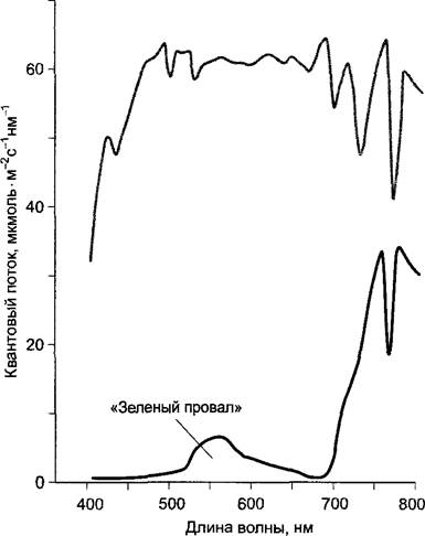
Under natural conditions, the amount of chlorophyll is not a factor limiting the rate of photosynthesis, because even at low light intensities, leaves with reduced chlorophyll content absorb enough photons to saturate the photosynthetic apparatus. High chlorophyll content in leaves may only play a role when it is necessary to absorb as completely as possible some small part of the spectrum required for photosynthesis, which is already carried out by other leaves (Fig. 6.85). Shade leaves generally contain a higher specific chlorophyll concentration per unit of leaf surface area than "sun leaves." They also have exceptionally large grana, which can stack up to 100 thylakoids. Shade-tolerant plants contain more pigment molecules serving the Electron Transport Chain (the "photosynthetic unit") and therefore have larger antennae and a reduced chlorophyll a : b ratio (i.e., relatively more chlorophyll b to better utilize the green part of the spectrum, where chlorophyll absorbs quanta with low probability). Furthermore, photosystem II predominates in leaves in the shade. Shade leaves are often thinner than "sun leaves," which reduces shading of the chloroplasts on the lower side (see Fig. 7.73). The photoreceptor system sensitive to the red region of the spectrum, phytochrome, is involved in regulating leaf development (see 7.7.2.4).
Fig. 6.85. Energy distribution spectrum above a wheat canopy (gray curve) and within the canopy foliage in the shade (measured at a distance of 80 cm from the soil (black curve); plant height is 90–95 cm)
At low irradiance, the rate of photosynthesis is proportional to the photon flux (Box 6.2; Fig. 6.86), as long as other factors are not limiting. This also holds true at higher light intensities, so the curve of observed (net) photosynthesis versus light intensity remains linear until, eventually, with a further increase in light intensity, the rate of photosynthesis no longer increases (light saturation is reached). Typically, in this situation, the limiting factor is the plant's CO2 supply. The light saturation range for plants adapted to sunny habitats lies between 500 and 1500 µmol • m-2 • s-1, and for shade-tolerant plants, between 100 and 500 µmol • m-2 • s-1 (the photosynthetic curve reaches a plateau). In C4 plants (see 6.5.8), unlike C3 plants, light saturation does not occur even under intense illumination due to a more efficient CO2 supply to the Calvin cycle. In C4 plants, the overall process of photosynthesis is usually limited by light, since water shortage cannot exert a sufficient limiting effect via CO2 when stomata are partially or completely closed1.
1 This refers to drought within physiological norms. Under extreme drought, the photosynthetic productivity of C4 plants also decreases. — Editor's note.
Fig. 6.86. Light dependence of net photosynthesis in a C3 plant. Schematic graph based on typical CO2 uptake values as a function of photosynthetically active radiation (400–700 nm, PAR) at optimal temperature

Under even stronger illumination, the photosynthetic apparatus can be damaged, causing photosynthetic activity to drop again. In nature, this can happen if plants adapted to shade are suddenly exposed to direct sunlight, especially at low temperatures when the ENZYMATIC REACTIONS OF CO2 fixation are slowed down (for mechanisms protecting photosynthesis from light-induced damage, see 6.4.8; 6.5.6).
Box 6.2. Basic Concepts of Photobiology
The quantum (photon) flux (also referred to as quantum or photon flux density) is defined as the number of photons incident on a specific area per unit time (Units of Measurement: mol • m-2 • s-1 = E • m-2 • s-1, 1 Einstein = 1 mol of photons). Often, only the range from 400 to 700 nm is considered, i.e., the photosynthetically active radiation (PAR) region. Under full sunlight with a high sun angle and clear skies, PAR is typically 1,500–2,000 µmol • m-2 • s-1, in cloudy weather about 190–220, in plant shade about 25–50 (baseline data), and at twilight only 1 µmol • m-2 • s-1. Under a clear full moon, one can measure 3.2 × 10⁻⁴ µmol • m-2 • s-1, while starlight alone yields a photon flux of 1.2 • 10-6 µmol • m-2 • s-1. The primary objective of measurements is often to determine the amount of energy rather than the number of moles of photons, which is represented by the equivalent unit of energy flux (J • m-2 • s-1 = W • m-2); the energy of a single photon is determined using Equation 6.41, and the energy of 1 mole of photons using Equation 6.42. The number of photons per unit time (mol • s-1) is called the photon flux. Only for monochromatic light, such as that used in measuring action spectra, can energy and photon fluxes be converted into one another. For radiation containing photons of various wavelengths, one must first determine the spectral energy distribution.
A. Basic Concepts of Spectral Photometry
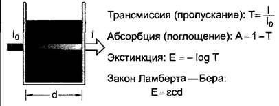
When light is passed through a sample, it is absorbed depending on the sample's structure (Fig. A). Transmission, or transmittance (T), is defined as the ratio of light intensity leaving the sample (I) to the light intensity entering the sample (I0). Transmission indicates what fraction of the light leaves the sample. The fraction of light absorbed by the sample is called absorption (A). Absorption and transmission are often expressed as percentages. Extinction (E) must not be confused with absorption. Extinction values are frequently used in photometry because, for substances in solution at a constant light path length (d), they are proportional to the concentration of the substance (c) (the Beer-Lambert law). The proportionality constant ε is called the molar extinction coefficient. It is measured in [l • mol-1 • cm-1]; the concentration of substances is given in [mol • l-1], and the light path length in [cm].
The light intensity at which CO2 consumption (and corresponding O2 production) is completely balanced by CO2 production from mitochondrial respiration (and corresponding O2 consumption) is called the light compensation point of photosynthesis (see Fig. 6.86), where net photosynthesis is zero. In "sun" leaves (and light-demanding plants), the light compensation point is about 10–15 µmol • m-2 • s-1, while in shade leaves (and shade-tolerant plants), it is about 1–10 µmol • m-2 • s-1. Consequently, light-demanding plants cannot grow under a dense leaf canopy, whereas shade-tolerant plants achieve a positive carbon balance even in deep shade (see 13.7.1).
6.5.11.2. Influence of Carbon Dioxide Concentration
The atmospheric СO2 concentration in 2000 was 370 ppm (0.037 % by volume); over the last 40 years, it has increased by an average of 1 ppm per year (see 13.7.6). This is primarily due to human activity: fossil carbon reserves are burned (currently about 6 •1012 kg annually, with the total amount of fossil carbon on Earth estimated at 3 500 • 1012kg). Due to the elevated С02 concentration, the atmosphere absorbs long-wave radiation in large quantities. Potential impacts on climate (greenhouse effect), vegetation, and complex influences on internal plant processes and ecosystems (see 13.7.6) must be intensively studied.
In С3-plants, photosynthesis under full insolation is presumably limited by the amount of available carbon dioxide (see Fig. 6.86). By increasing the ambient СO2 concentration, other things being equal, an increase in the photosynthetic activity of these plants can be achieved. This is done using the "СO2 Fertilization" method for greenhouse crops. The СO2 concentration in greenhouses with cucumbers and tomatoes is increased by 0.1%, and consequently, the yield increases by a third per season, provided that other nutrients and light are present in sufficient quantities (nutrient ecology, see 13.7.6).
Aquatic plants do not find it more difficult to absorb СO2 than terrestrial ones, since at a normal temperature of 15 °C, СO2 dissolves in water in the same percentage as it is present in the air (10 µmol); moreover, the slow diffusion of С02 in water is compensated by water movement (convection). Submerged plants, which lack stomata and have no distinct cuticle, either absorb only dissolved СO2 through their entire leaf surface, or (some plant species) additionally absorb Са(НСО3)2.
6.5.11.3. Influence of temperature
The primary photochemical reactions of photosynthesis are strongly temperature-dependent. Enzymatic processes also exhibit a significant temperature dependence (see 6.1.6.4), which can be described by the van 't Hoff rule: the reaction rate v (see equation 6.26) nearly doubles with a 10 °C increase in temperature (Q10 value):

It follows that at low light intensity (light being the limiting factor), photosynthesis depends less on temperature than at high intensity (СO2 being the limiting factor). With rising temperature, the increasing photosynthetic activity primarily reflects the high Rate of Enzymatic reactions. On the other hand, the declining photosynthetic activity at temperatures above the optimum is complex in nature: although the activity of RubisСО, the rate-limiting enzyme, increases with temperature, its affinity for СO2 decreases; at the same time, at high temperatures, СO2 dissolves less readily in water relative to O2, meaning that photorespiration is activated as temperature rises (see 6.5.6). Thus, net photosynthetic productivity decreases. At high temperatures, the photosynthetic apparatus is destroyed due to Enzyme inactivation and membrane damage. For plants of different habitats, the temperature limits and temperature optimum lie within characteristic ranges (ecophysiology — see 13.7.1).
Under The Influence of light, Earth's plants process a total of about 1 875 km3 of water to produce oxygen. Consequently, over nearly 8 million years, a total of about 1.5 • 109 km3 of liquid water has been subjected to photo-oxidation once. Since oxygenic photosynthesis has existed, Earth's water reserves have been split several hundred times. Nevertheless, only a very small fraction of the water used serves as a substrate for photosynthetic splitting in plants (see 6.4.4).
Water deficit does not mean a shortage of substrate; it acts indirectly: on one hand, it causes severe cell dehydration, destruction of enzymes and functional structures (e.g., membranes), and on the other hand, it leads to stomatal closure and reduced СO2 uptake. When stomata are closed, the leaf can only re-assimilate internal СO2 released during respiration.
Last update: 07/08/2026
Editorial and Educational Adaptation: This material has been compiled based on the primary/original source text. The project team performed an editorial review, corrected technical inaccuracies, structured sections, and adapted the content for an educational format.
What was processed:
- elimination of formatting defects (OCR errors, structural breaks, corrupted characters);
- editorial organization of content;
- standardization of terminology in accordance with academic sources;
- verification of factual statements against the original source text.
All mentions of the author, publication year, and origin of the primary text have been preserved in accordance with the source.