BIOCHEMISTRY - L. Stryer - 1984
VOLUME 2
PART II GENERATION AND STORAGE OF METABOLIC ENERGY
CHAPTER 19. PHOTOSYNTHESIS
19.12. ATP can also be formed via cyclic electron flow through photosystem I
There is an alternative pathway for electron flow originating from P700, the reaction center of Photosystem I. An electron with a high potential in bound ferredoxin can be transferred not to NADP +, but to cytochrome b563. This electron then flows back to the oxidized form of P700 via cytochrome c552 and plastocyanin. In other words, a cyclic flow of electrons takes place. ATP is generated as electrons return to the reaction center through cytochrome b563 and plastocyanin. Therefore, this process is called cyclic Photophosphorylation (Fig. 19.16). In this mechanism, ATP generation is not accompanied by the simultaneous formation of NADPH. Photosystem II does not participate in cyclic photophosphorylation, and consequently, no O2 is produced from H2O. Cyclic photophosphorylation Functions actively when the concentration of NADP is lower than required to accept electrons from reduced ferredoxin. Such a state arises when the [NADPH]/[NADP+ ] ratio is high.
Class="center">Fig. 19.16. Electron flow in cyclic photophosphorylation. Photon absorption is accompanied by Electron transfer from P700, the reaction center of photosystem I, to bound ferredoxin. The electrons return to P700

19.13. A transmembrane proton gradient across thylakoid membranes drives ATP synthesis
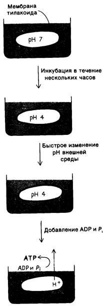
In 1966, André Jagendorf demonstrated that METABOLISM/14.html">Chloroplasts synthesize ATP in the dark when an artificial pH gradient is established across the thylakoid membrane. To create such a transient pH gradient, chloroplasts were immersed for several hours in a buffer at pH 4. The chloroplasts were then rapidly mixed with a buffer at pH 8 containing ADP and Pi. As a result, the stromal pH instantly rose to 8, whereas the pH of the thylakoid lumen remained at 4. The dissipation of this generated pH gradient across the thylakoid membrane was accompanied by a burst of ATP synthesis (Fig. 19.17). This remarkable experiment provides strong evidence in support of the chemiosmotic hypothesis of ATP synthesis (Section 14.5).
Fig. 19.17. ATP synthesis by chloroplasts in the presence of a pH gradient
Indeed, the Mechanism of ATP synthesis in chloroplasts is remarkably similar to that in Cell/35.html">Mitochondria. In both photophosphorylation and Oxidative Phosphorylation, ATP formation is driven by the proton-motive force. Furthermore, the enzyme assembly catalyzing ATP formation in chloroplasts, designated as the CF1–CF0 complex (where C stands for chloroplast and F for factor), closely resembles the mitochondrial F1–F0 complex. CF1 catalyzes The formation of ATP from ADP and Pi. It contains pairs of five types of subunits and has a mass of 325 kDa. CF0, which contains the proton channel, consists of Three types of subunits. Protrusions on the outer surface of the thylakoid membrane represent the CF1 components of these ATP-synthesizing complexes.
Electron transfer through the asymmetrically oriented Photosystems I and II generates a large proton gradient across the thylakoid membrane. The medium in the thylakoid lumen becomes markedly acidic, with the pH approaching 4. The light-induced transmembrane proton gradient is approximately 3.5 pH units. As discussed earlier (Section 14.5), the proton-motive force Δp is composed of the pH gradient and the Membrane Potential. In chloroplasts, almost the entire magnitude of Δp is generated by the pH gradient, whereas in mitochondria, THE CONTRIBUTION OF the membrane potential is more significant. The reason for this difference is that the thylakoid membrane is fully permeable to Cl- and Mg2+. Light-induced H+ transport into the thylakoid lumen is accompanied either by the influx of Cl- in the same direction or by the efflux of Mg2+ (one per 2H+) in the opposite direction. Thus, electroneutrality is maintained, and no membrane potential is generated. The proton-motive force across the thylakoid membrane, equal to 0.2 V, is equivalent to approximately 4.8 kcal per mole of protons. About three protons flow through the CF1–CF0 complex per molecule of ATP synthesized, which corresponds to a free Energy Consumption of 14 kcal per mole of ATP. When the pH gradient is less than two units, ATP synthesis does not occur because the driving force is too small.
CF1 is located on the stroma-facing surface of the thylakoid membrane, and consequently, the synthesized ATP is released into the stroma. Similarly, NADPH produced by photosystem I is released into the stroma. Thus, ATP and NADPH, the products of the light reactions of Photosynthesis, are localized
in such a way as to supply the subsequent dark reactions, during which CO2 is converted into CARBOHYDRATES.
19.14. Tracing the carbon pathway using pulse radioactive labeling
In 1945, Melvin Calvin and his coworkers began a series of studies that shed light on the dark reactions of photosynthesis. They used the unicellular green alga Chlorella in their work because this Organism is easily and reproducibly cultured. It was later found that their results could be extended to A wide variety of photosynthetic organisms, ranging from photosynthetic Bacteria to higher plants.
The goal of their work was to elucidate the pathway responsible for CO2 fixation into carbohydrates. The experimental strategy was to trace the transformations of CO2 using the 14C label. Radioactive 14CO2 was introduced into an illuminated suspension of Algae undergoing photosynthesis with ordinary CO2. After a certain period, the algae were killed by pouring the suspension into alcohol, which blocked all enzymatic reactions.
The radioactive compounds from the algae were separated and identified by two-dimensional paper Chromatography. The paper chromatograms were then placed under a press with X-ray film, which became dark in the areas where a radioactive spot was located on the paper. In his Nobel lecture, Calvin noted that their initial data referred "to the amount, position, and intensity of the radioactivity-induced blackened areas. Unfortunately, the paper does not print the names of these compounds itself, and for the next ten years, our main occupation consisted of correctly determining The Nature of the compounds in these blackened areas of the film."
Last update: 06/08/2026
Editorial and Educational Adaptation: This material has been compiled based on the primary/original source text. The project team performed an editorial review, corrected technical inaccuracies, structured sections, and adapted the content for an educational format.
What was processed:
- elimination of formatting defects (OCR errors, structural breaks, corrupted characters);
- editorial organization of content;
- standardization of terminology in accordance with academic sources;
- verification of factual statements against the original source text.
All mentions of the author, publication year, and origin of the primary text have been preserved in accordance with the source.