BIOCHEMISTRY - L. Stryer - 1984
VOLUME 2
PART II GENERATION AND STORAGE OF METABOLIC ENERGY
CHAPTER 19. PHOTOSYNTHESIS
19.9. Photosystem I Generates NADPH via Reduced Ferredoxin
The reaction center of Photosystem I (Fig. 19.12) is a chlorophyll *a* molecule situated in a specific environment. Its absorption maximum is shifted from 680 to 700 nm, which is why it is designated as P700 (where P stands for pigment).
Class="center">Fig. 19.12. Generation of NADPH by photosystem I
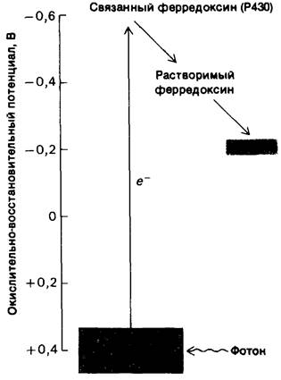
Many chlorophyll molecules absorb light and transfer the excitation energy to P700. Excited P700 transfers an electron to bound ferredoxin (P430), a membrane-associated form of ferredoxin, which is an Fe4-S4-type iron-sulfur protein with a molecular mass of 11.6 kDa. In the dark, the Redox Potential of P700 is +0.4 V. Light excitation of P700 causes a redistribution of electrons, shifting the redox potential to approximately -0.6 V. Thus, light drives an electron in photosystem I from a potential of about +0.4 V to a potential of -0.6 V. The energy of a red photon, which is 1.8 eV (electron-volts), is sufficient to raise the electron potential by 1.0 V. The transfer of the excited electron from P700 to bound ferredoxin creates an electron deficiency in P700. For P700 to function again as a reaction center, its oxidized form must first reacquire an electron; the source of these electrons will be discussed shortly.
Bound ferredoxin subsequently transfers its electron to the soluble form of ferredoxin. The iron atom in the active center of ferredoxin is alternately oxidized and reduced. Reduced ferredoxin transfers its electron to NADP+ to form NADPH. This reaction is catalyzed by ferredoxin-NADP reductase, which contains FAD as a prosthetic group. Note that the reduction of NADP+ to NADPH involves the transfer of two electrons, whereas ferredoxin is a single-electron carrier. Consequently, electrons from two reduced ferredoxins must converge to form a single molecule of NADPH.

19.10. Photosystem II Generates a Strong Oxidizing Agent That Splits Water
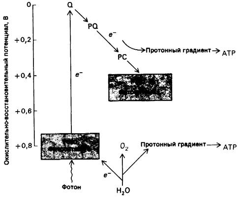
Relatively little is known about the reaction center (P680) and the primary electron acceptor of Photosystem II (Fig. 19.13). The redox potential of this reaction center is approximately +0.8 V.
Upon light excitation, a very strong oxidizing agent Z+ (the oxidized form of the reaction center or its derivative) and a weak reducing agent Q- localized within this center are generated. Z+ extracts electrons from H2O, yielding O2. Manganese plays an essential role in this process.
4Z+ + 2H2O → 4Z + 4H+ + O2.
Fig. 19.13. Excitation of photosystem II leads to O2 evolution and electron transfer to photosystem I. These processes generate a proton gradient across the thylakoid membrane (PQ denotes plastoquinone, PC denotes plastocyanin)
19.11. Electron Transfer from Photosystem II to Photosystem I Establishes a Proton Gradient
Photosystems I and II are linked by a series of electron carriers that serve a dual role.
1. Through this connecting link, electrons pass from photosystem II to photosystem I. These electrons are required to regenerate the reduced form of P700, the reaction center of photosystem I.
2. The transfer of electrons through this connecting link creates a proton gradient across the thylakoid membrane. Protons released during O2 evolution also contribute to The formation of this gradient. The pH within the thylakoid lumen becomes more acidic. The resulting proton gradient drives ATP synthesis (Section 19.13).
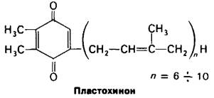
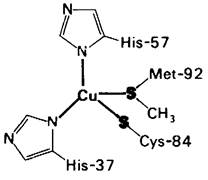
From the photoactivated reaction center of photosystem II, an electron is transferred to Q, a tightly bound plastoquinone molecule. This quinone is very similar to ubiquinone (Section 14.4), a component of the Mitochondrial Electron Transport chain, and to vitamin K (Section 11.11). From Q, the electron is then transferred to mobile plastoquinones and subsequently to cytochrome b559. The next member of the Electron Transport Chain is cytochrome c552 (formerly called cytochrome f), which, unlike mitochondrial cytochrome c, is an integral membrane protein. The ultimate electron carrier from photosystem II to photosystem I is plastocyanin, a 10.5-kDa protein containing a single copper atom coordinated to four groups: a Cysteine, a Methionine, and two Histidine side chains (Fig. 19.14). This spatial coordination differs from the planar geometry typical of low-molecular-weight Cu2+ complexes. The localized strain at the Cu atom likely facilitates electron transfer by allowing copper to cycle readily between the +1 and +2 oxidation states. The Cu atom is located near The surface of the plastocyanin molecule, shielded only by a histidine side chain, enabling electron transfer to Cu via a direct mechanism (Section 14.16). By transferring an electron from reduced plastocyanin to the oxidized form of P700, the reaction center is once again able to act as an electron donor that produces NADPH upon illumination. The overall reaction driven by the photoactivation of both photosystems is as follows:
2H2O + 2NADP+ → O2 + 2NADPH + 2H+.
Fig. 19.14. Structure OF THE coordinated copper ion in plastocyanin
In other words, light drives the transfer of electrons from H2O to NADP+ (Fig. 19.15).
Fig. 19.15. Electron flow in noncyclic Photophosphorylation. This is a true electron flow from H2O to NADP+ yielding NADPH.

Last update: 06/08/2026
Editorial and Educational Adaptation: This material has been compiled based on the primary/original source text. The project team performed an editorial review, corrected technical inaccuracies, structured sections, and adapted the content for an educational format.
What was processed:
- elimination of formatting defects (OCR errors, structural breaks, corrupted characters);
- editorial organization of content;
- standardization of terminology in accordance with academic sources;
- verification of factual statements against the original source text.
All mentions of the author, publication year, and origin of the primary text have been preserved in accordance with the source.