Antibiotics (Properties, Applications, Interactions) - M.P. Cherenko 1999
Surgical Infection. General Issues
CONCEPTS, Classification, AND PATHOGENS
Humanity is surrounded by a vast world of microorganisms as an integral part of the biocenosis. While many of these exist in peaceful coexistence with The Human Body (saprophytic commensals) or even in mutually beneficial relationships—such as certain gut microbes (mutualism)—many others can cause disease under specific conditions (pathogenic microorganisms). There are over 500 such infectious diseases. Their causative agents include numerous microbes, Viruses, Fungi, and Protozoa, with microbes and viruses playing The most significant role.
Long before microbiologists discovered this human-hostile microbial world, physicians (such as Girolamo Fracastoro, 1478–1559; D. Samoylovich, 1744–1805, and others) hypothesized their existence based on observations of epidemics of so-called pestilential or mass diseases (plague, cholera, smallpox, etc.) known since ancient times. Some of these diseases, particularly cholera and plague, assumed pandemic proportions, claiming hundreds of thousands and millions of lives.
Physicians suspected that these diseases were transmitted from person to person through direct contact between the sick and the healthy, as well as via air, food, and Water. These hypothetical entities or disease carriers were called contagions or sticky miasmas (from the Greek miasma, meaning pollution). In 1841, C. F. Hufeland introduced the term “infection” (infectium, Latin for contamination) specifically for these conditions, particularly Sexually Transmitted Diseases. This term aptly captures The Essence of the phenomenon. Later, following the discovery by scientists (L. Pasteur, R. Koch, A. Yersin, D. Ivanovsky, and others) of most pathogens responsible for both mass epidemics and wound suppuration, the term “infection” came to be applied to all forms of pathology caused by microorganisms (Bacteria, viruses, fungi, and protozoa). In modern terms, “infection” refers to a form of interaction between micro- and macro-organisms that results in The Development of disease in the latter. The development of an infection requires a pathogen (microbe), its entry into the body, and specific environmental conditions within the host. Equating an infection with its causative agents (microbes, viruses, etc.) is scientifically incorrect.
Among the numerous types of infections, a major group is known as surgical infection. Surgical infection refers to infectious pathologies that are either causally linked to surgical (mechanical) intervention and tissue wounding (infectious wound complications, notably suppuration and wound toxicosis) or require, or may at some point require, mechanical intervention for Treatment. These include infections that cause localized destruction of Tissues and Organs accompanied by the accumulation of toxic purulent exudate and necrotic Cells (e.g., abscesses, Phlegmon, carbuncles, Osteomyelitis, mastitis, appendicitis, Peritonitis, etc.).
Surgical infections are caused by various pathogens, predominantly microbes—either singularly or in multi-microbial associations; thus, this type of infection is polymicrobial. It lacks a clearly defined incubation period and, provided Sanitary and hygienic standards are maintained, does not pose a threat of epidemic spread. Non-surgical infectious diseases, which are typically cyclic in nature, are caused by a specific pathogen (microbe, virus, etc.) and therefore represent monoinfections. They are dangerous due to their systemic toxic effects rather than local tissue destruction (although certain forms can occasionally lead to this, such as bowel perforation in typhoid fever or dysentery). Cyclic infectious diseases require specific anti-epidemic measures to limit their spread.
Surgical infection, fundamentally rooted in purulent infection, is widespread, frequently complicates surgical Procedures, and has become increasingly resistant to Antibiotics. According to researchers, it acts as a major barrier to the further development of surgery. Patients with surgical infections account for at least one-third of admissions in general surgical departments and represent the second leading cause of hospital mortality. The incidence of surgical wound infection following internal organ surgeries ranges from 10–25% or more, significantly exceeding the infection rate of not only clean but also contaminated wounds (which is under 5%).
Along with an increase in the number of infectious complications and diseases, There is a significant shift in THE SPECTRUM OF infections—specifically, a rise in the proportion of Gram-negative pathogens (such as Pseudomonas aeruginosa, coliforms, etc.) and mixed infections involving conventional pyogenic bacteria combined with non-spore-forming anaerobic bacteria (bacteroides).
The general causes behind the spread of surgical infections and the shift in their profile include: violations of aseptic protocols in surgical facilities, particularly in operating rooms and dressing stations; The Emergence of multi-resistant strains of pathogenic microflora within surgical wards (hospitalism); complex, long-duration surgical procedures; a decline in the body's immunobiological reactivity due to environmental degradation; a substantial increase in the number of elderly and geriatric patients undergoing surgery, who present with numerous comorbid conditions, primarily cardiovascular, oncological, and urological; a rising prevalence of Diabetes Mellitus and other Metabolic Disorders among surgical patients; the widespread use of various endoprostheses and xenomaterials; an overestimation of The Role of antibiotics in infection prophylaxis alongside flawed administration techniques; the rapid, large-scale Introduction of new surgical techniques into clinical practice; a low technical proficiency in performing many surgical procedures, particularly emergency operations; and the Structure/175.html">Implementation of intensive care units, monitoring systems, and modern medical devices (endotracheal tubes, cannulae, catheters for various purposes, etc.).
Surgical infections are classified into the following forms: by clinical course—acute and chronic; by The Nature of the pathogen—acute purulent and putrefactive, acute specific, and acute anaerobic clostridial and non-clostridial. Acute specific and anaerobic clostridial infections originate as wound toxic infections, whereas other types are bacterial-toxic (both wound-related and non-wound-related).
Chronic infections are divided into chronic non-specific purulent and chronic specific forms.
The causative agents of acute and chronic purulent and putrefactive infections include staphylococci, streptococci, meningococci, gonococci, Escherichia coli, Pseudomonas aeruginosa, non-clostridial anaerobes such as Proteus, Bacteroides, and others.
Specific acute infections such as tetanus, anthrax, wound diphtheria, and rabies are caused by the tetanus bacillus, the anthrax bacillus, the diphtheria bacillus, and the rabies virus, respectively.
Acute anaerobic infections are caused by spore-forming clostridia: Cl. perfrinqenes, Cl. oedemotiens seu novyi, Cl. septicum, and Cl. hystoliticum.
Chronic non-specific purulent infections are caused by the same microorganisms that trigger acute purulent infections.
Causative agents of chronic specific infections include the bacilli of tuberculosis and Syphilis, actinomycete fungi, and others.
A detailed Description of the morphological, cultural, enzymatic, toxic, and antigenic Properties of the aforementioned primary pathogens of surgical infections can be found in specialized literature. Here, we will outline only certain properties of these pathogens and their clinical transmission routes.
Among the causative agents of the largest group of surgical infections—namely, purulent and putrid infections—Gram-positive staphylococci and streptococci, as well as Gram-negative bacteria such as Escherichia coli, Pseudomonas aeruginosa, Proteus, and Bacteroides, predominate.
Staphylococcus (Staphilococcus aureus) is one of the most common pathogens of purulent infection. Certain conditions in 95— 100 % of cases are caused specifically by this microbe (including furuncles, carbuncles, mastitis, osteomyelitis, and whitlow). Two other staphylococcal species—Stahp. cpidermidis and Staph, saprophyticus—play a minor role in the development of pathologies. It is a spherical microbe with a diameter of 0.5—1.5 µm, classified as a facultative anaerobe (meaning it can survive in oxygen-free environments), and tends to form irregular clusters resembling bunches of grapes. Widely distributed in nature, it inhabits the Skin surface, inanimate objects, air, and the intestines.
Staphylococcus is highly resistant to environmental factors in a dry medium (surviving for 3—6 months), and is destroyed at 80 °C within 10—15 minutes. It is highly sensitive to certain antiseptics, particularly brilliant green.
Staphylococcus produces over 25 exotoxins, Proteins, and pathogenic Enzymes possessing proteolytic and saccharolytic properties. The main exotoxins include hemolysins—which exhibit hemolytic, lethal, and dermonecrotic activity—and enterotoxin. It also produces fibrinolysin, hyaluronidase, coagulase (which coagulates plasma), and other pathogenic enzymes. Most strains produce penicillinase (B-lactamase), which destroys penicillin.
Staphylococcus possesses a high mutation capacity, which, driven by the widespread use of antibiotic therapy, has led to the emergence of strains highly resistant to antibiotics, particularly those of the first generation. Staphylococcus frequently causes infections in association with other microbes, especially streptococci and Escherichia coli.
Streptococcus (Stieptoccus pyoqenes) is a Gram-positive coccus of spherical or oval shape, with a diameter of 0.8—1.0 µm. In culture, it appears in pairs or chains. It is a facultative anaerobe, though some forms are strict anaerobes. They produce potent exotoxins—such as hemolysin, leukocidin, lethal toxin, and others—as well as endotoxin. There are numerous streptococcal species—totaling 21. The primary group is Group A, which includes hemolytic streptococcus (the main pathogenic variant) and conditionally pathogenic a-streptococcus. Among other groups, particularly Group D, fecal streptococcus is of major clinical significance. The pneumococcus also belongs to the streptococcus family. Pathogenic enzymes of streptococcus include hyaluronidase, fibrinolysin, streptokinase, and deoxyribonuclease. The first increases tissue permeability, while the second induces Fibrinolysis.
Anaerobic streptococci—Pepto-streptococcus putridus and Panaerobius—cause puerperal Sepsis. They are isolated from purulent and necrotic tissues characterized by a putrid odor.
Streptococcus is the primary CAUSATIVE AGENT OF numerous purulent and non-purulent infections, namely: erysipelas, tonsillitis, scarlet fever, endocarditis, sepsis, osteomyelitis, lymphangitis, lymphadenitis, Glomerulonephritis, and others. It is frequently part of microbial associations alongside other cocci, particularly staphylococci. The primary route of transmission is airborne. Post-infection Immunity following streptococcal disease is weak and short-lived, and sometimes even accompanied by hypersensitivity. Streptococci are resilient in both dry and low-Temperature environments. Except for enterococci, they are destroyed at 56 °C within 30 minutes.
Meningococcus is a Gram-negative coccus with a diameter of 0.6–0.8 µm. It is an aerobe and facultative anaerobe. Endotoxin is released during its autolysis. It does not produce exotoxins. It causes Purulent meningitis. Transmission occurs via airborne droplets. The source of infection includes bacterial carriers and convalescents.
Gonococcus is a diplococcus with a diameter of 0.6–1 µm. It is a Gram-negative anaerobe. It does not produce exotoxins. Endotoxin is released upon autolysis. Transmission occurs sexually. It affects the mucous membranes of the Urethra, cervix, and conjunctiva.
Escherichia coli is a rod-shaped bacterium with rounded ends, 1.5–4 µm in length; Gram-negative. It inhabits the human intestine and is an opportunistic microorganism. It is found in soil, water, air, and on various objects. It is a facultative anaerobe. It produces a neurotropic exotoxin and releases an enterotropic endotoxin upon lysis. It exhibits active enzymatic properties. It breaks down peptone, producing amines, ammonia, hydrogen sulfide, mercaptans, indole, skatole, etc. It ferments CARBOHYDRATES with The formation of acids.
Pathogenic serotypes can cause epidemic enterocolitis, predominantly in children under three years of age. They are pathogenic to animals (rabbits, mice, guinea pigs). They cause food poisoning in cases of massive contamination of food or water. It is the causative agent of shigella infections, putrid phlegmon, peritonitis, wound infections, Urinary Tract infections, etc. It frequently associates with Other types of pyogenic infections. The resistance of the bacillus is low.
Proteus is a facultative anaerobe 1–3 µm in length. It does not form spores. There are five species of Proteus, the main ones being Proteus vulgaris and Proteus mirabilis. It is opportunistic. Recently, it has frequently been encountered in food poisoning in children and purulent processes in adults (otitis media, cystitis, wound infections in association with staphylococcus and Pseudomonas aeruginosa).
Pseudomonas aeruginosa, also known as the pyocyanic bacillus, is a Gram-negative bacterium 1.5–3 µm in length. It is an obligate aerobe. It ferments glucose with acid production and produces hydrogen sulfide. It is found in soil, water, and plants, and is frequently isolated from burn surfaces, wounds, and the urethra. It causes local and systemic purulent processes (otitis media, pyelitis, cystitis, wound infections, sepsis). These infections lead to physical exhaustion and are more commonly observed in children. It produces an antagonistic antibiotic, pyocyanase. It is unstable to disinfectants. At 60 °C, it is destroyed within 1 hour. It is sensitive to polymyxin and neomycin.
Bacteroides are Gram-negative, obligate anaerobic small rods. They do not form spores, inhabit the intestine, and less frequently, the urinary and respiratory tracts. Among the 22 species, B. fragilis and B. melaninogenicus are most frequently observed. They are pathogenic in association with other microbes—Staphylococcus and Escherichia coli—causing putrid infections. They are sensitive to chloramphenicol, erythromycin, and clindamycin.
CAUSATIVE AGENTS OF ACUTE SPECIFIC INFECTIONS
Diphtheria bacillus (family Corynebacterium). An aerobe or facultative anaerobe 1–8 µm in length, featuring club-shaped thickenings at the ends (hence the name *coryne*—club,
*diphthera*—membrane). The source of the microbe includes diphtheria patients and bacterial carriers. It produces potent exotoxins—histotoxins, hemolysin, and dermonecrotoxin. At the site of penetration, a membrane forms containing A large number of bacteria. The action of the toxin causes necrosis and diphtheritic inflammation of the mucous membranes. Upon their absorption, damage to The Nervous system, myocardium, and parenchymatous organs occurs.
Bacillus anthracis—the causative agent of anthrax. It causes diseases in humans and animals. The name originates from the disease outbreaks in Siberia in 1786–1788 and 1875. It is a large rod with truncated and concave ends, 5–10 µm in length. It occurs singly and in pairs, and in smears from culture sediment, it forms long chains. It is Gram-positive. It forms spores in the center of The Cell. Spores are not formed at temperatures above 42 °C and below 18 °C. It is an aerobe and facultative anaerobe. It produces inflammatory and lethal toxins, which are involved in the Pathogenesis of the infection, as well as an immunogenic (protective) toxin. Vegetative forms are unstable and are destroyed at 80 °C within 3–5 minutes. Spores are killed in an autoclave at 120 °C in 15 minutes.
The source of infection for humans is an animal sick with anthrax (meat, hide, wool, bones, even from animals that died long ago). The incubation period is 3–5 days. Forms of the disease include cutaneous, intestinal, pulmonary, and septic. Permanent immunity is of an antimicrobial nature.
Tetanus bacillus (Clostridium tetani) is a rod 4–10 µm in length that forms terminal spores, giving the microbe a drumstick appearance. It is Gram-positive and motile. It produces a potent exotoxin consisting of two components—tetanospasmin and tetanolysin. When administered orally, the toxin is inactivated and is therefore non-toxic. Immunity is antitoxic. The bacillus inhabits the intestines of animals and humans and enters the soil with their excrement, where it can persist for a long time at considerable depths (up to 1 m). Well-manured soils contain particularly large amounts of it.
Rabies (Lyssa, rabies) is caused by a virus that inhabits the intestines of rodents and predators. Periodic outbreaks are observed following a surge in rodent population numbers. Humans become infected through bites or even licking of damaged skin or mucous membranes by infected animals—dogs, foxes, wolves. It spreads along nerves and, upon reaching the Spinal Cord and Brain, damages their Neurons.
CAUSATIVE AGENTS OF ACUTE ANAEROBIC (GAS) INFECTIONS
This group of pathogens mainly comprises four species of clostridia. Gas gangrene clostridium (Clostridium perfringens) is a bacterium with rounded ends, 1–1.5 µm in length. It is Gram-positive, encapsulating, and spore-forming. It is an anaerobic bacterium. It can occur both in monoculture and (predominantly) in associations with other clostridia, as well as with staphylococci, streptococci, and Escherichia coli. The main toxin of this clostridium (lecithinase) exhibits necrotic, neurotoxic, hemotoxic, and lethal effects. Pathogenicity enzymes include hyaluronidase, fibrinolysin, collagenase, and DNase. Cl. perfringens is the most frequent causative agent of anaerobic infection. Clostridium oedematiens (malignant edema clostridium) is a large bacterium 4–20 µm in length with subterminal spores. It is Gram-positive and motile. It produces (strains A and B) lethal and necrotic toxins. Alpha-toxin does not cause hemolysis; instead, it disrupts vascular permeability (capillary poison) and causes tissue edema. Beta-toxin is produced by strains B and D.
It possesses necrotic and hemolytic effects. It produces indole and hydrogen sulfide. Clostridial spores survive for up to 25 years without losing virulence. Boiling destroys them in 3 minutes, and direct sunlight kills the bacillus within a day.
Clostridium septicum is a rod 3–10 µm in length, less frequently found in association than the previous two.
Clostridium histolyticum is a rod 3–5 µm in length. It is Gram-positive, anaerobic, and spore-forming. It does not ferment carbohydrates. It produces abundant hydrogen sulfide. It synthesizes alpha-toxin (lethal and necrotic), beta-toxin (collagenase), hematoxin (proteinase), and epsilon-hemolysin. The main toxin is the alpha-toxin. It is very rare (found in 2–3% of patients).
In anaerobic clostridial infections, associations of anaerobes with other microbes are invariably cultured from the wound material in the examined samples.
All species of clostridia cause infection upon entering a wound. As inhabitants of the Large Intestine, they most frequently contaminate wounds located on the lower extremities, buttocks, and Perineum. The infection may also complicate surgical wounds of the gastrointestinal tract, as well as amputation stumps of the lower extremity resulting from atherosclerotic gangrene. The spread of infection is promoted by Blood loss, physical exhaustion, hypothermia, psychological trauma, etc.
ROUTES OF PATHOGENIC MICROORGANISM ENTRY INTO THE HUMAN BODY AND THEIR MECHANISMS OF AGGRESSION. HUMAN DEFENSE MECHANISMS. PATHOGENESIS OF INFECTION
Routes of microorganism entry into tissues and Body Cavities. The site of entry of an infectious agent into the body is called the portal of entry. Microflora causing surgical infection enters the body via various routes—contact, airborne, and dust-borne from the exterior, predominantly through damaged skin and mucous membranes, and less frequently from endogenous sources. Breaches in the integrity of the General Integument occur mainly As a result of mechanical trauma (wounds, abrasions, scratches, bites, etc.), physical agents (thermal Burns, frostbite, electrical, and radiation injuries), as well as chemical agents (burns caused by acids, bases, and other chemicals). Mechanical injuries can range in size from extensive (wounds of various origins, open fractures, etc.) to small, so-called microtraumas, to which individuals—especially during work—do not always pay due attention. Similar to mechanical microtraumas, injuries can result from bites and stings by various insects—mosquitoes, wasps, bees, bumblebees, horseflies, ticks, etc. Alongside mechanical skin damage, insects facilitate The entry of microflora and the development of infection through the toxic effect on tissues, which causes local changes, specifically a vascular reaction (Hemorrhage, itching) and scratching of the skin at the site of the bite or sting.
Along with the aforementioned artificial portals of entry in tissues of mechanical and other origins, microflora can, under certain conditions, also penetrate the body through natural channels and ducts—the digestive and urinary tracts, respiratory tracts, and the ducts of sebaceous, sweat, salivary, lacrimal, mammary, and other glands. These conditions can be either local or general. The entry and proliferation of microflora are facilitated by skin contamination, hyperhidrosis, impaired patency of anatomical tracts, channels, and ducts, and the hindered outflow of their secretions due to tumor processes, obstruction by calculi (salivary, urinary), foreign bodies, or condensed endogenous secretions, such as the blockage of a sebaceous gland duct (comedone), as well as dystrophic and functional disorders of these organs (e.g., Atrophy of the bronchial ciliated epithelium in smokers, which leads to a weakening of its drainage function and bronchial clearance). Only in rare cases do the portals of entry remain unknown. In such instances, one speaks of cryptogenic (from *kryptos*—hidden, *krypta*—crypt/crevice) penetration of microflora, or a cryptogenic infection. Most frequently, the portals of entry are endogenous sources, such as the crypts of the Tonsils, Gums, Teeth, scars, and particularly granulomas around foreign bodies.
General conditions that are significant for the penetration of microorganisms via natural pathways into the body include impaired systemic Circulation, Respiration, and METABOLISM (diabetes mellitus, hypoproteinemia, hypovitaminosis, etc.), anemia, immunodeficiency states, etc.
The Mechanism of aggression in surgical infection pathogens does not differ from the aggression mechanism of pathogenic microflora in general. Among the main virulence factors of pathogenic microorganisms are their count, invasiveness, virulence, and capsule-forming ability. Invasiveness is the capacity to penetrate the tissues of an infected—or more precisely, microbially contaminated—Organism. The spread of microorganisms within the body is facilitated by certain enzymes and enzyme-like toxins. These include hyaluronidase, which breaks down hyaluronic acid (a polysaccharide and the main ground substance of Connective Tissue) that normally resists the penetration of foreign bodies into tissues. This enzyme is produced by many pathogenic microbes, including staphylococci and anaerobic clostridia.
Spreading factors also include enzymes such as fibrinolysin, which is characteristic of many purulent infection agents, and neuraminidase, produced by numerous pathogens (such as streptococci, diphtheria bacilli, and anaerobic clostridia).
A major role in the penetration of microflora into the body is played by their toxins, particularly exotoxins (protein substances) that block cellular defense mechanisms. Exotoxins are produced by streptococci, staphylococci, *Pseudomonas aeruginosa*, tetanus and anthrax bacilli, anaerobic clostridia, etc. The invasion of microbes causing certain surgical infections—primarily wound toxicoses (such as tetanus and anaerobic clostridial infection)—is promoted by capsule formation. High virulence (the degree of pathogenicity) also determines high invasiveness. Under conditions of widespread (and frequently improper) antibiotic use and the passage of microbial strains among hospital patients (hospitalism), the virulence of pathogenic microbes sharply increases, which significantly raises the likelihood of microbial penetration into the body and their subsequent development within it. The virulence of microorganisms often increases when a wound or another entry site is contaminated by an association of different microbial species, such as anaerobes and aerobes, due to the mutual enhancement of their pathogenic properties (so-called microbial synergism). The virulence of many pathogenic microflora species is related to the resistance of their toxins to certain body environments (for example, the toxins of staphylococci and gas-producing clostridia are not destroyed by gastric juice), as well as to Mutations in A number of microorganisms, notably staphylococci, which lead to the emergence of enzymes that destroy antibiotics (such as beta-lactamase).
Throughout human coexistence with the hostile micro-world of living entities—microbes, viruses, fungi, and protozoa—a complex and highly efficient defense system has evolved. Successfully combating surgical infection is impossible without understanding this defense system, through which the human host protects itself against the aggressive action of microbes and viruses.
The defense system is multi-layered and comprises both innate (predominantly non-specific) and acquired (mainly specific) mechanisms. Defense against microbes depends on the functioning of The Immune System.
The first and most crucial line of defense is the body's general integument—the skin and mucous membranes along with their structural elements. When intact, the skin and mucous membranes are impermeable to microbes. Only *Staphylococcus aureus* can occasionally penetrate the Hair follicles and Sebaceous Glands of healthy, undamaged skin. The barrier function of the skin is clearly illustrated by burn injuries, where integumentary damage leads to infection. Through the Structural elements of the integument, glands secreting lactic, hydrochloric, and Fatty acids, as well as mucin, Lysozyme, and the action of ciliated epithelium, coughing, sneezing, and the secretion of mucus and urine, microorganisms landing on the body surface or entering bodily cavities and channels are mechanically (via flushing and removal) and chemically neutralized.
If microflora penetrates this primary (skin and mucosal) barrier—which typically happens when it is compromised—the second line of defense comes into play: the blood-cellular barrier, or the defense system primarily composed of inflammation and phagocytosis.
Integumentary damage (both mechanical and microbial) triggers a local inflammatory response, largely mediated by the kinin system and other vasoactive substances. These initiate the humoral-cellular defense system against infection. The Main Components of this defense include humoral, chemically destructive agents (lysozyme, Proteolytic Enzymes, The Complement System, C-reactive and cationic proteins, fibrinogen, interferon, etc.) alongside blood and connective tissue cells—neutrophils (microphages), the mononuclear phagocyte system (macrophages), and killer cells.
Blood chemical agents affect infection pathogens both directly (bactericidally and bacteriostatically) and by promoting phagocytosis (opsonization leads to chemotaxis, phagocyte adhesion to microbes, and their subsequent engulfment and Digestion). Neutrophils play a leading role in the phagocytosis of pyogenic bacteria (extracellular parasites), whereas macrophages combat microbes and viruses that have invaded and parasitized host cells.
Although the exact mechanism of microbial destruction via phagocytosis is not fully understood, a number of its key stages have been elucidated. The fusion of a phagosome containing a microbe inside a microphage (forming a phagolysosome) is accompanied by The production of peroxidases and hydrogen peroxide (the myeloperoxidase system), which exert a lethal effect on the ingested microbial cell both directly and through halogenation (chlorination and iodination). In addition to this oxygen-dependent mechanism, oxygen-independent factors also operate within the phagolysosome: bactericidal and bacteriostatic substances are released, including cationic proteins with proteolytic activity, hydrolytic enzymes (such as cathepsin and lysozyme), lactic acid, lactoferrin, and other agents. The process inside the phagolysosome concludes with leukocyte degranulation, destruction of the microbe, and the expulsion of breakdown products.
Effective execution of phagocytosis and the intracellular antimicrobial system requires adequate tissue blood perfusion and blood oxygenation.
The third strategic line of defense consists of the specific mechanisms of acquired immunity, which are activated when a pathogenic microorganism manages (via various pathways, including mutation) to evade the destructive action of the preceding lines of defense.
The catalog of human bodily defense barriers against infection demonstrates a vast arsenal developed through evolution, which is to a certain extent sufficient to destroy invading pathogens. Daily clinical experience confirms this. For example, during the Great Patriotic War, about 20% of soft-tissue wounds that did not undergo primary surgical debridement (all of which were contaminated with pathogenic microflora) healed without suppuration. A striking example is seen in open railway transport injuries: while 100% of these cases are contaminated with anaerobic microflora, anaerobic infection develops in only 1%. The same applies to soft-tissue purulent infections—although virtually everyone carries the causative agents of these infections on their skin, disease occurs in only a small fraction of individuals. Yet, despite possessing a powerful defense system against infection, surgical infections remain one of the most frequent forms of surgical pathology: approximately 30% of patients in general surgical hospitals present with surgical infections (V.I. Struchkov, 1981). American surgeons similarly highlight the high proportion of surgical infections. This indicates a high capacity of infections to overcome human defense barriers, developed through long-term interaction with the human body. Pathogenic microbes acquire particularly dangerous resistance and virulence as a result of so-called passage through transmission from a sick person to healthy ones—that is, hospitalism.
High virulence is characteristic of microorganisms whose natural habitats closely resemble the environment they invade (for example, microbes that vegetate in the intestines or Oral Cavity of humans or animals). Microbial virulence can increase sharply if a wound is contaminated by a microbial association, or polyinfection, due to the enhancement of one species' aggressive traits by another (microbial synergism).
In the pathogenesis of surgical infections, particularly purulent ones (such as wound infections), microbial load and virulence are of paramount importance. It has been established that the critical threshold of pyogenic infection pathogens capable of triggering disease development is 105–106 microbial bodies per 1 gram of tissue. At high levels of microbial contamination, the probability of infection development increases dramatically. In most cases, infection develops when microbial counts reach 108–1012 cells per 1 gram of tissue. Despite pathogenic microflora possessing mechanisms to overcome host defenses and initiate an infectious process, these factors are not fatal in themselves. For the aggressive properties of microflora penetrating tissues to manifest, there must be defects in the host's defense system alongside various auxiliary local and general factors.
During trauma involving damage to the skin or mucous membranes, pathogenic microflora virtually bypass this first barrier unhindered. Particularly dangerous regarding infection development is integumentary damage that creates a wound tract opening into the vascular lumen, through which pathogenic microbes directly enter the bloodstream. This sometimes occurs during therapeutic and diagnostic procedures (such as vascular catheterization and cannulation) performed with a breach of asepsis. Such a direct route of pathogen entry—bypassing the frontline defenses—under certain conditions (massive microbial invasion, high microbial virulence, increased host susceptibility, or lowered immunity) can lead to generalized surgical infection, specifically angiogenic sepsis. A similar threat of angiogenic sepsis resulting from pathogen entry into the bloodstream is posed by infectious lesions of Veins and Arteries (thrombophlebitis and vasculitis), especially when infection spreads to Blood Vessels from adjacent tissues, frequently accompanied by destruction of the vessel wall (arrosion).
Aside from the aforementioned massive contamination by potentially pathogenic microbes, infection development is facilitated by the anatomical and PHYSIOLOGICAL CHARACTERISTICS OF tissues at the site of entry, primarily reduced blood perfusion and oxygenation. Well-perfused tissues, body regions, and organs (such as the HEAD, Muscles, face, neck, hands, Liver, Kidneys, Spleen, and Lungs) are less frequently infected than adipose tissue, tendons, bones, and joints.
A major role in the pathogenesis of infection is played by localized Circulatory Disorders of various origins (atherosclerosis and resultant vascular occlusion, tight bandaging, excessive tension on wound edges during suturing and tight closure, The Use of electrocoagulation for hemostasis), as well as the presence of hematomas and Foreign bodies in the wound—especially substances that enhance microbial virulence (such as soil, pus, and calcium, magnesium, or iron salts).
An extraordinary role in infection development, particularly in wounds, is played by the presence of extensive damaged tissue within the wound, areas of contusion, radiation, or the injection site of medications (especially hypertonic solutions).
General conditions predisposing to infection include: acquired or congenital immunodeficiency, anemia, hypoproteinemia, hypovitaminosis, cardiovascular diseases (Hypertension, atherosclerosis, circulatory failure, etc.), obesity, alcoholism, advanced age (with its characteristic decline in immunity and numerous comorbid conditions such as diabetes mellitus, malignant tumors, blood dyscrasias, liver cirrhosis, and hypercortisolism, including that associated with prolonged treatment for conditions like Bronchial Asthma or collagenoses), along with other metabolic disorders. Additional contributing factors include poor skin hygiene, malnutrition, fatigue, hypothermia, nervous exhaustion, preoperative hospitalism, emergency surgical intervention, delayed first aid and primary wound debridement, and prolonged surgical duration exceeding 3 hours. It should be noted that many drugs, particularly antibiotics, by suppressing The activity of commensal microflora in the intestine and other cavities (such as the oral cavity), diminish resistance to pathogens (dysbiosis), notably fungi (candidiasis) and anaerobes (bacteroides).
The localization of surgical infections is largely determined by the functional load on specific body regions, which primarily leads to increased microtrauma (such as the hands, feet, and fingers).
The site of initial clinical manifestation and localization of an infection is referred to as the primary infectious focus. In most cases, it coincides with the portal of entry, except for cryptogenic or endogenous infections.
LOCAL AND GENERAL REACTIONS TO INFECTION. Clinical Features AND Diagnosis
The development of an infection, whether in a wound or as a surgical infectious disease, is accompanied by local and general clinical manifestations and symptoms, as well as a series of systemic bodily changes. Local signs of external forms of infection are the classic manifestations of inflammation: skin redness or hyperemia (rubor), tissue Swelling or tumefaction (tumor), elevated local temperature (calor), pain (dolor), and loss of function (functio laesa) of the affected organ. Infection is accompanied by tissue hyperemia along the wound edges, along with the accumulation and discharge of serous exudate in the early stages, transitioning later to purulent, putrid-purulent, or even serosanguinous/hemorrhagic exudate. Purulent exudate contains not only protein-rich fluid leaking through blood vessel walls from the circulation, but also a large number of leukocytes and breakdown products from destroyed cells and microbes. Inflammation in suppurating wounds or surgical infectious diseases causes tissue infiltration by exudate and its accumulation in the tissues surrounding the microbial entry site (inoculation). The exudate leads to cellular necrosis in the center of the focus and their liquefaction, thereby forming a pus-filled cavity in the tissues—an abscess or phlegmon.
Although extravascal exudate surrounding the primary infection focus promotes tissue destruction, by compressing and blocking Blood and Lymphatic vessels it limits microbial penetration and prevents the infection from spreading beyond its boundaries. Alongside this mechanical containment of microbes and their toxins, leukocytes migrating with the exudate into the focus form a so-called leukocyte wall around it, which acts as an antibacterial barrier through phagocytosis.
The second barrier that prevents the spread of infection and purulent inflammation is the proliferation (due to Hypoxia in infiltrated tissues) of a connective tissue capsule around the focus, the so-called pyogenic membrane (membrana piogenica). It transforms the local focus of infection into a localized form — an abscess or abscessing phlegmon.
However, in many cases of purulent infection, local defense mechanisms are for various reasons insufficient (they fail to develop in time due to high microbial virulence or massive invasion, or because of impaired immune defense). In such cases, microbes and their toxins spread through tissues or organs via lymphatic and blood vessels, as well as by contact (per contuitatem). The contact route is typical for the spread of infection from Internal Organs to surrounding tissues and organs, particularly to the serous membranes of body cavities (abdominal, thoracic, cranial, and joint cavities). Thus, in appendicitis or cholecystitis, the infectious process frequently spreads to the Peritoneum, causing peritonitis; similarly, an inflammatory process in the lungs (Pneumonia, lung abscess, etc.) extending to the Pleura becomes complicated by Pleurisy.
The spread of infection along anatomical pathways occurs when the infection affects tissues and organs enclosed by fascial leaves or fascial spaces. If purulent exudate accumulates and pressure rises within these spaces, the microflora spreads far from the primary focus. For instance, in deep neck phlegmons, the infection spreads through fascial spaces to the Mediastinum, causing mediastinitis. This type of infection spread leads to the development of massive ("boundless") phlegmons which, without intensive treatment, can cause systemic infection and death of the patient. With timely and energetic treatment, even a very widespread infectious inflammatory process (phlegmon) can be localized and eliminated. When infection develops in the skin and subcutaneous tissue, the classical symptoms of inflammation are clearly visible. In addition to these, signs such as fluctuation of the abscess upon Palpation and pink streaks on the skin extending upward from the abscess — ascending lymphangitis — as well as inflammatory edema of regional Lymph Nodes, and less frequently, signs of inflammation and thrombosis of venous vessels, are very often observed. In putrefactive and anaerobic non-clostridial, and especially clostridial infections, along with the inflammatory process, a sharply pronounced, sometimes prevailing, alterative necrotic component is frequently revealed. Tissues such as skin, subcutaneous tissue, fascia, and muscles over a wide area become necrotic and acquire a blackish-purple or dark gray color.
If the focus of infection is localized deep within tissues or internal organs, detecting it through a simple physical examination can be difficult, necessitating auxiliary diagnostic Methods to evaluate both the suspected site of the abscess and the patient's general condition. In these cases, along with clinical signs, auxiliary methods are used to study metabolism, blood composition, and the functional parameters of internal organs (liver, kidneys), as well as the hemogram (infection increases the level of leukocytes, particularly neutrophils, raises the number of immature forms, and accelerates the ESR). Plasma protein levels (fibrinogen, C-reactive protein, cationic proteins, IMMUNOGLOBULINS) and proteolytic enzymes (their levels increase) are determined. Microbiological (bacteremia) and immunological blood tests are performed. To detect deep foci, various Instrumental Diagnostic Methods are also employed, such as X-ray imaging, radiology, Ultrasonography, computed tomography, Magnetic Resonance imaging, and thermography.
General systemic symptoms in the event of infection are sharply pronounced: fever, elevated body temperature ranging from mild, subfebrile values to high readings exceeding 38 °C (febrile temperature), and chills. The latter are caused by toxic effects on the Central Nervous System, particularly the hypothalamic thermoregulatory centers. Manifestations of toxemia include decreased or loss of appetite, headache, weakness, sweating, and others. Intoxication also causes a number of other Changes in the body: tachycardia, increased Cardiac Output and frequently arterial pressure, as well as an elevated basal metabolic rate, pulmonary hyperventilation, increased fluid loss, and an elevated WHITE BLOOD CELL count (ranging from 8,000–10,000 to 15,000 per 1 mm³ or even more, depending on the size of the focus and the body's reaction to the infection), an increase in stab neutrophils, and occasionally the appearance of juvenile leukocyte forms (a shift of the leukocyte formula to the left — an increase in immature forms of neutrophil leukocytes). Intoxication is caused by both microbial exo- and endotoxins and by products of inflammation and tissue destruction. Sharply pronounced general changes in the patient's body caused by local purulent surgical infection, or more precisely by intoxica
tion, are called purulent-resorptive fever. Alongside intoxication, some patients may experience episodic bacteremia, but this primarily occurs when the infectious agent spreads through the bloodstream and lymphatics throughout the body, indicating generalized infection or the development of sepsis, which must be differentiated from purulent-resorptive fever. Purulent-resorptive fever rapidly regresses following radical surgical intervention at the focus, which does not happen in sepsis.
The clinical course of infection largely depends on the CHARACTERISTICS OF THE patient's bodily reaction (reactivity). It can be adequate regarding the volume of the focus—that is, proportional to it—manifesting as both local and general signs of inflammation. Such a reaction is called normergic. It occurs in the majority of patients. However, due to endogenous and exogenous causes, particularly environmental influences, the reaction in some patients may be either weakly pronounced (hypergic) or excessive (hyperergic). A hypergic reaction is characterized by minor local inflammatory changes and almost no general manifestations, whereas a hyperergic reaction is accompanied by violent local inflammatory changes, sometimes with tissue necrosis, high body temperature, and severe intoxication.
Clinical experience shows that a normergic reaction best corresponds to an optimal state of immunity; a hyperergic reaction is observed more frequently in cases of insufficiently developed immunity and less frequently when immunity is good (more often seen in elderly and senile individuals). Although a hyperergic reaction is considered a sign of high-level immune mechanisms, as a manifestation of allergy, hypersensitivity to infection (often due to sensitization to this microflora), and overly active combat against it (formation of toxic antigen-antibody complexes, etc.), it is also dangerous due to the potential for sharp, uncontrolled disruption of the Functions of various organs and the body as a whole (severe anaphylactic Shock, pulmonary edema, bronchospasm, etc.). Therefore, patients with hypergic and hyperergic reactions to infection require the Use of immunomodulators in their treatment.
The treatment of surgical infection is one of the most pressing and complex problems in surgery due to the widespread nature of this infection and the insufficient efficacy of antimicrobial measures, particularly antibiotics. Treatment of infection must be comprehensive, individualized, targeted, energetic, and timely. It involves the use of both surgical and Conservative methods. Depending on these factors and the Nature of the nosological form of the infection (wound infection, cavity infection, infectious lesions of internal organs, etc.), therapy differs significantly in form (conservative or surgical) as well as in content and scope. The patient's age-related characteristics, the type of pathogen, and the stage and severity of the inflammatory process must be taken into account.
TREATMENT OF PATIENTS WITH INFECTION
Regardless of the nature of therapeutic measures, the goals of local therapy are: a) to reduce or eliminate pain; b) to limit the spread of the inflammatory process and tissue destruction; c) to establish a good outflow of exudate from the focus, which helps reduce intoxication and The amount of microflora in the infection zone; d) to inhibit the development or reduce the amount of microflora in the focus of infection.
In the initial stage of purulent infection — serous inflammation — local treatment is carried out using conservative measures, the main one being immobilization of the affected organ, application (for external localization) of dressings and compresses with 95% ethanol, which through tissue dehydration reduces their edema, pain, and circulatory disorders; irradiation of the infection focus with ultra-high frequency (UHF) waves or defocused laser beams, which possess anti-inflammatory effects. If the infection focus is localized on the extremities, particularly the fingers, good efficacy is provided by a conduction novocaine block using 0,25–0,5% novocaine solution with antibiotics according to Vishnevsky, performed proximally to the focus at the level of uninflamed tissues (at the Base of the finger in subcutaneous felon of the terminal or middle phalanx, or at the level of the distal forearm in early forms of tendinous felon and hand phlegmons), or a short novocaine block around the infection focus, but avoiding inflamed tissues (e.g., retromammary block in mastitis). Simultaneously with such a block, a Vishnevsky balsam oil dressing is applied to the focus. The affected organ is elevated (using a sling or support) to reduce blood inflow and improve outflow. In recent years, therapeutic efficacy of a continuous galvanic current has been reported, with the affected organ or the entire body placed between its electrodes (A.V. Alekseenko, V.V. Tarabanchuk, 1991). Parallel to local conservative treatment, general therapy with antimicrobial agents — antibiotics or other drugs — is administered in most cases (orally or via injection in appropriate doses, taking into account the sensitivity of the infection to the antibiotic).
It must be emphasized that conservative local therapy alone is capable of arresting only the initial forms of inflammation (mainly serous) in soft tissues (superficial felon, infiltrate in the wound area, mastitis, erythematous and erythematous-bullous erysipelas, etc.). It is continued only if, after 12–24 hours, there are tangible signs of patient improvement and regression of local symptoms. If there is no such effect, but also no deterioration, it may be extended in some cases for a maximum of another day or two. If the patient's condition still does not improve, particularly if pain and other local symptoms persist, Surgical treatment must be resorted to. In the initial stage of treatment, it is especially important to timely determine The Need for surgical intervention.
Surgical intervention is paramount in the local treatment of surgical infection. It is either used when conservative therapy initiated in the early stage of infection proves ineffective, or applied immediately as a primary measure when a clearly formed focus of purulent infection is already present. Surgical intervention most commonly consists of a wide incision of an abscess, phlegmon, felon, mastitis, etc., with the removal of necrotic tissues and pus, or complete excision of the focus, removal of the infected organ (Appendix, Gallbladder, Ovary, etc.) or a soft tissue focus if it is small and anatomical conditions do not limit The Scope of the Procedure. Less frequently, in infections with the formation of limited cavities filled with pus (amebic liver abscesses, superficial abscess, lung abscess, subdiaphragmatic abscess, or even superficial abscessing phlegmon), aspiration (puncture) of the abscess or a small incision with the Introduction of a drain for passive or, more often, active suction of purulent exudate is used, accompanied by simultaneous cavity irrigation with antiseptics through this or another drain introduced into the cavity, along with the administration of antibiotics into the cavity (in pulmonary and other deep abscesses).
Incision of the purulent focus is the most important element in eliminating pain, halting tissue destruction, preventing the spread of infection, and reducing intoxication. For a long time, treatment was limited to this. Subsequent management of the purulent wound was carried out conservatively until complete cleansing and healing. At best, secondary sutures were applied to accelerate the closure of the cleansed granulating wound. However, this method has the disadvantage that the wound healing process takes a very long time; the defect is closed by granulations and only subsequently covered slowly by epithelium.
Recently, surgeons have shifted to the so-called active surgical method of treating purulent wounds, which largely eliminates this disadvantage and accelerates the closure of the wound defect. Its essence lies in the fact that the purulent focus is completely excised whenever possible, and the wound is closed with a primary suture (with continuous-irrigation and drainage washout of the wound cavity) after introducing a drain and irrigating the cavity with antiseptics. If the infection focus is large and cannot be excised completely, it is widely opened, all pus-saturated and necrotic tissues are removed, the wound is intensively (permanently or fractionally) irrigated to accelerate cleansing, and then, upon the onset of active granulation, it is closed with early secondary sutures. Along with mechanical intervention and pus irrigation, Applications of proteolytic enzymes and various types of sorbents (Debrisan — made of dextran polymers; tissue-based) to the infection focus are widely used, alongside treatment with UVR and ultrasound (gravitational cavitation). Open-air treatment is conducted in aero-therapeutic isolated and general (ATG) chambers.
Sometimes, to stop the further progression of purulent surgical infection in the Abdominal cavity, particularly in the peritoneum or pelvic tissues in cases of intestinal integrity disruption (injury or perforation due to a pathological process, etc.), along with opening the purulent focus and removing its source (bowel perforated by a tumor, purulent oophorosalpingitis, etc.), procedures are performed such as exteriorizing the proximal end of the bowel (after removing the damaged or affected loop) from the abdominal cavity, or creating a colostomy on the sigmoid colon in case of rectal injury. This prevents anastomotic leakage under peritonitis conditions and infection of pelvic tissues when fecal matter passes through an unstitched rectal wound.
Surgical intervention for anaerobic surgical infection differs from that for purulent infection. Anaerobic infection is initially treated exclusively by surgical means, and the surgical procedure is always significantly more extensive than in purulent infections: complete removal of the focus within healthy tissues, even if it requires removing an organ, such as a leg or an arm. If complete removal of affected tissues is impossible, they are widely incised. Wounds in anaerobic infection are not closed with sutures after surgery until complete elimination of the infection is certain. They are drained with tubular drains followed by irrigation with oxidizing solutions — hydrogen peroxide or potassium permanganate. An auxiliary therapeutic measure in the postoperative period for anaerobic infection is Hyperbaric Oxygenation.
Local treatment of abscesses after incision in the classic version requires taking into account both the nature of the infectious pathogen and the Anatomical and physiological Features of the localization, as well as the phase of the wound process — cleansing, proliferation, and scarring. Depending on the type of pathogen, an appropriate antiseptic or antibiotic is chosen (if indicated postoperatively).
The anatomical and physiological features of the focus, along with the topographical relationships between it and blood Vessels and nerves, dictate the appropriate choice of incision site and type of drain. Incisions for purulent infection should be made through the center or at the lowest point of the focus to ensure optimal drainage of pus. At the same time, general rules for making incisions must be observed.
T. Kocher recommended making them parallel to the direction of Cleavage lines (Langer's lines). In this case, maximum cosmetic effect is achieved. In some cases, this rule is departed from. Thus, it is undesirable to make incisions directly over major blood vessels in their projection, nor direct incisions over purulent bone fistulas in osteomyelitis. In such cases, the incision is made slightly to the side and parallel to the vessel or bone, creating (undermining) a skin-subcutaneous flap directed toward the abscess (thus providing access to its center). This prevents vessel erosion, the formation of a surgical ulcer, and osteomyelitis recurrence.
Surgical intervention on an infection focus is usually accompanied by drainage of the purulent wound cavity or any other purulent cavity. The drains used for this purpose vary in material composition and MECHANISM OF ACTION. The most common type of drain for superficial purulent cavities is a polyvinyl chloride or rubber tube with a diameter of 0,5–0,7 cm featuring 2–3 side holes at the intra-cavity end (Fig. 62), and less frequently, a rubber or gauze (dry or medicated) strip. Through these drains, purulent exudate and remnants of necrotic tissues drain passively from the open cavity under The Influence of gravity. They simultaneously serve as spacers between the edges of the purulent wound, keeping it open for the required period.
Tubular drains of various chemical compositions — polyvinyl chloride, silicone, or rubber — can be closed, siphon, or semi-closed irrigation drains (permanent or fractional). If a purulent cavity is not opened, for example in pleurisy or a subdiaphragmatic abscess, its drainage can be performed using a closed drain introduced via a skin puncture with a trocar. If the external end of the drain (covered with a cut rubber glove finger placed over it) is lowered into a Glass jar containing an antiseptic, such a drain is called a siphon drain (Fig. 63). If two drains are introduced into the cavity, an antiseptic solution for irrigation is continuously or periodically administered (usually drop by drop) through one, while the washout fluid containing exudate and necrotic tissue debris passively drains through the other. In the semi-closed form, outflow occurs partially into the dressing through an opening in the wound — such a drain is called an inflow-outflow drain (Fig. 64).
Class="center">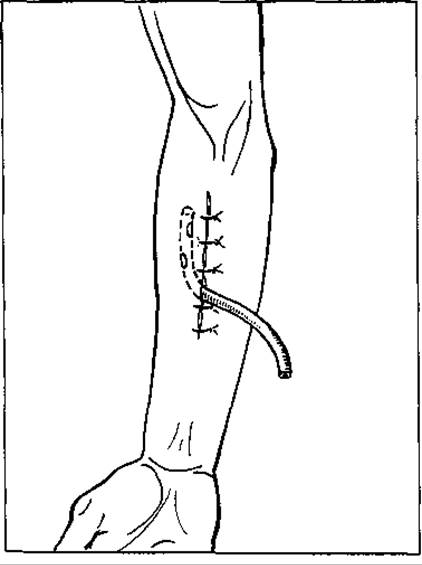
Fig. 62. Open passive wound drainage
In dual-lumen drains of special design, irrigation and outflow from the cavity occur through the drainage tube itself. Alongside such active-passive, continuous or intermittent, inflow-outflow drains, closed tubular drains are widely used. They are designed for the aspiration of purulent exudate and the introduction or instillation of antibiotics or antiseptics into purulent cavities (such as lung or liver abscesses), as well as closed drains for active continuous vacuum aspiration of purulent exudate from various cavities. An example is the Redon vacuum drain, in which one end featuring side openings is inserted into the purulent cavity, while the other is connected to a hermetically sealed low-pressure vessel that aspirates the purulent exudate, thereby causing the purulent cavity to collapse. The vessel can be replaced by a rubber bulb (pear syringe): compressing its walls creates a vacuum inside, which drives the suction of the exudate until the bulb expands back to its original shape as it fills (Fig. 65).
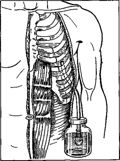
Fig. 63. Closed siphon drainage of the pleural cavity using Bülau's method
The second type of closed vacuum aspiration drain, which allows for precise regulation of negative pressure within the system, is used in the treatment of purulent pleurisy. It consists of a non-collapsible silicone or rubber tube inserted into the lower section of the pleural cavity, with its outer end connected to Bobrov's apparatus. This apparatus comprises two interconnected glass jars or three interconnected glass ampoules, where The transfer of aseptic liquid from the upper jar to the lower one creates a vacuum and ensures the suction of exudate (Fig. 66).
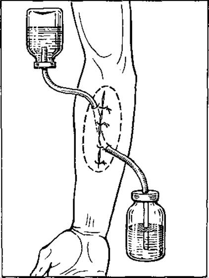
Fig. 64. Inflow-outflow (irrigation) wound drainage
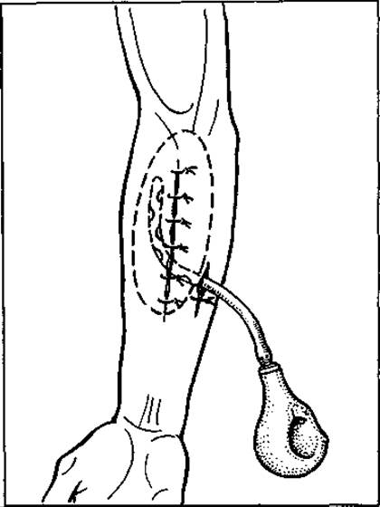
Fig. 65. Vacuum wound drainage using Redon's method
Although modern purulent surgery relies primarily on tubular drains, and less frequently on glove-finger-tubular drains, gauze drains are sometimes still used alongside tubular ones despite rapidly losing their drainage properties once soaked. They are inserted not so much for drainage as to limit foci of infection within the abdominal cavity, achieve hemostasis during capillary bleeding in abdominal purulent foci, and maintain the patency of large external purulent cavities to prevent the premature formation of isolated chambers within them.
Drains, especially non-thermolabile ones made of rubber or glass, must not be placed directly adjacent to blood vessels to avoid causing thrombosis or pressure sores (erosion). They should be isolated from vessels by soft tissues.
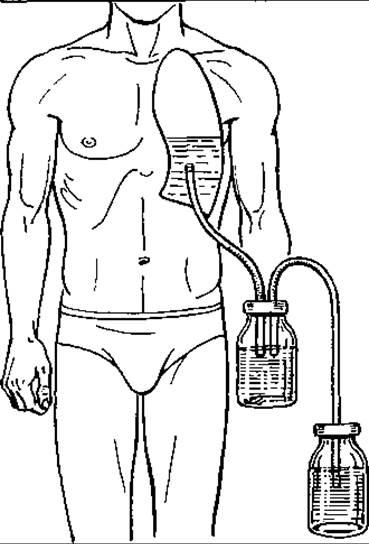
Fig. 66. Vacuum drainage with regulated negative pressure using Bobrov's apparatus
The most commonly used tubular drains are single- and dual-lumen silicone, polyvinyl chloride, and other thermolabile plastic variants that do not collapse, become obstructed, or clog with pus.
GENERAL MANAGEMENT OF PATIENTS WITH SURGICAL INFECTION
General treatment is essential for all patients with infections, regardless of the type—whether it is a wound complication (including postoperative) or a surgical infectious disease—because there is no such thing as a purely localized infection. Every infection is accompanied by systemic phenomena caused by intoxication. The sources of intoxication include microbes and their toxins, breakdown products of necrotic tissues, and non-specific metabolic byproducts, including so-called medium-molecular-weight Peptides. General treatment primarily aims to create optimal conditions for the patient's endogenous defense mechanisms against microbial toxic aggression (intoxication). The patient must be provided with physical and mental rest, and all adverse stimuli, especially pain, should be eliminated. Maximum attention is paid to the patient, explaining the nature of the disease and outlining the requirements that will help them recover as quickly as possible. Inpatients are provided with favorable conditions and a qualitatively and quantitatively balanced diet. This is a crucial component and prerequisite of general treatment.
The complex of general therapeutic measures can vary in composition and purpose. However, regardless of this, it must be directed toward: 1) halting the development and spread of infection; 2) reducing and eliminating systemic intoxication; 3) activating the body's defense forces and the patient's regenerative and reparative capacities; 4) correcting functional disorders of organs and systems caused by the infection; 5) accelerating healing and eliminating the local focus.
Detoxification is carried out through various approaches, chief among which are the destruction of microbes, reduction of toxin and microbial concentration, their dilution, neutralization of toxins via binding or inactivation, and excretion. General measures aimed at halting the development of microflora, destroying it, and stopping its spread primarily involve the administration of systemic antimicrobial agents, specifically ANTIBIOTICS AND CHEMOTHERAPEUTIC drugs (administered enterally or parenterally). Typically, they are prescribed either during the treatment of local infection without surgery or following surgical intervention (for certain forms of infection). Regardless of the latter, the threat of pathogenic microflora spreading often persists (due to a large focus of infection, dangerous localization, insufficient bodily resistance, etc.). For instance, in Hematogenous osteomyelitis, Purulent Arthritis, tendinous felon, extensive phlegmon or carbuncles, in diabetic patients, and in many other types of infection, local surgical intervention is supplemented by the administration of antibiotics or antimicrobial chemotherapeutic drugs (sulfonamides, fluoroquinolones such as ciprofloxacin and its analogs, etc.). The use of these antimicrobial agents must be based on knowledge of the microorganism species and their drug susceptibility. Since determining these parameters takes time, while acute infection requires immediate treatment, broad-spectrum antibiotics or alternative chemotherapeutic agents with high antibacterial activity are frequently employed.
Among antibiotics, the most frequently used include benzylpenicillin sodium salt, Semisynthetic Penicillins (such as ampicillin), semisynthetic Cephalosporins (cefazime), Aminoglycosides (gentamicin, kanamycin), and clindamycin. Among antimicrobial chemotherapeutic agents, metronidazole (active against anaerobic non-clostridial microflora), sulfonamides, and nalidixic acid derivatives (ciprofloxacin, fluoronorfloxacin, cifran, etc.) are commonly utilized.
It should be noted that streptococci, gonococci, pneumococci, meningococci, Klebsiella, Salmonella, Clostridia, and certain other microbes retain high susceptibility to penicillin-group drugs. Therefore, diseases caused by these pathogens (such as erysipelas, gonococcal salpingitis and salpingo-oophoritis, meningitis, pneumococcal peritonitis, etc.) should be treated immediately with penicillin-group drugs without waiting for antibiogram results.
Staphylococci and Escherichia coli, due to their capacity for mutation and transmissible resistance (The ability to produce beta-lactamase, an enzyme that inactivates penicillin and Other Antibiotics), are resistant to first-generation antibiotics. In these cases, performing an antibiogram is mandatory. Consequently, for Infections caused by staphylococci, E. coli, Pseudomonas aeruginosa, Bacteroides, etc., treatment should be initiated with broad-spectrum antibiotics combined with metronidazole (against Bacteroides). Once microbiological test results and antibiograms are obtained, targeted therapy should be applied or the treatment regimen adjusted accordingly.
It is worth noting that many species of pathogenic microbes causing purulent infections (staphylococcus, Pseudomonas aeruginosa, E. coli, non-clostridial anaerobic microflora) are also highly resistant to modern broad-spectrum antibiotics. Even the most active aminoglycoside (gentamicin sulfate) exhibits an efficacy of no more than 70–80% against staphylococcal infection. Therefore, in severe forms of infection, the latest semisynthetic antibiotics resistant to beta-lactamases produced by microbes (such as Mefoxin or cefoxitin—a fourth-generation cephalosporin; Augmentin—amoxicillin combined with clavulanic acid, a beta-lactamase inhibitor), as well as antibacterial agents from the fluoroquinolone group (ciprofloxacin, Cipro) and carbapenems (Tienam, etc.), must be utilized.
Ciprofloxacin and Tienam even surpass antibiotics in their antimicrobial potency. They are effective against virtually any surgical infection caused by both gram-positive and gram-negative microflora, including bacteroidal infections.
The body's detoxification is achieved through numerous means. It should begin with the simplest and most accessible methods: primarily, administering large volumes of fluid (in the absence of contraindications) and the intravenous infusion of isotonic sodium chloride or sodium lactate solutions, Ringer-Locke, Hartmann, or lactosol solutions, among others. In addition to sodium chloride (6–8 g/L), the latter solutions contain potassium chloride (0.2–0.3 g/L), calcium chloride (0.1–0.2 g/L), sodium bicarbonate (absent in Hartmann's solution), and sodium lactate (3.1–3.3 g/L, which is absent in Ringer's and Ringer-Locke solutions). Lactosol additionally contains magnesium chloride. Isotonic sodium chloride solution (0.9%) and lactosol are standard pharmaceutical products manufactured in medical supply plants, whereas all others are compounded in pharmacies.
These solutions reduce intoxication both by diluting toxins and by enhancing their renal excretion from the body.
For many patients, the administration of these solutions (along with local intervention and regional measures) is sufficient to eliminate the focus of infection and detoxify the body. However, in certain cases, these detoxification measures—which act simply through fluid dilution and the correction of hydro-ionic imbalances—are insufficient. Therefore, specialized infusion-Transfusion Therapy is prescribed, and in a number of instances, particularly in systemic infections, additional measures such as artificial hemodialysis (hemo- and lymphosorption, etc.) are also employed.
Among the special agents for infusion-based detoxification that bind and eliminate toxic substances from the body, vinyl compound preparations are the most widely used—specifically, polyvinylpyrrolidone, hemodes, and polydesis, as well as periston and neocompensan. In our practice, the primary agent of this class is a 6% solution of low-molecular-weight polyvinylpyrrolidone (neocompensan). This drug works by binding (adsorbing) toxins in the bloodstream and rapidly clearing them from the body, predominantly via the kidneys. It is eliminated within 4–9 hours (M.T. Terekhov, 1979). Hemodes also facilitates detoxification by improving microcirculation (relieving erythrocyte stasis) and alleviating tissue hypoxia. It is administered intravenously by drip infusion at 10 mL/kg of body weight for children and 400 mL per infusion for adults. A repeat infusion can be given no earlier than 12 hours later. The drug begins to take effect within 10–15 minutes. Another agent, polydesis, is a low-molecular-weight solution of polyvinyl alcohol. A 3% polydesis solution in an isotonic (0.9%) sodium chloride solution is used at a dose of 400–800 mL per day for the same indications as hemodes.
Alongside the aforementioned solutions, many other substances are employed for detoxification. These not only bind toxins but also exhibit additional therapeutic effects, such as hemodynamic, antithrombotic, or rheological properties. They are prescribed for patients whose intoxication is complicated by hypoproteinemia, hypotension, or Blood Coagulation disorders (thromboembolic conditions, erythrocyte aggregation).
Prominent among these are protein-based agents such as albumin (10–20% solution) and protein solution (10% solution), which contain up to 80% albumin and 20% globulins. They are administered in doses of 100 mL and 200–400 mL once daily. Protein preparations form complexes with toxins, bind and retain water within the vascular bed (1 g of albumin retains 20 mL of fluid intravascularly), elevate blood pressure, and enhance renal filtration capacity, thereby promoting The excretion of toxins.
For infections accompanied by hypercoagulation and microcirculatory disturbances that carry a risk of thromboembolism, rheopolyglucukin—a low-molecular-weight polyglucukin—is highly effective. It is administered at a dose of 400 mL per day, with its effect lasting for up to 8 hours. Other anticoagulants (such as heparin) and antiplatelet agents (such as acetylsalicylic acid) are also used in the management of such patients.
Immune protein preparations, including gammaglobulin and antistaphylococcal plasma, are likewise utilized for detoxification and antimicrobial purposes. Containing Antibodies against specific microorganisms and their Antigens, these preparations bind them into antigen–antibody complexes, neutralizing and deactivating them. They are particularly effective in cases of hypogammaglobulinemia.
High detoxifying properties are also characteristic of osmotic Diuretics, which include polyhydric alcohols such as mannitol and sorbitol. These are hexahydric alcohols administered intravenously as 10–20% (mannitol) and 20% (sorbitol) solutions at doses of 1–2 g of dry substance per 1 kg of body weight. Their onset of action occurs within 15 minutes, with peak diuresis observed at 2 hours. The majority of the drug is excreted by the kidneys within 24 hours (up to 80%), while the remainder is utilized by the body as an energy source. These medications are primarily indicated for ACUTE RENAL FAILURE.
In cases of exceptionally severe infectious intoxication, such as that seen in sepsis, artificial hemodialysis, peritoneal dialysis, hemo- and lymphosorption, ultraviolet and
laser blood irradiation, and even the connection of a xenogeneic spleen or liver (typically porcine) are employed. It must be borne in mind that body detoxification is a broad concept and cannot be reduced solely to the administration of the aforementioned drugs and procedures. The infusion agents listed are primarily directed at the consequences of the infection—that is, its metabolic products. Detoxifying effects are also possessed by all agents that act directly on the infectious pathogens (antibiotics and chemotherapeutic drugs) or correct body functions and metabolism, without the normalization of which detoxification processes cannot proceed fully. Therefore, alongside targeted special agents, all other general measures for treating surgical infection are equally crucial in patient management. Specifically, these include symptomatic medications (cardiovascular drugs, analgesics, sedatives, etc.), immunocorrectors and immunostimulants (tactivin, thymalin, thymogen, etc.), anabolic Hormones and preparations (androsterone, nerobol, retabolil, solcoseryl solution, etc.), as well as proteolysis and prostaglandin inhibitors.
Prevention OF SURGICAL INFECTION
The high health risks and enormous economic costs associated with patient treatment make the prevention of surgical infection a higher priority today than the search for ever-new and largely ineffective treatments.
Naturally, infection prevention is inherently complex due to its multifaceted nature, driven by a multitude of causative factors and favorable conditions. The previously cited list of major general causes contributing to the spread of surgical infection serves as a guiding framework for selecting preventive measures.
Maintaining a strict aseptic regimen in surgical hospitals, and above all in operating rooms and dressing suites, is an urgent priority. This entails, first and foremost, raising aseptic requirements for all personnel in surgical, anesthesiology, and intensive care units; enhancing professionalism in the execution of aseptic procedures; and strengthening oversight by the head of the surgical department (operating unit) and the senior operating nurse. Additional measures include airing out wards, disinfecting bedding, and performing wet cleaning. A high standard of hygiene within the department, staff hygiene (specialized workwear, screening and treatment of carriers), patient hygiene (showers with antimicrobial soap, complete isolation of patients with open infection foci from healthy individuals), minimizing the preoperative hospital stay, and avoiding bedside dressing all help reduce the threat of nosocomial infection and patient body surface contamination by multi-resistant microbes.
Thorough preoperative evaluation and examination of patients to identify any underlying anomalies, immune deficiencies, or other risk factors, followed by their correction, reduces the risk of postoperative infectious complications. Limiting invasive diagnostic procedures to strict indications, clipping rather than shaving hair in the operative field 30 minutes prior to surgery, disinfecting the skin of the operative site with antiseptics before the procedure, using waterproof surgical drapes for the surgical team, covering the prepared operative field with self-adhesive incise drapes through which the incision is made, using sharp instruments for tissue dissection, handling tissues with care, minimizing the number of ligatures, using monofilament non-absorbable or absorbable sutures in contaminated wounds, promptly replacing damaged gloves, and changing instruments after skin incision—all of these measures contribute to minimizing the risk of surgical site infections. Among the various types of surgical infections, wound infections and soft tissue injuries (microtraumas, burns, frostbite, insect bites and stings, etc.), as well as postoperative (wound and cavity) local and systemic infections, account for the largest share. Consequently, preventing these types of infection constitutes a paramount challenge. The prevention of accidental wounds and injuries consists of their timely diagnosis and the provision of first aid. Well-organized operations at trauma centers, first-aid posts, surgical offices, and ambulance stations are of immense importance both for infection prevention and, frequently, for the patient's ultimate prognosis. Primary surgical debridement forms the cornerstone of treatment for such wounds, ideally performed within the first 6–12 hours after injury, but no later than 24 hours, supplemented by adjunctive measures (wound cavity drainage, local chemical and physical treatments, aseptic and antiseptic dressings, physiotherapy, general supportive care). Alongside surgical debridement, patient immunization (passive, passive-active, or active) plays a major role in preventing wound-specific infections such as tetanus and rabies. The prevention of microtrauma infection is based on health education and prompt management of microtraumas—specifically, washing the injured area if contaminated, encouraging bleeding from puncture wounds by laterally compressing the soft tissues around the injury site, disinfecting the wound with an antiseptic (iodine solution, alcohol, or brilliant green), and subsequently sealing it with BF-2 medical glue or an adhesive bandage, or coating the affected area with an antiseptic liquid adhesive. Various special antimicrobial adhesive compositions that solidify upon application to the injured site have been proposed for treating microtraumas. They provide simultaneous disinfecting and sealing effects (e.g., M.V. Novikov's liquid, BF-2 glue, etc.). Radical excision of macroscopically damaged tissue, meticulous hemostasis (preferably without electrocoagulation), non-tight suture placement close to the wound edges (no more than 0.5 cm), gentle tissue handling, apodactylic technique, and strict adherence to all asepsis rules during the procedure play a vital role in preventing accidental wound infections. If surgical debridement is inevitably delayed for 24 hours or more, the patient should be prescribed prophylactic antibiotics (preferably semi-synthetic penicillins such as ampicillin or pentrexylic, etc.) or a sulfonamide drug (such as sulfadimetoxin) to inhibit infection development in the wound. In cases of delayed surgical debridement where signs of serous inflammation are already present (the so-called infected wound), the wound should not be closed to prevent suppuration (developing into a "purulent wound"); instead, it is drained and managed with moist-drying antiseptic osmotic dressings. Physiotherapeutic procedures, open-air treatment in aseptic chambers or aerotherapeutic units (ATUs), and general supportive measures (antibiotic therapy, etc.) are effective. Early wound closure, including plastic closure (using pedicle or free skin flaps), serves as an effective preventive measure against infection. An equally important measure, following the debridement of contaminated or infected wounds (in the serous inflammation stage), is the insertion of an inflow or inflow-outflow drainage system into the wound cavity infused with an antiseptic until all signs of inflammation completely subside.
For the prevention of cavity infections (peritoneal, pleural, cranial, articular), early diagnosis of acute diseases affecting the organs within these cavities is critical. This includes preventing contamination by luminal contents and material from affected organs and tissues, and removing residual blood and other substances (tissue fragments, fluids) during surgery through irrigation and aspiration of the operative cavities. In many cases, cavity drainage (using tubes or catheters) is performed both to evacuate residual exudate and to instill antiseptics or antibiotics through tubular drains. In preventing postoperative infection, one cannot overstate The Importance of preoperatively eliminating latent infection foci, anemia, hypoxemia, Protein deficiency (hypoproteinemia), electrolyte imbalances, and other metabolic disorders—particularly Carbohydrate Metabolism in diabetic patients.
An equally vital role is played by providing adequate surgical access, refined operative technique and anesthesia, and immediate prophylactic antibiotic therapy (administered 1–2 hours preoperatively) at the maximum daily dose (intravenously or intramuscularly) for interventions involving heavily contaminated wounds, infectious-inflammatory surgical processes in internal organs, surgeries on the colon and rectum, cardiovascular prosthetic procedures, and operations in diabetic patients. Prophylactic antibiotic therapy (Chemotherapy) is recommended in the following scenarios:
1) in accidental contaminated wounds that cannot undergo timely surgical debridement;
2) in all microbially contaminated surgical wounds;
3) in all surgeries involving endoprosthetics;
4) in operations performed on individuals with active infections or those who have recently recovered from an infectious illness;
5) in surgeries on patients with preexisting Heart valve disease (for the prevention of endocarditis);
6) in "clean-contaminated" procedures involving the opening of the intestinal lumen, biliary tract, etc.
Broad-spectrum antibiotics are used for prophylactic purposes.
Proper patient and wound care, adequate sanitary and hygienic conditions in hospital wards, and rational general medical management (vitamin-enriched Nutrition, correction of fluid-electrolyte balance and other metabolic disorders, oxygenation, and the use of immune, hormonal, and enzymatic preparations) are of paramount importance.
Special attention in preventing postoperative infection must be directed toward enhancing the professional qualifications of surgeons—particularly those on emergency duty—and intensive care unit personnel, as well as combating bacterial carriage, which is especially prevalent among surgical department staff.
In preventing infectious surgical diseases, well-maintained occupational and domestic sanitary-hygienic conditions, industrial safety regulations, prompt management of microtraumas, treatment of occult foci of latent infection and systemic chronic diseases (cardiovascular, metabolic, endocrine, allergic, and others), prevention and management of obesity, Physical Exercise, health-promoting sports, and body conditioning are of great significance.
Certainly, medical measures alone are not sufficient for the prevention of surgical infections. The latter can be effective only against the backdrop of people's economic well-being and favorable environmental living conditions.
Last update: 08/08/2026
Editorial and Educational Adaptation: This material has been compiled based on the primary/original source text. The project team performed an editorial review, corrected technical inaccuracies, structured sections, and adapted the content for an educational format.
What was processed:
- elimination of formatting defects (OCR errors, structural breaks, corrupted characters);
- editorial organization of content;
- standardization of terminology in accordance with academic sources;
- verification of factual statements against the original source text.
All mentions of the author, publication year, and origin of the primary text have been preserved in accordance with the source.