Sexually Transmitted Diseases - I. I. Mavrov 2005
Sexually transmitted diseases
Inguinal lymphogranuloma
Inguinal lymphogranulomatosis (lymphogranuloma venereum;
lymphogranulomatosis inguinalis) is a sexually transmitted disease. It is characterized by the appearance of a painless erosion or a small sore on the external genitalia, which rapidly epithelizes, followed by The Development of regional lymphadenitis, breakdown of the Lymph Nodes resulting in bleeding ulcers, and multiple fistulae.
Etiology. The disease is caused by chlamydiae. According to the current Classification, the causative agent belongs to the species Chlamydia trachomatis, serological types L1-L3. It is the most virulent representative of chlamydiae that primarily infect humans and inhabit the urogenital Organs; it is capable of replicating within the Cells of various Tissues. During a natural infection, it exhibits a pronounced lymphotropism, determining the lymphoproliferative character of the disease.
The size and staining Properties of the pathogens are similar to those of other chlamydiae. They can multiply in tissue cultures and chicken embryos (see insert VIII, 1-2). Cell susceptibility to chlamydial infection is not enhanced by pretreatment of cells with DEAE-dextran. Neuraminidase reduces the infectious activity of chlamydiae.
The pathogen particles contain Complement-fixing, thermostable chlamydial group Antigens (resistant to boiling) that are found in all other chlamydiae. Their composition includes one of three specific antigens (L1-L3), which can be detected using the immunofluorescence assay. Infectious particles are toxic.
The infection can be reproduced in laboratory conditions in mice and monkeys; in birds, it is difficult to achieve.
Routes of transmission. Inguinal lymphogranulomatosis is a sexually transmitted disease. The pathogen is spread through sexual contact. The portal of entry can sometimes be the eyes (Conjunctivitis with oculoglandular syndrome). The reservoir of infection consists of chronic patients with lesions of the genitalia and rectum, in whom the disease may be asymptomatic.
Epidemiology and general pathology. The highest incidence of inguinal lymphogranulomatosis is observed in tropical and subtropical zones (South and Southeast Asia, Central and South America). The intensification of communication networks occurring in recent decades increases the risk of emergence and spread of this disease in other Regions of the world.
The incubation period typically ranges from 3 to 30 days. The course of the disease generally comprises three stages: the appearance of primary signs of infection; involvement of regional lymph nodes; and severe symptoms of the illness.
Primary lesions in men frequently occur on the glans Penis, less often in the Urethra or on the Skin of the inguinal region; in women, in the Vagina or on the labia, less commonly on the cervix. They may appear as papules, pustules, a superficial sore, erosion, or manifest as signs of nonspecific urethritis (Fig. 30, a).
During orogenital or manual sexual contact, signs of the primary lesion may localize on the Tongue, in the rectum, on the fingers, etc. These manifestations are often short-lived and may go unnoticed by infected individuals. Patients typically seek medical attention In the second stage of the disease, which occurs several days or weeks after infection.
During this period, regional lymphadenitis or lymphadenopathy develops, most often unilateral. Multiple node involvement is frequently observed, the topography of which depends on the site of the initial infection. In men, the inguinal lymph nodes are usually the first to be affected; in women (with a primary vaginal lesion), retroperitoneal lymph nodes are more commonly involved, the enlargement of which goes unnoticed for a long time. Occasionally, skin rashes appear in the form of urticaria, scarlatiniform rash, and multiform, exudative, or nodular erythema.
In the bubonic form, the nodes enlarge and become painful, especially with the development of periadenitis. As a result of the inflammatory process, abscesses may form within them with tissue breakdown, frequently accompanied by fluctuation or, conversely, induration. Spontaneous rupture of the bubo is often observed, followed by remission or fistula formation.
Forms of the disease that run an acute course are accompanied by fever, pain in the JOINTS OF THE extremities, and may feature gastrointestinal disturbances, headache, malaise, and signs of general toxicosis. In some cases, the inflammatory process is sluggish (presenting as chronic adenopathy) over many months and years, with periodic exacerbations.
Without appropriate Treatment, the disease progresses, leading to the Third Stage, during which severe destructive changes develop not only in the lymph nodes but also in the surrounding tissues and organs. The Liver and Spleen enlarge.
In inguinal lymphogranulomatosis, particularly in cases of prolonged progression, characteristic disturbances in the quantitative content of serum globulins are observed in patients. Typically, the concentration of y-globulin and IgA increases. Cryoglobulins (normally absent) appear in the serum. Untreated infection transitions into chronic forms with persistence of the pathogen for many years. Immunity has not been sufficiently studied. Many chlamydial infections are characterized by the simultaneous combination of a latent infection with the presence of Antibodies and cell-mediated immunity reactions. Blood tests show mild leukocytosis, occasionally lymphopenia and monocytosis, anemia, and a slight left shift of the differential leukocyte count.
Clinical manifestations. Several days or weeks after infection, a small papule or vesicles appear on the external genitalia, in the anal region, in the rectum, or elsewhere, and quickly disappear. The lesion may ulcerate. Frequently (especially in women), it remains unnoticed and disappears within a few days.
Shortly thereafter, approximately in 2 weeks, regional lymph nodes enlarge, subsequently coalesce, and become painful. In men, the infection most frequently involves the lymph nodes located both above and below Poupart's ligament. The skin over these nodes becomes hyperemic, the nodes suppurate, and the resulting pus discharges through multiple fistulas. In women and homosexual men, perirectal lymph nodes are primarily affected, proctitis develops, and bloody muopurulent discharge appears from the rectum. Lymphadenitis may be observed in the area of the cervix.
During acute lymphadenitis, general symptoms of the disease are noted (headache, fever, signs of meningeal irritation, conjunctivitis, skin rashes, nausea, vomiting, arthralgia, etc.). In rare cases, meningitis, Arthritis, and pericarditis are observed.
In women, the proximal sections of the vagina and rectum are more frequently affected. As a result, rectal or rectovaginal fistulas are formed, strictures, scarring of tissues, damage to the lower digestive tract, and rectal obstruction occur (Fig. 30, b). In men, fistulas of the penis and rectum are more common. Deformation of the genitalia and elephantiasis are possible in both men and women.
Genitoanorectal syndrome is characterized by generalization of the infection, leading to serious lesions of the Central Nervous system (meningitis), Heart (endo- and myocarditis), Lungs (Pneumonia), skin (erythema nodosum and multiforme), eyes, and other organs.
Thus, inguinal lymphogranulomatosis differs significantly in its Pathogenesis and clinical manifestations from Other forms of Urogenital Chlamydiosis, which is largely determined by the biology and ecology of the causative agent. Its clinical symptoms may include a deep infiltrative ulcerative and sclerofibrotic process with signs of elephantiasis in the region of the external genitalia, Perineum, anus, and rectum. The scarring of ulcers leads to severe complications, such as strictures and atresia of the urethra and rectum. Papules and vegetations are also encountered, which complicate the clinical picture of inguinal lymphogranulomatosis.
Class="center">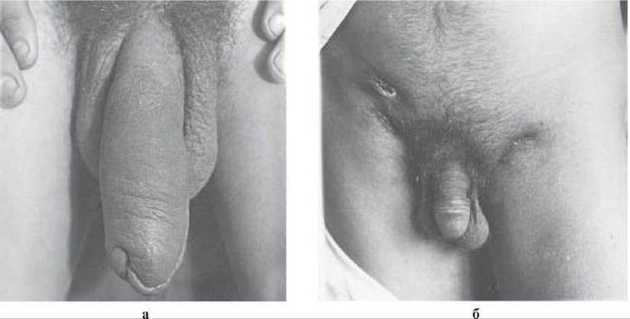
Fig. 30. Inguinal lymphogranulomatosis:
a) lymphostasis and edema in the area of the penis; b) ulceration and fistulae in the axillary region.
Inguinal lymphogranuloma should be differentiated from Chancroid, Tuberculosis of the inguinal lymph nodes, Syphilis, donovanosis (venereal granuloma), deep mycosis, tularemia, and genitoanorectal syndrome, which involves combined lesions of the female genitalia, rectum, and anus.
Diagnosis. Because the Clinical presentation of inguinal lymphogranuloma is heterogeneous and variable, identifying the causative agent is of paramount importance. Smears prepared and stained using the Romanowsky-Giemsa method are made from pus and biopsy specimens of the affected area; however, chlamydiae are rarely detected. To isolate the pathogens, suspected material is inoculated into the yolk sacs of developing chicken embryos, cell cultures, or mice (intracerebrally). Streptomycin may be added to the test material to eliminate accompanying Bacteria (penicillin and ether must not be used!). Pathogens are identified based on their Morphology and the results of serological reactions.
Most researchers consider the complement fixation test (CFT) to be the simplest serological method for detecting antibodies to the antigens of the CAUSATIVE AGENT OF inguinal lymphogranuloma. However, Specificity may be limited due to cross-reactivity with psittacosis antigens. The antigen is prepared from infected yolk sacs. The reaction becomes positive only several weeks (2–4) after infection, with a significant titer rise occurring over the subsequent three weeks. In clinically suspicious cases of rising titers or antibodies, a single positive reaction at a titer of 1:64 or higher is compelling evidence of an active infection. If the infectious process is resolved as a result of treatment, CFT titers decline.
The immunofluorescence assay can also be used for the serological diagnosis of inguinal lymphogranuloma; however, the antibodies are prone to cross-reacting with many other chlamydiae. More specific antibodies can be detected using counter-Immunoelectrophoresis, which utilizes a protein antigen derived from the chlamydial strains that cause inguinal lymphogranuloma.
Intradermal test for inguinal lymphogranuloma (Frei test). This utilizes an antigen obtained from the infected yolk sac of a developing chicken embryo or mouse Brain. The results of the intradermal administration (on the forearm area) of a suspension of embryo-grown pathogens (0.1 mL) are compared with a control material prepared from uninfected yolk sacs. Skin test results are evaluated after 48–72 hours. The reaction is considered positive if a papule at least 6 mm in diameter forms at the injection site (absent in the control reaction). During the course of the infection, the test becomes positive only after the affected lymph nodes have fused with the skin. False-positive reactions may occur. Delayed-type hypersensitivity may develop against the thermostable antigen of various chlamydiae possessing a group-specific lipopolysaccharide. Furthermore, the test remains permanently positive even after successful Treatment of the disease. The intradermal test for inguinal lymphogranuloma lacks specificity; therefore, its interpretation must always be correlated with the clinical picture.
Histological findings. Affected lymphatic areas exhibit granulomatous inflammation with central abscess formation, epithelioid cells, dendritic Langerhans cells, plasma cells, and lymphoid elements. While non-specific, this histological picture, when combined with the Clinical symptoms of the disease, can support the diagnosis.
Treatment. Tetracyclines, macrolides, rifampicins, and sulfonamides are indicated. Antibiotic and sulfonamide therapy should be administered for 21–23 days. Etiotropic therapy is highly effective, particularly in the Cytology/cytology/16.html">Early stages of the disease. For the management of later stages, etiotropic therapy is combined with pathogenetic and symptomatic agents, alongside surgical intervention. In some patients, a significant decrease in complement-fixing antibody titers is observed following treatment, which may indicate the elimination of the pathogen from the body. Patients treated for inguinal lymphogranuloma must undergo thorough follow-up examinations under dispensary surveillance.
Control measures for inguinal lymphogranuloma are the same as those for other Sexually Transmitted Infections. Identifying infection sources and contacts, early specific treatment, and patient monitoring are of essential importance.
Last update: 10/08/2026
Editorial and Educational Adaptation: This material has been compiled based on the primary/original source text. The project team performed an editorial review, corrected technical inaccuracies, structured sections, and adapted the content for an educational format.
What was processed:
- elimination of formatting defects (OCR errors, structural breaks, corrupted characters);
- editorial organization of content;
- standardization of terminology in accordance with academic sources;
- verification of factual statements against the original source text.
All mentions of the author, publication year, and origin of the primary text have been preserved in accordance with the source.