Human Anatomy - Kotsan I. Y. 2009
Tissues
A tissue (textus) is a collection of Cells and non-cellular structures that share a common origin and have similar Structure AND Functions. There are four main groups of tissues: epithelial, connective, muscular, and nervous.
Epithelial Tissue (textus epithelialis) consists of tightly packed cells with a minimal amount of Extracellular matrix. It covers the outer surface of the body, lines Body Cavities and Internal Organs, and forms the majority of glands. It performs protective, absorptive, excretory, and secretory functions. The protective function lies in the fact that, by forming the outer layer of the Skin (the epidermis) and lining all organs from the inside, these tissues protect deeper structures from mechanical damage, microbial invasion, and other harmful substances, while in the gastrointestinal tract, they protect the wall from being digested by digestive juices. Epithelial tissues facilitate the absorption of digested food in the gastrointestinal tract and the Elimination of Metabolic waste products—both unneeded and toxic (such as urine, the retention of which in the body leads to death). The secretory function involves participating in The formation of glands, their terminal units, and duct walls to produce secretions, such as digestive juices (Digestive System glands), sebum (Sebaceous Glands), and sweat (Sweat Glands).
Class="center">Table 2

Epithelial tissues develop from all three germ layers: the ectoderm, endoderm, and mesoderm. The skin epithelium is of ectodermal origin, whereas that of The Stomach and intestine is endodermal. There is also a group of mesoderm-derived epithelia, such as the epithelium of serous membranes (mesothelium), Gonads, and Kidneys.
The Classification of epithelial tissues depends on their Structural and functional features, as well as the organs they help form. Epithelia are divided into covering (lining) and glandular epithelia (Table 2).
Depending on the structure and arrangement of the cells, covering epithelium is classified into simple (single-layered) and stratified (multi-layered) (Fig. 5).
Simple Epithelium can be squamous, cuboidal, or columnar. Simple squamous epithelium lines serous membranes (the Pleura and Peritoneum); cuboidal epithelium forms the walls of Kidney tubules; and columnar epithelium lines the mucous membrane of the digestive organs (stomach and intestines).
Columnar epithelium can be single-rowed or multi-rowed (pseudostratified). Pseudostratified epithelium consists of cells of varying shapes and heights. Because The Cell heights differ, their nuclei lie at different levels, creating the appearance of multiple layers. A specialized type of simple columnar epithelium is ciliated epithelium. The cells of this epithelium feature cytoplasmic projections (cilia) on their apical surface, which move in a coordinated direction to transport mucus.
Simple ciliated epithelium lines the respiratory tract and Fallopian tubes.
Stratified Epithelium. Based on the shape of the cells in the superficial layer, stratified epithelium is predominantly squamous. Depending on the functional state (keratinization) of the cells in the upper layers, stratified epithelium is divided into two types: keratinized (the Superficial layer of the skin) and non-keratinized (the mucous membrane of the Nasal cavity and Pharynx). There is also a specialized form of stratified epithelium known as transitional epithelium (found in organs subject to volume changes), which transitions between multi-layered and two-layered states.
Covering epithelia share several characteristic morphological features:
1. Epithelial cells form continuous sheets that rest upon a basement membrane.
2. Epithelial sheets lack Blood Vessels. The cells are nourished via the diffusion of nutrients through the basement membrane, which separates the epithelium from the underlying loose Connective Tissue and serves as structural support.

Fig. 5. Diagram of epithelial tissue structure
1 — simple squamous epithelium; 2 — simple cuboidal epithelium; 3 — simple columnar epithelium; 4 — ciliated epithelium; 5 — transitional epithelium; 6 — stratified (non-keratinized) squamous epithelium
3. There is very little extracellular matrix between the cells, and they are tightly connected via specialized Intercellular junctions (desmosomes, tight junctions, etc.).
4. Epithelial tissues possess a high regenerative capacity, consistently containing cells capable of mitosis.
Glandular Epithelium releases its products onto the epithelial surface into hollow organs or directly into the BLOOD AND Lymph, thereby forming exocrine and Endocrine glands.
Glands can be unicellular (for example, mucus-secreting goblet cells in the intestine) or multicellular, formed by the invagination of the epithelial layer. They vary greatly in shape and function, including tubular, alveolar, and mixed (tubulo-alveolar) glands. Additionally, glands may be simple, branched, or compound.
Simple tubular glands are tube-like invaginations (such as intestinal glands).
Branched tubular glands consist of several simple tubules opening into a common excretory duct (for example, Glands of the pyloric stomach and uterine glands).
Compound tubular glands are formed by multiple branched tubular units that open into a common excretory duct (such as lacrimal glands, the Liver, and Testes).
Simple alveolar glands resemble small bubbles or saccules (such as sebaceous glands).
Branched alveolar glands consist of several simple alveolar glands that open into a common excretory duct (e.g., tarsal glands of the eyelids).
Compound alveolar glands are formed by several branched alveolar glands sharing a common excretory duct (e.g., Pancreas, mammary gland).
Simple alveolar-tubular glands are tubular structures with expanded ends (found in the pyloric region of the stomach).
Branched alveolar-tubular glands consist of several simple alveolar-tubular glands (e.g., urethral glands).
Compound alveolar-tubular glands are composed of branched alveolar-tubular glands (e.g., Prostate Gland).
In some cases, the described glands have a more complex structure. For instance, simple tubular glands may have coiled ends (e.g., sweat glands).
Connective tissue (textus connectivus).
Connective Tissues (Tissues of the Internal Environment) include the following: connective tissue proper, Cartilage, bone, blood, and lymph. Connective tissues perform trophic, protective, and supportive functions.
The trophic, or nutritive, function is primarily performed by blood and lymph, and partially by loose connective tissue. They participate in supplying the body with nutrients and oxygen, as well as in removing metabolic wastes and carbon dioxide.
These tissues also perform a protective function, which involves producing substances that destroy microbes entering the body, synthesizing antitoxins and immune bodies, and utilizing the ability of certain cells to engulf and digest microbes and foreign substances (phagocytic property).
The supportive function is mainly carried out by Cytology/practical/45.html">Dense connective tissue, cartilage, bone, and partially by loose connective tissue. Individual types of Internal Environment Tissues participate in forming both the Hard and Soft skeletons. This includes not only Bones and Their Joints, but also ligaments, fasciae, interosseous and intermuscular septa, the framework of internal organs, etc.
Connective tissues are extremely diverse in their structure. The common morphological feature of these tissues is that they consist of cells and a large amount of intercellular matrix, which comprises fibrous structures and a ground amorphous substance. Fibers provide the strength and elasticity of tissues. Based on their appearance and physicochemical properties, fibers are classified into Collagen, elastic, and reticular.
Collagen fibers (fibra collagenosa) are formed from the protein collagen. They are thick, strong, poorly stretchable, arranged in bundles, and constitute the mechanical framework of connective tissue.
Elastic fibers (fibra elastica) are built from the protein Elastin. Unlike collagen fibers, they are thin, resilient, and highly stretchable, allowing connective tissue to recoil and spring back.
Reticular fibers (fibra reticularis) are thin and short; when densely aggregated, they form a delicate, dense network, hence their name (reticulum - net).
The ground amorphous substance of connective tissue is a gelatinous mass rich in mucopolysaccharides that fills the spaces between cells and fibers.
The cellular composition of connective tissue is very diverse. Connective tissue cells include fibroblasts, histiocytes, fat cells, plasma cells, adventitial cells, and others.
Classification of connective tissues. Connective tissues are subdivided into connective tissue proper, cartilage, bone, blood, and lymph (Table 3).
Table 3
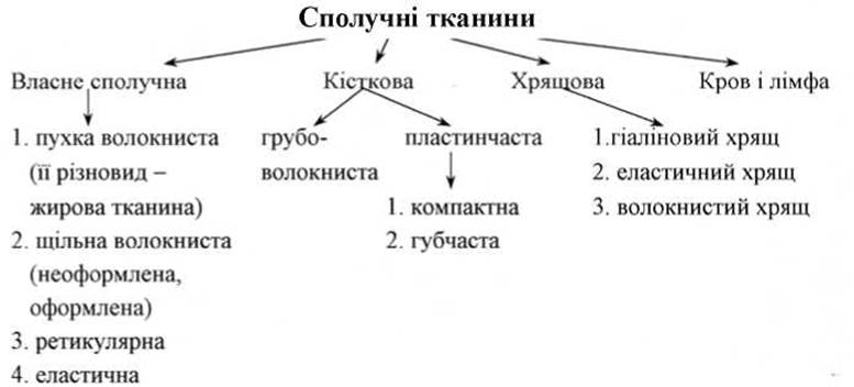
Connective tissue proper (textus connectivus propria) includes the following tissues: loose fibrous (its variant is adipose tissue), dense irregular fibrous, dense regular fibrous, elastic, and reticular.
Loose Fibrous connective tissue (textus connectivus fibrosus laxus) is widely distributed in the bodies of humans and animals (Fig. 6). It is located between organs, accompanies blood vessels, is part of all organs, and forms the stroma in many of them. The cells of this type of connective tissue can accumulate fat in their Cytoplasm and transform into fat cells, allowing the tissue itself to become adipose tissue. The main cells of loose connective tissue are fibroblasts and histiocytes. In addition to fibroblasts and histiocytes, loose connective tissue contains other cells (pigment cells, fat cells, plasma cells, lymphocytes). The intercellular matrix of loose connective tissue contains collagen, elastic, and reticular fibers, which are arranged loosely and in various directions.
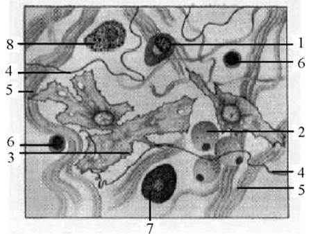
Fig. 6. Loose fibrous connective tissue
1 — plasmacyte; 2 — lipocyte, or adipose tissue; 3 — fibroblast; 4 — elastic fiber; 5 — collagen fiber; 6 — lymphocyte; 7 — tissue basophil; 8 — macrophagocyte
Dense irregular connective tissue (textus connectivus fibrosus compactus irregularis) is characterized by a relatively high Abundance of densely packed connective tissue fibers, a low content of ground substance, and a sparse population of cellular elements interspersed between the fibers (Fig. 7). In this tissue, the fibers run closely together and interlace with one another. Dense irregular connective tissue forms the Middle layer of the skin, known as the dermis.
In dense regular connective tissue (textus connectivus fibrosus compactus regularis), the fibers run parallel to each other and are bundled together. Cells known as fibrocytes are located in the intercellular matrix between the fiber bundles (Fig. 8). This tissue contributes to the formation of ligaments, tendons, membranes, and fasciae.
Elastic connective tissue (textus connectivus elasticus) consists of thick, rounded, or flattened fibers that frequently branch to form a wide-meshed, elongated network. This tissue forms the structural framework of large blood vessels, the Trachea, Bronchi, and other structures.
Reticular connective tissue (textus connectivus reticularis) forms the connective tissue framework of the Bone Marrow, Spleen, and Lymph Nodes. It is composed of reticular fibers and reticular cells, which under certain conditions can transform into macrophages (Fig. 9).
The distinguished Russian scientist and Nobel laureate Élie Metchnikoff was the first to draw attention to the fact that many organs in animals and humans contain macrophage cells that react uniformly to various harmful agents. He proposed grouping these cells into the macrophagic system. Fibroblasts and endothelial cells perform analogous functions. At the suggestion of the German scientist L. Aschoff, these formations were grouped into the reticuloendothelial system, now referred to as the mononuclear phagocyte system. The structural units of this system destroy microorganisms, neutralize toxic substances, and produce immune bodies.
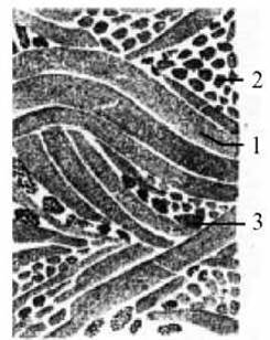
Fig. 7. Dense irregular connective tissue
1 — longitudinal bundles of collagen fibers; 2 — collagen fibers in cross-section; 3 — nuclei of fibrocytes

Fig. 8. Dense regular connective tissue: A - tendon: 1 - collagen fibers; 2 - tenocyte (tendon cell); 3 - layer of loose connective tissue; B - ligament: 1 - elastic fibers.

Fig. 9. Reticular connective tissue: 1 - reticulocyte.
Cartilage tissue (textus cartilagineus) consists of chondrocytes and an intercellular substance known as cartilage matrix, which possesses increased density.
Cartilage cells are oval or round in shape, occurring individually or in groups within specialized lacunae. The cartilage matrix (intercellular substance) is composed of collagen and elastic fibers along with an amorphous ground substance. Due to the density of this intermediate substance, cartilage tissue achieves high mechanical strength and also exhibits elasticity.
Cartilage tissue makes up the bulk of cartilaginous structures. Externally, cartilage is covered by the perichondrium, which consists of two layers: an outer fibrous layer and an inner chondrogenic layer that generates cartilage cells. The perichondrium performs a trophic function (nutrients diffuse into the cartilage from its blood vessels) and a regenerative function (regeneration of the cartilage occurs at the expense of its inner chondrogenic layer).
Depending on The structure of the intercellular matrix, cartilage tissue is divided into three main types: hyaline, elastic, and fibrocartilage (Fig. 10).
Hyaline cartilage (cartilago hyalina) is the most widespread in the Human and Animal body. It covers the articular surfaces of bones, forms the anterior ends of the Ribs, the laryngeal cartilages (except the epiglottis), the large bronchi, and a portion of the nasal septum. Hyaline cartilage is firm, resilient, translucent, and has a bluish tint. In its natural state, hyaline cartilage has a structureless, homogeneous intercellular substance containing delicate connective tissue fibers. The cells of hyaline cartilage reside in small lacunae, completely filling them.
Elastic cartilage (cartilago elastica) is built on the same principle as hyaline cartilage, but it is opaque and contains A large number of elastic fibers alongside collagen fibers. Due to these elastic fibers, it has a yellowish color that clearly distinguishes it from hyaline cartilage. Elastic cartilage is predominantly found in the auricle, external acoustic meatus, auditory tubes, and epiglottis.

Fig. 10. Cartilage tissue
I — hyaline cartilage; II — elastic cartilage; III — fibrocartilage; 1 — chondrocytes or cartilage cells; 2 — cartilage matrix
Fibrocartilage, or white fibrocartilage (cartilago fibrosa), occupies an intermediate structural position between dense regular connective tissue and hyaline cartilage. It differs from the latter in that the collagen fibers of its matrix are assembled into bundles, giving it a clearly defined fibrous structure. The presence of an abundance of collagen fibers imparts exceptional strength to fibrocartilage. Fibrocartilage forms the intervertebral discs and the Pubic Symphysis (interpubic disc). It is also found in the sternoclavicular and temporomandibular joints, as well as at the attachment sites of certain Muscle tendons and ligaments to bones.
Bone tissue (textus osseus) forms the human Skeleton, determines body shape, protects organs located within the cranial, thoracic, and pelvic cavities, and participates in mineral and Lipid METABOLISM. The Red bone marrow contained within bones serves as the central hematopoietic organ and performs biological defense functions.
Bone tissue consists of cells and intercellular substance.
Three main cell types are distinguished in bone tissue: osteoblasts, osteocytes, and osteoclasts.
Osteoblasts are young cells responsible for forming bone tissue. They vary in shape, appearing as cubic, pyramidal, or polygonal, and contain a round or oval eccentrically positioned Nucleus. Their cytoplasm features a well-developed granular Endoplasmic reticulum, Mitochondria, and a stacked Golgi apparatus. Osteoblasts gradually change and mature into osteocytes.
Osteocytes are the primary cells of bone tissue, characterized by a distinct stellate (star-like) shape. Their nuclei are round or oval, and sometimes positioned eccentrically within the cell. In mature tissue, osteocytes are highly specialized cells that have lost The ability to divide. Due to the high density of the extracellular matrix, each individual cell resides within a specific lacuna that precisely matches its shape. These lacunae are interconnected by tiny canaliculi through which the cellular processes of osteocytes communicate, giving the entire bone tissue structure a syncytial character. Bone canaliculi make contact with the perivascular space, facilitating the exchange of substances between osteocytes and blood via tissue fluid.
Osteoclasts are very large, multinucleated cells containing anywhere from 2 to 50 nuclei. Functionally, an osteoclast is a specialized macrophage that plays an active role in the resorption of calcified cartilage and bone, thereby contributing to their continuous remodeling and renewal.
An essential role in bone tissue is played by the intercellular ground substance, which consists of an amorphous matrix and collagen fibers. Depending on the arrangement of these fibers, bone tissue is classified into coarse-fibered (woven) and lamellar bone.
Coarse-fibered bone tissue is characterized by collagen fibers gathered into thick, rough bundles distributed randomly throughout the amorphous matrix, intersecting one another in various directions; bone cells (osteocytes) are likewise scattered haphazardly among them. The skeleton of lower vertebrates and embryonic higher vertebrates is built of coarse-fibered bone tissue. During The Development of the latter, coarse-fibered bone is eventually replaced by lamellar bone. In adult humans, coarse-fibered bone tissue persists permanently only at the attachment sites of muscle tendons to bones and in the region of closing Skull sutures.
Lamellar bone tissue has a more complex structure, is functionally superior, and is significantly stronger than coarse-fibered bone. Virtually all bones in vertebrates and humans are constructed primarily of lamellar bone tissue. Its structural basis is formed by bone lamellae composed of dense bundles of collagen fibrils (fibers).
Depending on the arrangement pattern of these bone lamellae, bone substance is categorized into spongy (cancellous) and compact bone.
Spongy bone substance has a simpler structural Organization. Its lamellae form trabeculae of varying thickness that intersect with one another in multiple directions.
The spaces between the trabeculae are filled with red bone marrow. Spongy substance forms, for instance, the epiphyses of long tubular bones.
Compact bone substance, found in the diaphyses of tubular bones, features a more intricate structure. Here, the arrangement of lamellae is dictated by The pathway of blood vessels that permeate the bone in large numbers. These vessels run through channels known as Haversian canals (Fig. 11). The bone lamellae are arranged in concentric circles around these canals, expanding outward to form cylinder-in-cylinder structures. Each such system of lamellae with a central canal is called an osteon. The osteon serves as the structural unit of bone.
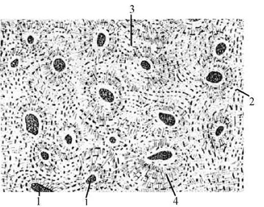
Fig. 11. Cross-section of a bone
1 — Haversian canals; 2 — external circumferential lamellae; 3 — interstitial lamellae; 4 — Haversian system
Blood (haema, sanguis) is a fluid connective tissue that performs trophic, respiratory, protective, excretory, and regulatory functions.
The trophic (nutritive) function of blood lies in delivering nutrients absorbed in the digestive tract to the body's cells.
Its respiratory function involves transporting oxygen from the Lungs to the tissues and carbon dioxide from the tissues back to the lungs.
The protective function is ensured by the ability of certain Blood Cells to perform phagocytosis (engulfing harmful substances and microbes), as well as the blood's capacity to produce protective Proteins (Antibodies) that neutralize toxins and pathogens.
The excretory function consists of transporting metabolic end-products generated within Cells and Tissues to the organs of excretion.
The regulatory function of blood is associated with The transport of Hormones and other BIOLOGICALLY ACTIVE SUBSTANCES that regulate the functional activity of individual organs. The regulation of bodily functions via the blood is known as humoral regulation.
The total volume of blood in The Human Body is approximately 7% of body mass, amounting to about 5–6 liters.
Blood consists of plasma and formed elements (cells): erythrocytes, leukocytes, and thrombocytes (Fig. 12).
Blood Plasma (plasma sanguinis) is the fluid intercellular matrix. It accounts for 55–60% of total blood volume. Blood plasma is composed of 90% Water, up to 7% proteins, and 3% other organic and inorganic substances. Plasma circulates nutrients, hormones, Enzymes, and Metabolic waste products destined for elimination. Among Plasma Proteins, of particular importance are fibrinogen, which is involved in blood clotting; albumin, which transports poorly soluble substances; and globulins, which produce protective antibodies during infectious diseases, among others. Protein-free blood plasma is called serum, which is widely used in medicine for prophylactic and therapeutic purposes.
Erythrocytes (erythrocytes), or red blood cells, are highly specialized anucleated cells. They contain a specific blood pigment, Hemoglobin, which facilitates the transport of oxygen and carbon dioxide. There are approximately 4.0–4.5 million erythrocytes per 1 mm3 of blood in women and 4.5–5 million in men. Erythrocyte counts can vary: high-altitude residents have more than lowland dwellers, and athletes have more than untrained individuals. Erythrocytes are produced in the red bone marrow and have a lifespan of 3–4 months. Their breakdown occurs primarily in the spleen. Approximately 1/100 of all erythrocytes are destroyed daily, and the entire population of blood erythrocytes is replaced every 3 months.

Fig. 12. Human blood
1 — small lymphocytes; 2 — eosinophilic leukocytes; 3 — group of Blood Platelets (thrombocytes); 4 — neutrophilic leukocytes; 5–8 — erythrocytes in various positions; 9 — large lymphocyte; 10 — basophilic leukocyte; 11 — monocyte; 12 — medium lymphocyte
Leukocytes (leucocytus), or white blood cells. Unlike erythrocytes, leukocytes possess a nucleus. Leukocytes are significantly fewer in number than erythrocytes. Normally, there are 6,000 to 8,000 leukocytes per 1 mm3 of blood. Throughout the day, the leukocyte count in the blood fluctuates due to Digestion and physical exertion.
Leukocytes are classified into granular and agranular. Granular leukocytes include neutrophils, basophils, and eosinophils, while agranular leukocytes include monocytes and lymphocytes. Based on their origin and development, lymphocytes are divided into two main groups, namely: B lymphocytes and T lymphocytes.
The percentage ratio between individual types of leukocytes is called the leukocyte differential (leukocyte formula). In A number of diseases, the character of the leukocyte differential changes. Therefore, counting Various Forms of leukocytes is of diagnostic importance. In the blood of a healthy person, this ratio is as follows: lymphocytes — 25–30%; monocytes — 6–8%; eosinophils — 1.5–5.5%; basophils — 0.5–1.5%; neutrophils — 60–70%.
Granular leukocytes are produced in the red bone marrow, whereas agranular ones are produced in the lymph nodes, spleen, and Thymus (breast) gland. The lifespan of leukocytes averages from 3 days to 6 months. All types of leukocytes are capable of amoeboid movement and exiting blood vessels into tissues. The primary function of leukocytes is to protect the Organism from microbes and other foreign bodies through phagocytosis and antibody production. B lymphocytes play a significant role in humoral Immunity (i.e., clearing the body of Bacteria, Viruses, etc.). T lymphocytes are of great importance in cell-mediated immunity (i.e., the destruction of genetically altered cells of one's own body, such as foreign tissues).
Thrombocytes (thrombocytus), or blood platelets, are anuclear bodies of round, oval, or spindle shape. It has been proven that thrombocytes are not cells, but rather fragments of cytoplasm pinched off from bone marrow cells. There are 150,000 to 300,000 of them per 1 mm3 of blood. It has been established that thrombocytes are involved in blood clotting. The lifespan of thrombocytes is 5–6 days.
Lymph (lympha) is a transparent fluid of greenish-yellow or gray color. In its composition, lymph resembles blood plasma, but unlike it, contains a lower amount of proteins. Lymph contains white blood cells — lymphocytes — along with a small number of monocytes and eosinophils. There are 2,000 to 20,000 lymphocytes per 1 mm3 of lymph. Red blood cells are absent in lymph.
Lymph serves as a nutritive medium and, simultaneously, a medium into which cells excrete their metabolic waste products. In addition, lymph plays a protective role in the body. The total volume of lymph in the body is difficult to measure, but it is estimated to be 1–2 liters. Lymph formation occurs through the fluid part of blood plasma filtering from blood capillaries into tissues, and then from tissues into lymphatic capillaries.
Muscle Tissues (textus muscularis). The primary property of these tissues is contractility. The contraction of Muscle tissue ensures body movement in space, displacement of organs, and changes in their volumes. Muscle tissue is divided into smooth, striated, and cardiac.
Smooth muscle tissue (textus muscularis nonstriatus) forms part of the walls of hollow internal organs (intestines, Uterus, Urinary Bladder, etc.), blood vessels, and the excretory ducts of glands (Fig. 13a).
The structural unit of smooth muscle tissue is the myocyte, an elongated cell. The contractile apparatus of these cells consists of myofibrils — filaments 1–2 µm in diameter arranged in parallel. Myocytes contract involuntarily (independently of human will), slowly, fatigue very slowly, and possess the ability to regenerate, meaning they recover quickly after damage.
Striated muscle tissue (textus muscularis striatus) participates in the formation of skeletal Muscles (hence it is also called Skeletal Muscle), is part of the Tongue, pharynx, upper Esophagus, and forms the external anal sphincter. Functionally, it is voluntary, as its contraction and relaxation are subject to human will. However, the contraction of Respiratory Muscles, which are built of striated muscle tissue, can occur both voluntarily (consciously) and involuntarily (as happens during Sleep). Striated muscle tissue is formed of striated muscle fibers of complex structure, in which light and dark segments (discs) with different light-refracting properties alternate (Fig. 13b).
A muscle fiber is an elongated multinucleated myosymphysis (myosyntab / multinucleated muscle symplast) formed As a result of the fusion of muscle cells. In shape, muscle fibers resemble very long cylinders with rounded ends. The length of the fibers ranges from 1 to 40 mm, and can reach 10–12 cm. The fiber diameter varies from 0.01 to 0.1 mm. Each muscle fiber has a membrane called the sarcolemma (sarcos — flesh, lemma — husk/sheath), a cytoplasm called the sarcoplasm, and a large number of nuclei (about a hundred).
The nuclei are usually oval, sometimes slightly elongated, with a low Chromatin content. In mammalian and human muscle fibers, the nuclei are located closer to the periphery of the fiber, thus occupying a superficial position.
Electron Microscopy examinations of the fiber sarcoplasm have revealed a well-developed system of membrane-bound tubules and cisterns. Due to its structural similarity to The endoplasmic reticulum of other cells, it is called the sarcoplasmic reticulum. Glycogen is synthesized within it, and it stores Calcium Ions.
The fiber sarcoplasm also contains a large number of mitochondria, which are referred to here as sarcosomes. Their high content of oxidative enzymes determines the crucial role of sarcosomes, much like the mitochondria of other cells, in oxidative Metabolism and Energy supply to the fiber.
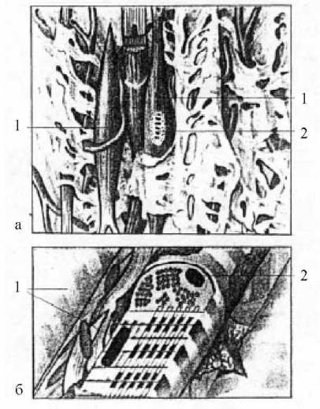
Fig. 13. Muscle tissues
a — smooth muscle tissue: 1 — smooth muscle cells (myocytes); 2 — myocyte nucleus; b — striated (skeletal) muscle tissue: 1 — myofibrils; 2 — nucleus
The functionally most important Structural elements of the fiber are myofibrils, which determine its contractility. In striated muscles, unlike smooth ones, they are non-homogeneous; this accounts for their cross-striation. Myofibrils within a muscle fiber form a bundle that extends from one end of the fiber to the other.
Each myofibril consists of alternating discs (plates) that differ in their physicochemical and optical properties. Some of them appear optically anisotropic under illumination and therefore look dark; they are designated by the letter A. Others — isotropic — are light and designated by the letter I. The discs, in turn, are divided into two parts by a membrane. In disc A, this membrane is designated as M (mesophragma), and in disc I, as Z (telophragma). In all fibrils, dark discs are located at the same level as dark ones, and light discs at the same level as light ones. Due to this, the fiber appears cross-striated.
Each myofibril is divided into segments called sarcomeres. Since the boundaries of a sarcomere are the telophragms running in the middle of disc I, each sarcomere comprises two halves of disc I and is positioned in the middle by disc A.
Using an Electron microscope, it has been established that each myofibril consists of protofibrils of two types: thick (16 nm) and thin (5–7 nm). Thick protofibrils are formed of longitudinally oriented protein molecules of Myosin, while thin ones are formed of another contractile protein, Actin. Muscle fiber contraction occurs due to the sliding of actin protofibrils between myosin protofibrils (sliding filament theory). The sarcomere shortens like a complex spyglass telescope. Its volume remains constant, while its transverse dimension increases. The sliding filament theory was proposed by A. Huxley in 1957. Other explanations for the Mechanism of muscle contraction also exist.
Depending on fiber thickness and Myoglobin content, red, white, and intermediate striated fibers are distinguished. Red fibers are rich in myoglobin and mitochondria, yet they are quite thin, with myofibrils arranged in groups. Intermediate fibers are thicker than red ones, but their myoglobin and mitochondrial content is lower. White fibers are the thickest, contain little myoglobin and mitochondria, but have a high myofibril count distributed evenly rather than in bundles.
Structure and function of fibers are inextricably linked. Thus, white fibers contract rapidly but also fatigue quickly. Red fibers are more endurance-oriented, contracting more slowly and for longer periods. In humans, skeletal muscles contain all types of fibers, with one type or another predominating depending on the muscle's function.
Cardiac muscle tissue (textus muscularis striatus cardiacus) resembles smooth muscle in its function and striated muscle in its structure. It consists of cardiac myocytes (cardiomyocytes) and cardiac conducting myocytes.
Each cardiac myocyte contains a single centrally located nucleus, with bundles of myofibrils concentrated at the periphery. Their transverse striation is identical to that of skeletal muscle myofibrils. Arranged in chains, the myocytes form muscle fibers. The boundaries between myocytes are formed by the sarcolemma and intercellular matrix. These unique structures lie transversely across the muscle fibers and are called intercalated discs. Electron microscopic examination has shown that myofibrils do not pass through these discs. It is believed that intercalated discs divide muscle fibers into individual segments—symplasts—which contribute to the relative independence of cardiomyocytes. This is a crucial feature of cardiac tissue that enables it to contract continuously throughout the life of the organism. Intercalated discs not only connect cells to one another but also participate in transmitting excitation from one cell to the next. Together with lateral cytoplasmic processes, intercalated discs ensure that the myocardium contracts as a single functional unit.
Some of The Heart's muscle fibers are made up of conducting myocytes, which differ significantly in structure from regular cardiomyocytes. For instance, a conducting myocyte contains multiple nuclei, and its myofibrils are far fewer in number than in cardiomyocytes, running in various directions and intersecting with one another. The aggregate of cardiac conducting myocytes forms a cardiac conducting muscle fiber. These fibers constitute the conduction system, which coordinates contractions across different regions of The cardiac muscle tissue.
Nervous Tissue (textus nervosus) is the most highly specialized tissue in the human body. It is the primary component of The Nervous system, which regulates and coordinates all body processes and mediates communication between the organism and its external environment. Nervous tissue has the capacity to perceive stimuli, process them into nerve impulses, analyze them, and transmit them to an effector organ.
Nervous tissue consists of Nerve Cells, or Neurons, and an intercellular substance known as neuroglia (Fig. 14). Neurons perform the functions of excitation and Nerve Impulse Conduction. Neuroglial cells, or gliocytes, provide structural support, trophic support, secretion, and protection, thereby ensuring optimal conditions for neuronal activity.
A nerve cell, or neuron, consists of a cell body and processes.
The cell body features a membrane, cytoplasm, a nucleus, Organelles, and specialized structural elements found exclusively in nerve cells (neurofibrils and tigroid substance).
There are two types of processes: dendrites and axons.
Dendrites are short processes that branch out profusely and immediately close to the cell body into numerous ramifications. Their number can vary. Dendrites conduct nerve impulses toward the cell body, which is located in the Brain or Spinal Cord.
Axons are long processes. A cell always has a single axon. It is characterized by a significant length, measuring in centimeters and sometimes reaching 1–1.5 m. The axon conducts nerve impulses away from the nerve cell body to an effector organ or another nerve cell.
Neurons may have one, two, or multiple processes. The region where a process emerges from the cell is slightly tapered and is called the cell pole. Consequently, a cell with a single process is unipolar; a cell with two processes is bipolar; and a cell with multiple processes is multipolar. True unipolar neurons do not exist in humans; instead, there are so-called pseudounipolar neurons, which develop from bipolar neurons through the fusion of their processes into a single one. Unipolar cells are typically round, bipolar cells are oval, and multipolar cells are polygonal.

Fig. 14. Nervous tissue
1 — nerve cells; 2 — nerve cell nuclei; 3 — nerve cell processes
Nerve cell processes are also referred to as nerve fibers. There are two Types of Nerve fibers: myelinated and unmyelinated.
Myelinated nerve fibers possess two sheaths: an outer sheath—the unmyelinated (or Schwann) sheath—and an inner myelin sheath. The Schwann (unmyelinated) sheath participates in regulating the metabolism and growth of the nerve fiber's axis cylinder and acts as an electrical insulator. The myelin sheath provides trophic and insulating functions while increasing the conduction velocity of the nerve impulse. Along the course of a myelinated fiber, the myelin sheath is periodically interrupted (where adjacent Schwann cells meet), forming the nodes of Ranvier. These are biologically active regions where mitochondria, ions, and metabolic products accumulate. In myelinated fibers, nerve impulses propagate saltatorily from one node of Ranvier to the next, unlike unmyelinated fibers, where the impulse travels continuously along the membrane of the axis cylinder.
Unmyelinated nerve fibers lack a myelin sheath. The conduction velocity of nerve impulses in these fibers is much lower. In a myelinated fiber, the conduction velocity is 50–120 m/s, whereas in an unmyelinated fiber, it is 1–2 m/s (up to 10 m/s).
Nerve fibers terminate in specialized structures called nerve endings. Depending on their function, they are divided into sensory nerve endings (receptors) and motor nerve endings (effectors).
Receptors perceive stimuli, convert them into nerve impulses, and transmit them to their own cell body or to other cells. They are categorized into exteroreceptors, which receive stimuli from the external environment (receptors of the skin, retina, and nasal mucosa); interoreceptors, which are activated by stimuli originating within the internal environment of the body (receptors of internal organs and blood vessels); and proprioceptors, which respond to Changes in the spatial position of individual body parts (receptors in muscles, ligaments, and tendons).
Effectors transmit nerve impulses from nerve cells to effector organs (muscles and glands).
A special group of nerve endings is formed by so-called synapses. A synapse is the junction where excitation passes from a nerve fiber to a muscle, glandular, nerve, or other cell. Each neuron can possess thousands of synapses, which are classified as axo-axonic, axo-dendritic, axo-somatic, dendro-dendritic, and others. A synapse consists of two membranes—presynaptic and postsynaptic—with a synaptic cleft between them. Within the presynaptic membrane are specialized vesicles containing biologically active substances known as Neurotransmitters (norepinephrine, acetylcholine, serotonin, dopamine, etc.). Neurotransmitters mediate the transmission of impulses from neuron to neuron, and from neurons to muscle elements, secretory cells, or other targets.
Depending on their function, neurons are subdivided into the following groups:
1. Sensory (afferent) neurons, which conduct impulses from the periphery to the center;
2. Motor (efferent) neurons, which carry impulses from the center to organs, prompting them to activity;
3. Interneurons (association neurons), which connect sensory and motor neurons to one another and carry out the analysis and synthesis of nerve impulses;
4. Secretory neurons that possess the property of neurosecretion. The Formation of the secretion is associated with the tigroid substance and the Golgi apparatus. Neurosecretory granules move along the axon away from the cell body, but instead of entering synaptic regions like other transported substances, they are released into the blood or CEREBROSPINAL FLUID (similar to hormones).
Basic Latin terms
Tissue — textus
Epithelial tissue — textus epithelialis
Connective tissue — textus connectivus
Collagen fibers — fibra collagenosa
Elastic fibers — fibra elastica
Reticular fibers — fibra reticularis
Proper Connective Tissue — textus connectivus propria
Loose fibrous connective tissue — textus connectivus fibrosus laxus
Dense irregular fibrous connective tissue — textus connectivus fibrosus compactus irregularis
Dense regular fibrous connective tissue — textus connectivus fibrosus compactus regularis
Elastic connective tissue — textus connectivus elasticus
Reticular connective tissue — textus connectivus reticularis
Cartilage tissue — textus cartilagineus
Hyaline cartilage — cartilago hyalina
Elastic cartilage — cartilago elastica
Fibrocartilage — cartilago fibrosa
Bone tissue — textus osseus
Lamellar bone tissue — textus osseus lamellaris
Blood — sanguis, haema
Erythrocytes — erithrocytus
Leukocytes — leucocytus
Platelets — thrombocytus
Lymph — lympha
Muscle tissue — textus muscularis
Smooth muscle tissue — textus muscularis nonstriatus
Striated muscle tissue — textus muscularis striatus
Cardiac muscle tissue — textus muscularis striatus cardiacus
Nervous tissue — textus nervosus.
Last update: 08/08/2026
Editorial and Educational Adaptation: This material has been compiled based on the primary/original source text. The project team performed an editorial review, corrected technical inaccuracies, structured sections, and adapted the content for an educational format.
What was processed:
- elimination of formatting defects (OCR errors, structural breaks, corrupted characters);
- editorial organization of content;
- standardization of terminology in accordance with academic sources;
- verification of factual statements against the original source text.
All mentions of the author, publication year, and origin of the primary text have been preserved in accordance with the source.