LEHNINGER PRINCIPLES OF BIOCHEMISTRY - VOL. 2. BIOENERGETICS AND METABOLISM - 2014
PART II. BIOENERGETICS AND METABOLISM
Class="center">Alcoholic Fermentation, which underlies the wondrous and seemingly spontaneous process of converting simple grape juice into exhilarating wine, has captured the attention of natural scientists since ancient times.
Arthur Harden, Alcoholic Fermentation, 1923
14. GLYCOLYSIS, GLUCONEOGENESIS, AND THE PENTOSE PHOSPHATE PATHWAY
Glucose occupies a central position in the METABOLISM of plants, animals, and many microorganisms. The glucose molecule is rich in energy and is therefore used by Cells as a "fuel." The Complete oxidation of glucose to carbon dioxide and Water is accompanied by a standard free-energy change of -2840 kJ/mol. In cells, glucose is stored in the form of high-molecular-weight polymers such as starch or Glycogen, which allows for the accumulation of A large number of hexose units while maintaining a comparatively low osmolarity of the Cytosol. When the energy demands of The Cell increase, glucose is released from intracellular stores and used to produce ATP via aerobic or anaerobic pathways.
Glucose is not only an excellent source of energy, but also a versatile precursor from which numerous intermediates are formed in biosynthetic reactions. For instance, the bacterium Escherichia coli uses glucose to produce the carbon skeletons of all Amino Acids, NUCLEOTIDES, Coenzymes, Fatty acids, and other products necessary for its growth. To fully understand the metabolic processes involving glucose, one would need to consider hundreds or even thousands of different reactions. In animal cells and vascular plant cells, glucose undergoes three MAIN TYPES OF transformations: 1) storage in the form of Polysaccharides or sucrose; 2) oxidation via Glycolysis to three-carbon compounds (Pyruvate) with the generation of ATP and various metabolic intermediates; 3) oxidation via The pentose phosphate (phosphogluconate) pathway to ribose-5-phosphate, which is required for the synthesis of Nucleic Acids, and NADPH, which participates in biosynthetic reduction reactions (Fig. 14-1).
Fig. 14-1. Major PATHWAYS OF GLUCOSE utilization in the cell. Glucose undergoes other transformations as well, but in most cells, The most significant amounts of glucose are funneled through these three most important directions.

Organisms that cannot obtain glucose from other sources must synthesize it themselves. Photosynthetic organisms generate glucose from atmospheric CO2, converting it first to a triose and subsequently to glucose. Nonphotosynthetic cells produce glucose from simpler three- and four-carbon precursors via Gluconeogenesis (the reverse of glycolysis), which effectively utilizes many of the same glycolytic Enzymes.
In this chapter, we will examine specific reactions of glycolysis, gluconeogenesis, and the Pentose Phosphate Pathway, and discuss the Functional Significance of each. We will also look at the various fates of pyruvate produced by glycolysis; these include fermentation processes used by many organisms under anaerobic conditions to generate ATP, and by humans for the industrial production of ethanol, lactic acid, and other important products. Furthermore, we will review the metabolic pathways that supply various sugars from mono-, di-, and polysaccharides to feed into glycolysis. The Structure/133.html">Discussion of glucose metabolism continues in Chapter 15, which covers the anabolic and catabolic processes linking glucose to glycogen. We will illustrate the mechanisms of Metabolic Regulation using the Synthesis and degradation of CARBOHYDRATES as Examples. The biosynthetic pathways for producing Extracellular matrix polysaccharides and cell walls from glucose are discussed in Chapter 20.
14.1. Glycolysis
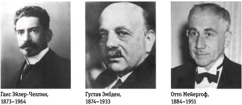
In The process of glycolysis (from the Greek glykys, meaning sweet, and lysis, meaning rupture or splitting), a glucose molecule is sequentially degraded through enzyme-catalyzed reactions to yield two molecules of the three-carbon compound pyruvate. A portion of the Free energy released in this process is conserved in the form of ATP and NADH. Of all metabolic pathways, The sequence of reactions constituting glycolysis was the first to be elucidated, and it is arguably the best understood. From Eduard Buchner's discovery of fermentation in Yeast extracts in 1897, up to the elucidation of the complete metabolic pathway in yeast (by Otto Neuberg, Otto Warburg, and Hans von Euler-Chelpin) and in Muscle (by Gustav Embden and Otto Meyerhof) in the 1930s, the main thrust of biochemical research focused on the reactions of glycolysis in yeast extracts and Muscle tissue. The shift in scientific outlook brought about by this discovery was eloquently captured by Jacques Loeb in 1906:
"By Buchner's discovery, biology was freed from yet another myth. The decomposition of sugar into CO2 and alcohol cannot be considered a manifestation of a 'vital principle,' any more than the Hydrolysis of cane sugar by invertase. The history of this question is instructive because it warns us against considering certain problems beyond our understanding simply because they have not yet found their explanation."
The Study of glycolysis stimulated The Development of Enzyme Purification Methods, helped reveal The Role of coenzymes such as NAD, and uncovered the central metabolic function of ATP and other phosphorylated compounds. Glycolytic enzymes from numerous organisms have been purified and studied in detail.
Glycolysis is a nearly universal central pathway of Glucose Catabolism; in most cells, the bulk of carbon-compound transformations proceeds through this route. In certain mammalian Cells and Tissues (e.g., erythrocytes, the renal medulla, the Brain, and sperm), glycolytic breakdown of glucose is the sole source of metabolic energy. Certain plant tissues adapted for starch storage (such as potato tubers), as well as A number of aquatic plants (e.g., watercress), derive most of their required energy via glycolysis, and many anaerobic organisms rely entirely on it.
Fermentation is a general term denoting the anaerobic breakdown of glucose or other organic nutrients to release energy conserved as ATP. Because living organisms first evolved in an oxygen-free atmosphere, the anaerobic degradation of glucose is arguably the most ancient biological mechanism for extracting energy from organic molecules. This sequence of chemical transformations has been remarkably well-preserved throughout evolution. The glycolytic enzymes of vertebrates bear a striking resemblance to their homologues in yeast and spinach, both in Amino Acid Sequence and tertiary structure. Glycolysis differs among species only in the fine details of its regulation and the ultimate fate of the pyruvate produced. At the same time, the thermodynamic principles and types of regulatory mechanisms governing glycolysis are the same as those for all Metabolic Pathways in the cell. Thus, glycolysis plays a vital role in the chemistry of life and serves as a convenient model process for studying the general Principles of Metabolism.
Before examining the individual steps of glycolysis in detail, let us take An Overview of the process as a whole.
Glycolysis takes place in two phases
The conversion of a six-carbon glucose molecule into two molecules of three-carbon pyruvate consists of a sequence of 10 reactions, the first five of which are referred to as the preparatory phase (Fig. 14-2, a). First, glucose is phosphorylated at the hydroxyl group on C-6 (reaction (1)). The resulting D-glucose-6-phosphate is converted into D-fructose-6-phosphate (reaction (2)), which is in turn phosphorylated—this time at C-1—yielding D-fructose-1,6-bisphosphate (reaction (3)). In both phosphorylation reactions, ATP serves as the donor of the phosphoryl group. Because all sugar derivatives formed during glycolysis are D-isomers, we have chosen to omit the prefix D from the compound names here, except where emphasizing their stereochemical properties is important.
Fig. 14-2. The Two phases of glycolysis. In the preparatory phase (a), each molecule of glucose is converted into two molecules of glyceraldehyde-3-phosphate; both of these molecules undergo further reactions in the payoff phase. (b) The final product of the second phase of glycolysis is pyruvate. In The first phase of glycolysis, two molecules of ATP are consumed per molecule of glucose, whereas four molecules of ATP are generated In the second phase, resulting in a net yield of two molecules of ATP per molecule of glucose converted to pyruvate. The reaction numbers in the scheme correspond to those in the text; the enzymes catalyzing the respective reactions are listed on the right. Note that each phosphoryl group, designated here as  , carries two negative charges (-PO2-3).

Fructose-1,6-bisphosphate is cleaved to yield two three-carbon molecules: dihydroxyacetone phosphate and glyceraldehyde-3-phosphate (reaction (4); it is this "lysis" step that gives the entire process its name). Subsequently, isomerization of dihydroxyacetone phosphate yields a second molecule of glyceraldehyde-3-phosphate (reaction (5)), concluding the first phase of glycolysis. Below, we will examine in chemical detail why the isomerization step (reaction (2)) is so crucial for the phosphorylation and subsequent C—C bond Cleavage in reactions (3) and (4). Note that prior to the cleavage of glucose into three-carbon units, two molecules of ATP are consumed; shortly thereafter, this ATP investment is not only recovered but surpassed. Thus, in the preparatory phase of glycolysis, the energy trapped in ATP raises the potential energy of intermediates, and the carbon chains of all degraded hexoses are funneled into a single common product: glyceraldehyde-3-phosphate.
The second (payoff) phase of glycolysis is accompanied by a net release of energy (Fig. 14-2, b). Each molecule of glyceraldehyde-3-phosphate is oxidized and phosphorylated with inorganic phosphate (not ATP!) to form 1,3-bisphosphoglycerate (reaction (6)). Energy is released during the conversion of two molecules of 1,3-bisphosphoglycerate into two molecules of pyruvate (reactions (7)-(10)).
As a result of the coupled phosphorylation of four ADP molecules, a large portion of this Energy is stored as ATP. However, the net yield of ATP during glycolysis is not four, but only two molecules per glucose molecule, because two molecules of ATP are consumed in the preparatory phase. In addition, The energy released in the Second Stage of glycolysis is conserved in the form of two NADPH molecules per glucose molecule.
Glycolysis involves three main types of transformations: 1) the Cleavage of the carbon Skeleton of glucose to yield pyruvate; 2) the phosphorylation of ADP to ATP driven by high-energy phosphorylated compounds generated during glycolysis; and 3) The transfer of a hydride ion to NAD+ to form NADH.
Fates of the Pyruvate Molecule.
With a few interesting exceptions among Bacteria, in all other organisms the pyruvate produced by glycolysis undergoes further metabolism via one of three major Catabolic pathways. In aerobic organisms and in tissues under aerobic conditions, glycolysis serves merely as The First stage of the complete degradation of glucose (Fig. 14-3). The Oxidation of Pyruvate yields the acetyl group of acetyl-coenzyme A, and the carboxyl group of pyruvate is converted into CO2. Subsequently, the acetyl group is fully oxidized to CO2 in The Citric Acid Cycle (Chapter 16). These processes are coupled to the transfer of electrons to O2 with The formation of H2O in Mitochondria. The energy released during electron transfer drives the synthesis of ATP in mitochondria (Chapter 19).
Fig. 14-3. Possible catabolic fates of pyruvate formed by glycolysis. Pyruvate also serves as a precursor in many anabolic reactions not shown here.

A second major pathway for pyruvate is its reduction to lactate via Lactic acid fermentation. During strenuous muscle activity under oxygen deprivation (Hypoxia), NADH cannot be rapidly recycled back to NAD+; however, NAD+ is essential as an electron acceptor for the continued oxidation of pyruvate. Under such conditions, pyruvate is reduced to lactate, accepting electrons from NADH, thereby regenerating the NAD+ required to sustain glycolysis. Certain cell types and tissues (such as retinal cells and erythrocytes) convert glucose to lactate even under aerobic conditions; furthermore, some microorganisms also produce lactate via glycolysis under anaerobic conditions (Fig. 14-3).
In the third major catabolic pathway, pyruvate is converted into ethanol. The conversion of pyruvate into ethanol and CO2 in certain plant tissues, specific invertebrate species, Protozoa, and microorganisms (such as baker's yeast) under oxygen deprivation or anaerobic conditions is known as alcoholic fermentation (Fig. 14-3).
While The oxidation of pyruvate is a crucial catabolic pathway, pyruvate also participates in anabolic reactions. For instance, it serves as the carbon skeleton precursor for The amino acid Alanine. We will explore these reactions of pyruvate in subsequent chapters.
ATP and NADH Formation in Glycolysis.
A portion of the energy stored in the glucose molecule is channeled into ATP during glycolysis, while another fraction is retained in pyruvate. The overall equation for glycolysis can be written as follows:
Glucose + 2NAD+ + 2ADP + 2Pi —> 2 pyruvate + 2NADH + 2H+ + 2ATP + 2H2O (14-1)
The breakdown of each glucose molecule to pyruvate is accompanied by the formation of two molecules of ATP from ADP and Pi and two molecules of NADH via the reduction of NAD+. The hydrogen acceptor in this reaction is NAD+ (see Fig. 13-24), which binds to a Rossmann fold motif (see Fig. 13-25). The reduction of NAD+ is preceded by the enzymatic transfer of a hydride ion (:H-) from the aldehyde group of glyceraldehyde 3-phosphate to the nicotinamide ring of NAD+, yielding the reduced coenzyme NADH. Another hydrogen atom from the substrate molecule is released into the solution as H+.
Glycolysis can be broken down into two separate reactions: an exergonic reaction (the conversion of glucose to pyruvate)
Glucose + 2NAD+ —> 2 pyruvate + 2NADH + 2H+ (14-2)
∆G'° = -146 kJ/mol
and an endergonic reaction (the synthesis of ATP from ADP and Pi)
2ADP + 2Pi —> 2ATP + 2H2O (14-3)
∆G2° = 2 • 30.5 kJ/mol = 61.0 kJ/mol
Summing equations 14-2 and 14-3 gives the overall free-energy change for glycolysis (∆G'°total):
∆G'°total = ∆G1'° + ∆G2'° = -146 kJ/mol + 61.0 kJ/mol = -85 kJ/mol
Under standard cellular conditions, glycolysis is irreversible and proceeds to completion, driven by a large negative free-energy change.
Energy Contained in Pyruvate.
Glycolysis releases only a minor fraction of the energy stored in a glucose molecule. The resulting two pyruvate molecules still retain a substantial share of the glucose molecule's energy, which can be extracted through oxidative reactions in The Citric Acid cycle (Chapter 16) and Oxidative Phosphorylation (Chapter 19).
The Role of Phosphorylated Intermediates.
Each of the nine intermediates in the conversion of glucose to pyruvate is phosphorylated (Fig. 14-2). Phosphoryl groups appear to perform three Functions.
1. Because The Plasma Membrane generally lacks carriers for phosphorylated sugars, phosphorylated intermediates cannot leave the cell. Once the initial phosphorylation has taken place, there is no longer a need to expend energy to retain the intermediates inside the cell, despite the steep concentration gradient across the membrane.
2. Phosphoryl groups play a vital role in preserving metabolic energy. The energy released during the cleavage of a phosphoanhydride bond (e.g., in ATP) is partially conserved through the formation of phosphoric acid esters (e.g., glucose-6-phosphate). High-energy phosphorylated compounds generated during glycolysis (1,3-bisphosphoglycerate and phosphoenolpyruvate) transfer their phosphoryl group to ADP molecules, resulting in ATP formation.
3. The binding energy of phosphate groups in the active sites of enzymes lowers the activation energy and increases the Specificity of enzymatic reactions (Ch. 6). The phosphate groups of ADP, ATP, and glycolytic intermediates form complexes with Mg2+ ions, and the substrate-binding sites of many glycolytic enzymes are specific for these complexes. Most glycolytic enzymes require Mg2+ ions for their catalytic activity.
ATP is consumed in the preparatory phase of glycolysis
In the preparatory phase of glycolysis, two molecules of ATP are consumed, and the phosphorylated six-carbon sugar is split into two phosphorylated three-carbon sugars. The realization that the intermediates of glycolysis are indeed phosphorylated hexoses was not arrived at immediately and was, in a sense, accidental. In 1906, Arthur Harden and William Young were testing their hypothesis that inhibitors of Proteolytic Enzymes should stabilize enzymes in yeast extract during glucose fermentation. They added Blood serum (known to contain proteolytic Enzyme Inhibitors) to yeast extract and indeed observed enhanced glucose metabolism. However, a control experiment designed to demonstrate the absence of stimulating activity in boiled serum showed that even after boiling, the serum exerted the same stimulatory effect on glycolysis. Careful Analysis of the boiled serum's contents revealed that the stimulatory effect was due to the presence of inorganic phosphate. Harden and Young soon discovered that glucose added to the yeast extract was converted into hexose bisphosphate (the "Harden-Young ester," later identified as fructose-1,6-bisphosphate). This discovery initiated a long series of investigations into the functions of organic phosphoric acid esters in biochemistry, which ultimately shaped our modern understanding of The Central Role of phosphoryl group transfer.

(1) Phosphorylation of glucose.
The first reaction of glycolysis is the activation of glucose via phosphorylation at the C-6 carbon atom, yielding glucose-6-phosphate; ATP serves as the phosphoryl group donor:

This reaction, which is irreversible under intracellular conditions, is catalyzed by hexokinase. Recall that Kinases are enzymes that catalyze the transfer of a terminal phosphoryl group from ATP to a nucleophilic acceptor molecule (see Fig. 13-20). Kinases belong to the transferase class of enzymes (see Table 6-3). In the case of hexokinase, the acceptor is a hexose (usually D-glucose, although in some tissues hexokinases also catalyze the phosphorylation of other common hexoses, such as D-fructose and D-mannose).
Like many Other Enzymes, hexokinase requires Mg2+ ions for its activity because the true substrate for the enzyme is not ATP4-, but the MgATP2- complex (see Fig. 13-2). The Mg2+ ions shield the negative charges of the ATP phosphoryl groups, making the terminal phosphorus atom more accessible to nucleophilic attack by the -OH group of glucose. Upon glucose binding, the hexokinase molecule undergoes a significant conformational change (induced fit), and upon ATP binding, the two Protein domains move closer together by ~8 Å (see Fig. 6-22, Vol. 1). As a result of this shift, the bound ATP molecule is brought into close proximity with the bound glucose molecule, which prevents surrounding water from entering. Otherwise, water could gain access to the Active Site and attack (hydrolyze) the phosphoanhydride bonds of ATP. Like the other nine glycolytic enzymes, hexokinase is a soluble protein located in the cytosol.
Hexokinase is present in almost all organisms. The Human Genome encodes four hexokinases (I–IV), all of which catalyze the same reaction. Enzymes that catalyze the same reaction but are encoded by different genes are called Isoenzymes (see Box 15-2). One of the forms found in hepatocytes, hexokinase IV (also known as glucokinase), differs from all other hexokinase forms in its kinetic and regulatory properties, which is of great physiological significance (see Sect. 15.3).
(2) Conversion of glucose-6-phosphate to fructose-6-phosphate.
The enzyme phosphoglucose isomerase (glucose phosphate isomerase) catalyzes the reversible isomerization of the aldohexose glucose-6-phosphate and the ketohexose fructose-6-phosphate:
The Mechanism of this reaction involves the formation of an enediol intermediate (Fig. 14-4). The reaction proceeds readily in both directions, which is reflected in the small change in free energy. Isomerization plays a crucial role in the overall process of glycolysis because the rearrangement of carbonyl and hydroxyl groups at the C-1 and C-2 carbon atoms is required for the next two reactions. The phosphorylation reaction (reaction (3)) requires the creation of an alcohol group at the C-1 atom, whereas the cleavage of the bond between the C-3 and C-4 atoms (reaction (4)) requires a carbonyl group at the C-2 atom (p. 22).
Fig. 14-4. Reaction mechanism. The reaction catalyzed by phosphoglucose isomerase. Ring opening and closure (steps (1) and (4)) involve an active-site His residue (omitted here for simplicity). The proton (highlighted in pink), originally located at the C-2 atom, becomes more mobile due to electron delocalization toward the carbonyl oxygen and the adjacent hydroxyl oxygen. Following proton transfer from the C-2 atom to a Glu residue (a weak acid) in the active site, it readily exchanges with protons from the solution, meaning it is not strictly necessary for the exact same proton to attach to the C-1 atom in step (3). MECHANISM OF ACTION of phosphoglucose isomerase

(3) Phosphorylation of fructose-6-phosphate to fructose-1,6-bisphosphate.
In the second of the two "priming" reactions of glycolysis, Phosphofructokinase-1 (PFK-1) catalyzes the transfer of a phosphoryl group from ATP to fructose-6-phosphate, yielding fructose-1,6-bisphosphate:

Key Conventions.
Compounds containing two phosphate or phosphoryl groups attached to different positions in the molecule are called bisphosphates (or bisphospho compounds); examples include fructose-1,6-bisphosphate and 1,3-bisphosphoglycerate. Compounds with two interconnected phosphate groups, as in a pyrophosphoryl group, are called diphosphates, such as adenosine diphosphate (ADP). The same rules apply to the nomenclature of compounds with three phosphate groups: a distinction is made between trisphosphates (e.g., Inositol-1,4,5-trisphosphate) and triphosphates (e.g., adenosine triphosphate, ATP). ■
The enzyme that converts fructose-6-phosphate to fructose-1,6-bisphosphate is designated PFK-1 to distinguish it from another enzyme, PFK-2, which catalyzes the Formation of fructose-2,6-bisphosphate from fructose-6-phosphate in a different metabolic pathway. Under intracellular conditions, the reaction catalyzed by PFK-1 is irreversible; it represents the first major regulatory checkpoint of glycolysis. Although glucose-6-phosphate and fructose-6-phosphate can undergo other metabolic transformations, fructose-1,6-bisphosphate is committed exclusively to glycolysis.
The phosphofructokinases of certain bacteria, protozoa, and apparently all plants utilize pyrophosphate (PPi) rather than ATP as the phosphoryl donor for the synthesis of fructose-1,6-bisphosphate:
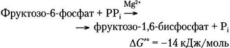
Phosphofructokinase-1 is an enzyme subject to complex Allosteric Regulation. Its activity increases whenever ATP levels in the cell drop or its breakdown products—ADP and especially AMP—begin to accumulate. Conversely, the enzyme is inhibited when the cell has an adequate supply of ATP as well as other fuel molecules, such as fatty acids. In some organisms, fructose-2,6-bisphosphate (not to be confused with fructose-1,6-bisphosphate, which is produced by the action of PFK-1) acts as a potent allosteric activator of PFK-1. Ribulose-5-phosphate, an intermediate of the pentose phosphate pathway discussed later in this chapter, also indirectly activates phosphofructokinase. The various levels of regulation at this stage of glycolysis are discussed in more detail in Chapter 15.
(4) Cleavage of fructose-1,6-bisphosphate.
The enzyme fructose bisphosphate aldolase, often referred to simply as aldolase, catalyzes a reversible aldol Condensation reaction (see Fig. 13-4). Fructose-1,6-bisphosphate is cleaved to yield two triose phosphates: the aldosetriose glyceraldehyde 3-phosphate and the ketosetriose dihydroxyacetone phosphate:

There are two classes of aldolases. Class I aldolases, found in plants and animals, operate via the mechanism shown in Fig. 14-5. Class II enzymes, present in Fungi and bacteria, do not form a Schiff base intermediate. A zinc ion in the Active Site of these enzymes is coordinated with the carbonyl oxygen at the C-2 atom; Zn2+ polarizes the carbonyl group and stabilizes the enolate derivative formed during C-C bond cleavage.
Figure 14-5 Reaction mechanism. The reaction catalyzed by aldolase I. The reverse of an aldol condensation is shown here. Note that the cleavage of the bond between C-3 and C-4 depends on the presence of a carbonyl group at the C-2 atom. A and B represent amino acid residues.

Although the cleavage of fructose-1,6-bisphosphate has a positive standard free-energy change, at low intracellular concentrations of the reactants the actual free-energy change is small, allowing the aldolase reaction to proceed in either direction. As we will see later, in gluconeogenesis aldolase catalyzes the reverse reaction (see Fig. 14-16).
Figure 14-6 Fate of carbon atoms during the conversion of glucose to glyceraldehyde 3-phosphate. (a) Origin of the carbon atoms in the two three-carbon products of the Reactions Catalyzed by aldolase and triose phosphate isomerase. The final product of these two reactions is glyceraldehyde 3-phosphate (2 molecules). (b) In the glyceraldehyde 3-phosphate molecule, the carbon atoms originate from one of two specific carbon atoms of the glucose molecule. Note that the numbering of the carbon atoms in glyceraldehyde 3-phosphate differs from that of the carbon chain in the glucose from which it is formed. In the glyceraldehyde 3-phosphate molecule, the carbon of the most complex functional group (the carbonyl group) is assigned position number one, C-1. This change in numbering is important to keep in mind when interpreting the results of experiments using isotopically labeled glucose (see Problems 3 and 5 at the end of the chapter).

(5) Interconversion of triose phosphates.
In subsequent steps of glycolysis, only one of the two triose phosphates produced by aldolase can participate directly, namely glyceraldehyde 3-phosphate. The other product, dihydroxyacetone phosphate, is rapidly and reversibly converted into glyceraldehyde 3-phosphate by the fifth glycolytic enzyme, triose phosphate isomerase:

The mechanism of this reaction is analogous to that of the phosphoglucose isomerase reaction (step 2) catalyzed (see Fig. 14-4). As a consequence of the triose phosphate isomerase action, the C-1, C-2, and C-3 atoms in the original glucose molecule are chemically indistinguishable from the C-4, C-5, and C-6 atoms, respectively (Fig. 14-6)—both "halves" of the glucose molecule are converted into glyceraldehyde 3-phosphate.
This reaction completes the preparatory phase of glycolysis: the phosphorylation of a hexose molecule at positions 1 and 6 and its cleavage to yield two molecules of glyceraldehyde 3-phosphate.
The payoff phase of glycolysis produces ATP and NADH
The second (recovery) phase of glycolysis (the "payoff" stage, Fig. 14-2, b) involves phosphorylation reactions in which a portion of the free energy from the original glucose molecule is captured in the form of ATP. As shown above, one molecule of glucose yields two molecules of glyceraldehyde 3-phosphate, which undergo further transformations in the second phase of glycolysis. The conversion of two molecules of glyceraldehyde 3-phosphate into two molecules of pyruvate is accompanied by the formation of four molecules of ATP from ADP. However, per single molecule of glucose, the net yield of ATP is only two molecules, because two molecules of ATP were consumed during the preparatory phase of glycolysis to phosphorylate the two terminal carbons of the hexose molecule.
(6) Oxidation of glyceraldehyde 3-phosphate to 1,3-bisphosphoglycerate.
The first reaction of the second phase of glycolysis is the oxidation of glyceraldehyde 3-phosphate to 1,3-bisphosphoglycerate, catalyzed by glyceraldehyde 3-phosphate dehydrogenase:

This is the first of two glycolytic reactions that result in energy capture in the form of ATP. In this reaction, the aldehyde group of glyceraldehyde 3-phosphate is oxidized, but instead of a free carboxyl group, a mixed anhydride of phosphoric and carboxylic acids is formed. An anhydride of this type, called an acyl phosphate, is characterized by a very high Standard Free Energy of hydrolysis (∆G′° = -49.3 kJ/mol; see Fig. 13-4, Table 13-6). The acyl phosphate group at the C-1 atom of 1,3-bisphosphoglycerate retains a large fraction of the energy released from the oxidation of the aldehyde group of glyceraldehyde 3-phosphate.
During the reaction, glyceraldehyde 3-phosphate remains covalently bound to the enzyme (Fig. 14-7). The aldehyde group of glyceraldehyde 3-phosphate reacts with the -SH group of a Cys residue in the active site of the enzyme; this reaction resembles hemiacetal formation (see Fig. 7-5), but in this case, a thiohemiacetal is formed. Interaction of this catalytically crucial Cys residue with heavy Metal Ions, such as Hg+, leads to irreversible inhibition of the enzyme.
The cellular concentration of NAD+ (≤10-5 M) is significantly lower than The amount of glucose metabolized within a few minutes. If the NADH produced at this stage of glycolysis were not continuously oxidized and recycled, glycolysis would quickly grind to a halt. We will discuss NAD+ recycling shortly.
Figure 14-7 Reaction mechanism. The reaction catalyzed by glyceraldehyde 3-phosphate dehydrogenase.

(7) Transfer of a phosphate group from 1,3-bisphosphoglycerate to ADP.
The enzyme phosphoglycerate kinase catalyzes the transfer of the high-energy phosphate group from the carboxyl group of 1,3-bisphosphoglycerate to ADP, yielding ATP and 3-phosphoglycerate.

Note that the name of the enzyme, phosphoglycerate kinase, corresponds to the reverse reaction, in which a phosphoryl group is transferred from ATP to 3-phosphoglycerate. Like all enzymes, phosphoglycerate kinase catalyzes the reaction in both directions. True to its name, this enzyme functions in gluconeogenesis (see Fig. 14-16) and photosynthetic assimilation of CO2 (see Fig. 20-4). During glycolysis, the reaction catalyzed by this enzyme proceeds in the direction of ATP synthesis, as described above.
Glycolysis reactions (6) and (7) are coupled because they share a common intermediate, 1,3-bisphosphoglycerate, which is formed in the first (endergonic) reaction and donates its acyl phosphate group to ATP in the second (exergonic) reaction. The overall equation for these two reactions is as follows:
Glyceraldehyde 3-phosphate + ADP + Pi + NAD+ ⇄ 3-phosphoglycerate + ATP + NADH + H+ ∆G′° = -12.5 kJ/mol
Thus, the overall reaction is characterized by a negative change in standard free energy.
As discussed in Chapter 13, the actual free energy change ∆G depends on the standard free energy change ∆G′° and the mass-action ratio Q — The ratio of the product of reactant concentrations to the product of reactant concentrations (see Equation 13-4). For reaction (6), we can write:

Note that [H+] is not included in the parameter Q because, in biochemical calculations, [H+] is considered constant (10-7 M) and is incorporated into ∆G′° (p. 15).
Fig. 14-8. Reaction catalyzed by phosphoglycerate mutase.

If Q < 1, then ln Q < 0. The consumption of the product of reaction (6) (1,3-bisphosphoglycerate) in reaction (7) keeps the steady-state concentration of 1,3-bisphosphoglycerate quite low; consequently, for the overall energy-coupled process, Q is a small value. The term ln Q is therefore negative, making the free energy change (∆G) a large negative value. Thus, we have demonstrated in yet another way how reactions (6) and (7), which share a common intermediate, are coupled.
The ultimate result of these two coupled reactions, which are reversible under cellular conditions, is that the energy released during the oxidation of the aldehyde group to a carboxyl group is conserved through the coupled formation of ATP from ADP and Pi. ATP synthesis resulting from phosphoryl transfer from the substrate 1,3-bisphosphoglycerate is termed substrate-level phosphorylation, as distinguished from the mechanism of oxidative phosphorylation. Substrate-level phosphorylation involves soluble enzymes and chemical intermediates (in this case, 1,3-bisphosphoglycerate), whereas Oxidative phosphorylation is associated with transmembrane proton translocation mediated by membrane-bound enzymes (Chapter 19).
(8) Conversion of 3-phosphoglycerate to 2-phosphoglycerate.
The enzyme phosphoglycerate mutase catalyzes the reversible transfer of a phosphoryl group between the C-2 and C-3 carbons of the glycerate backbone; Mg2+ ions play an essential role in this reaction:

The reaction proceeds in two steps (Fig. 14-8). First, the phosphoryl group attached to a His residue in the enzyme is transferred to the C-2 hydroxyl group of 3-phosphoglycerate, yielding 2,3-bisphosphoglycerate (2,3-BPG). Next, the phosphate group at the C-3 position of 2,3-BPG is transferred back to the same His residue, producing 2-phosphoglycerate and regenerating the phosphorylated enzyme. Phosphoglycerate mutase becomes phosphorylated by the phosphate group from 2,3-BPG; a catalytic amount of the latter is required to initiate the catalytic cycle, and thus 2,3-BPG is continuously replenished during the reaction.
(9) Dehydration of 2-phosphoglycerate to form phosphoenolpyruvate.
This is the second glycolytic reaction that generates a compound capable of high-potential phosphoryl group transfer (the first is reaction (6)). The enzyme enolase catalyzes the reversible dehydration of 2-phosphoglycerate to yield phosphoenolpyruvate (PEP):
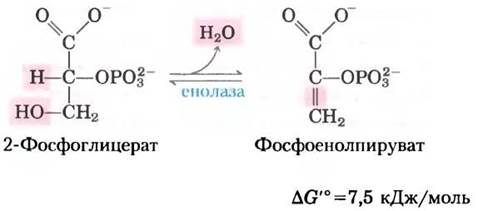
The mechanism of the enolase-catalyzed reaction involves the formation of an enolic intermediate stabilized by Mg2+ ions (Fig. 6-23). Despite the relatively small standard free energy change for this reaction, the ∆G of hydrolysis of the phosphoryl groups of the substrate and product differs substantially: -17.6 kJ/mol for 2-phosphoglycerate (a low-energy phosphate ester) versus -61.9 kJ/mol for phosphoenolpyruvate (a high-energy phosphorylated compound) (see Fig. 13-3, Table 13-6).
(10) Transfer of a phosphoryl group from phosphoenolpyruvate to ADP.
The final step of glycolysis involves the transfer of a phosphoryl group from phosphoenolpyruvate to ADP, catalyzed by pyruvate kinase, which requires K+ and either Mg2+ or Mn2+ for activity:

In this substrate-level phosphorylation reaction, the product (pyruvate) is initially formed in its enol form, which then undergoes rapid non-enzymatic tautomerization to yield the keto form, the predominant species at pH 7:
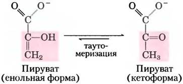
The overall reaction is characterized by a large negative standard free-energy change, largely driven by the spontaneous Conversion of the enol tautomer of pyruvate into the keto form (see Fig. 13-3). For the hydrolysis of phosphoenolpyruvate, ∆G'° = -61.9 kJ/mol. Nearly half of this energy is conserved in the phosphoanhydride bond of the ATP molecule (-30.5 kJ/mol), while the remainder (-31.4 kJ/mol) acts as a driving force that pulls the equilibrium toward ATP synthesis. Under intracellular conditions, the pyruvate kinase-catalyzed reaction is essentially irreversible and plays a crucial regulatory role (see Chapter 15).
The energetic gain in ATP becomes apparent when compiling the overall balance sheet of glycolysis
Let us construct the overall material balance for glycolysis. To do this, we must consider: 1) The Fate of the glucose carbon skeleton; 2) the consumption of Pi and ADP and the yield of ATP; and 3) the pathways of electron transfer in oxidation-reduction reactions. On the left side of the equation, we list all reactants consumed during glycolysis—namely, ATP, NAD+, ADP, and Pi (see Fig. 14-2)—and on the right side, all glycolytic products (keeping in mind that two molecules of pyruvate are produced per molecule of glucose).
Glucose + 2 ATP + 2 NAD+ + 4 ADP + 2 Pi —> 2 pyruvate + 2 ADP+2 NADH + 2 H++4 ATP+2 H2O
Subtracting identical terms from both sides of the equation yields the net equation for aerobic glycolysis:
Glucose + 2 NAD+ + 2 ADP + 2 Pi—> 2 pyruvate + 2 NADH + 2 H+ + 2 ATP + 2 H2O
The two NADH molecules generated in the cytosol during glycolysis are reoxidized to NAD+ under aerobic conditions by passing their electrons through the Electron Transport Chain, which in Eukaryotic cells resides in the mitochondria. The electron transport chain delivers these electrons to their final destination, the O2 molecule:
2 NADH + 2 H+ + O2 —> 2 NAD+ + 2 H2O
The transfer of electrons from NADH to O2 in the mitochondria provides the energy required to synthesize ATP via oxidative phosphorylation (Chapter 19).
Thus, glycolysis converts one molecule of glucose into two molecules of pyruvate (the carbon pathway). Two molecules of ADP and two molecules of Pi are converted into two molecules of ATP (the phosphoryl group pathway). Four electrons, in the form of two hydride ions, are transferred from two molecules of glyceraldehyde 3-phosphate to two molecules of NAD+ (the electron transfer pathway).
Glycolysis is under strict control
While studying glucose fermentation in yeast, Louis Pasteur discovered that The rate of glucose consumption and the total amount of glucose consumed under anaerobic conditions are many times greater than under aerobic conditions. Later studies on muscle tissue revealed a similarly striking difference between anaerobic and aerobic glycolytic rates. Today, the BIOCHEMICAL BASIS OF the "Pasteur Effect" is well understood. The ATP yield from anaerobic glycolysis (2 molecules per molecule of glucose) is much lower than that obtained from the complete oxidation of glucose to CO2 under aerobic conditions (30 or 32 ATP molecules per glucose molecule; see Table 19-5). Consequently, to generate the same amount of ATP, roughly 15 times more glucose must be broken down anaerobically as aerobically.
The amount of glucose degraded via glycolysis is regulated to maintain a nearly constant intracellular level (concentration) of ATP (along with glycolytic intermediates required for Biosynthesis). The necessary control over the glycolytic flux is achieved through a complex interplay among ATP consumption, NADH regeneration, and allosteric regulation of key glycolytic enzymes (including hexokinase, PFK-1, and pyruvate kinase); concentrations of key metabolites undergo rapid, continuous fluctuations to maintain the intracellular balance between ATP production and consumption. Over slightly longer time scales, glycolysis is modulated by Hormones (Glucagon, epinephrine, and Insulin) as well as by Changes in the Gene Expression of certain glycolytic enzymes. Of particular interest is the aberrant Regulation of glycolysis observed in malignant tumors. In 1928, the German biochemist Otto Warburg noted that glycolysis proceeds at substantially higher rates in almost all tumor tissues than in normal tissues, even in the presence of adequate oxygen. This observation forms The basis of several diagnostic and therapeutic approaches in oncology (Box 14-1).
Warburg is widely regarded as one of the most brilliant biochemists of the first half of the 20th century. He made monumental contributions across numerous fields, with renowned research in cellular Respiration, Photosynthesis, enzymology, and Intermediary Metabolism. Beginning in 1930, Warburg and his co-workers isolated and crystallized
seven enzymes involved in glycolysis. These same researchers developed an apparatus that revolutionized Traditional Methods FOR studying oxidative metabolism. Using the Warburg manometer, investigators could determine tissue oxygen consumption by measuring gas volume changes over time, subsequently using these data to calculate the concentrations of oxidative enzymes.
In the laboratory of Emil Fischer (recipient of the 1902 Nobel Prize in Chemistry), Warburg initially studied Carbohydrate Chemistry before winning the Nobel Prize in Physiology or Medicine himself in 1931. A remarkable number of Warburg’s students and associates were also awarded Nobel Prizes, including Otto Meyerhof (1922), Hans Krebs and Fritz Lipmann (1953), and Hugo Theorell (1955). Meyerhof's laboratory trained Lipmann as well as several other future Nobel laureates: Severo Ochoa (1959), André Lwoff (1965), and George Wald (1967).

Box 14-1. MEDICINE. High Glycolytic Rates in Tumor Tissues Underlie Chemotherapeutic Strategies in Certain Cancers and Facilitate Diagnosis
In many types of Human and Animal tumors, glucose uptake and glycolysis proceed roughly 10 times faster than in normal tissues. Most tumor cells exist in a state of hypoxia (i.e., oxygen deprivation) because early tumor growth outpaces the Development of the capillary network, preventing an adequate supply of oxygen. In tumor cells located more than 100–200 µm away from the nearest capillary, ATP production relies entirely on glycolysis (without subsequent pyruvate oxidation). The energy yield (two ATP molecules per glucose molecule) is far lower than that obtained by fully oxidizing pyruvate to CO2 in the mitochondria (~30 ATP molecules per glucose molecule; see Chapter 19). To generate an equivalent amount of ATP, tumor cells are forced to consume significantly more glucose than normal cells, converting it first to pyruvate and then to lactate, coupled with NADH recycling. It is likely that during the initial transformation of normal cells into tumor cells, first, ATP production becomes heavily dependent on glycolysis and, second, cells develop a tolerance to low extracellular pH driven by the accumulation of lactic acid, the end product of glycolysis. In general, the more aggressive the tumor, the higher its glycolytic rate.
This upregulation of glycolysis is achieved, in part, by increased production of glycolytic enzymes and the glucose transport Proteins GLUT1
and GLUT3 (see Table 11-3), which mediate glucose uptake across the plasma membrane. (Recall that GLUT1 and GLUT3 are insulin-independent.) Hypoxia-inducible Transcription factor-1 (HIF-1) is a regulatory protein that acts at the mRNA level, stimulating The production of at least eight glycolytic enzymes and glucose transporters under low-oxygen conditions (Fig. 1). By elevating the rate of glycolysis, tumor cells manage to survive anaerobically until new Blood Vessels can form. Another protein induced by HIF-1 is vascular endothelial growth factor (VEGF), a peptide hormone that stimulates rapid blood vessel growth (angiogenesis) within the tumor.
Fig. 1. Anaerobic Glucose metabolism in tumor cells yields considerably less ATP (two ATP molecules per glucose molecule) than complete oxidation to CO2 in healthy cells under aerobic conditions (~30 ATP molecules per glucose molecule). As a result, tumor cells must consume substantially larger amounts of glucose to meet their energy demands and produce the same ATP yield. Tumor tissues exhibit a marked upregulation in the Synthesis of glucose transporters and glycolytic enzymes. Pharmacological agents that inhibit hexokinase, glucose-6-phosphate dehydrogenase, or transketolase block glycolytic ATP production, thereby destroying Cancer cells by depriving them of essential energy.

In addition, there is evidence that the tumor suppressor p53, which is mutated in many types of cancer (Vol. 1, p. 670), regulates the synthesis and assembly of mitochondrial proteins required for the transfer of electrons to molecular oxygen. In cells carrying mutant forms of p53, Mitochondrial Electron Transport is impaired; consequently, these cells rely predominantly on glycolysis for ATP production (Fig. 1).
The high dependence of tumor cells on glycolysis compared to normal cells offers a promising strategy for antitumor Chemotherapy: glycolysis inhibitors can target and destroy tumor cells by restricting their ATP reserves. Currently, three effective hexokinase inhibitors are known that can be used as chemotherapeutic agents: 2-deoxyglucose, lonidamine, and 3-bromopyruvate. By preventing the synthesis of glucose-6-phosphate, these three compounds not only deprive tumor Cells of the ATP generated via glycolysis but also block the production of pentose phosphates in the pentose phosphate pathway, which likewise initiates with glucose-6-phosphate. Without pentose phosphates, cells are unable to synthesize nucleotides essential for DNA and RNA Synthesis, thereby halting growth and division. Another antitumor drug already approved for clinical use is imatinib (Gleevec; see Box 12-5). It inhibits a specific Tyrosine kinase, preventing the activation of hexokinase synthesis normally stimulated by this kinase. Currently undergoing preclinical trials as an antitumor agent is thiamine analog oxythiamine, which blocks The activity of transketolase, the enzyme that converts xylulose-5-phosphate into glyceraldehyde-3-phosphate (Fig. 1).
The high rate of glycolysis in tumor cells also has diagnostic value. Comparing glucose uptake rates across different tissue regions can help locate tumors. In positron emission tomography (Positron Emission Tomography, PET), patients are administered a safe, radiolabeled glucose analog that is taken up by tissues but not further metabolized. One such labeled compound is 2-fluoro-2-deoxyglucose (FDG), in which the C-2 hydroxyl group of glucose is replaced by 18F (Fig. 2). This compound is taken up by GLUT transport proteins and serves as a good substrate for hexokinase, yet it cannot be converted into the enediol intermediate in the reaction catalyzed by phosphoglucose isomerase (see Fig. 14-4); consequently, it accumulates as 6-phospho-FDG. The extent of this accumulation depends on the rate of uptake and phosphorylation, which, as noted earlier, is typically 10-fold or higher in tumor tissues than in healthy tissues. Upon radioactive decay of 18F, positrons are emitted (two per 18F atom) and detected by an array of sensitive sensors surrounding the patient's body, enabling precise localization of 6-phospho-FDG accumulation zones (Fig. 3).
Fig. 2. Hexokinase phosphorylates 18F-labeled 2-fluoro-2-deoxyglucose (FDG or FdG); the resulting labeled 6-phospho-FDG enters cells, where it can be detected via positron emission from 18F.
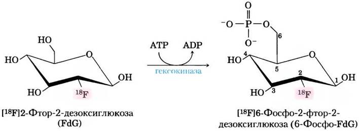
Fig. 3. Detection of tumor tissue using positron emission tomography (PET). An adult male patient underwent surgical removal of a primary Skin tumor (malignant melanoma). Left: whole-body computed tomography (CT scan) showing the layout of soft tissues and bones. Center: PET results following the administration of 18F-labeled 2-fluoro-2-deoxyglucose (FDG). Dark spots correspond to regions of active glucose uptake. As expected, high signal intensity is observed in the brain and Urinary Bladder, because the brain is the primary consumer of glucose in the body, and 18F-labeled 6-phospho-FDG is excreted via urine. Mapping the PET signal intensity to a pseudocolor scale (increasing from green through yellow to red) and overlaying this image onto the CT scan reveals the presence of cancer cells in the upper spine, Liver, and certain Muscles (right) due to metastasis of the primary malignant tumor.

Impaired Cellular Glucose Uptake in Type 1 Diabetes Mellitus
Glucose metabolism in mammals is limited by the rate of cellular glucose uptake and its subsequent phosphorylation by hexokinase. Glucose Transport from the bloodstream is mediated by the GLUT family of glucose transporters (see Table 11-3). Glucose transporters in hepatocytes (GLUT1, GLUT2) and brain Neurons (GLUT3) reside permanently in the plasma membrane. In contrast, the primary glucose transporter in Skeletal Muscle, cardiac muscle, and adipose tissue (GLUT4) is sequestered in small intracellular vesicles and translocates to the plasma membrane only in response to insulin signaling (Fig. 14-9). The mechanism of insulin signal Transduction was discussed in Chapter 12 (see Fig. 12-16). Thus, in skeletal muscle, Heart, and adipose tissue, glucose uptake and metabolism depend on adequate insulin production by pancreatic β-cells in response to rising blood glucose levels (see Fig. 23-27).
Fig. 14-9. Carbohydrate and Fat Metabolism in adipocytes during Type 1 diabetes. Normally, insulin triggers the insertion of GLUT4 glucose transporters into the plasma membrane through the fusion of GLUT4-containing vesicles with the membrane, thereby enabling glucose uptake from the bloodstream. When blood insulin levels drop, GLUT4 is internalized back into vesicles via endocytosis. In insulin-dependent diabetes (Type 1 diabetes), this normal sequence of events is disrupted. The absence of insulin prevents GLUT4-mediated glucose uptake; as a result, cells are starved of glucose while it accumulates in excess in the blood. The shortage of glucose for energy production forces adipocytes to break down triacylglycerols stored as lipid droplets and supply the resulting fatty acids to other tissues for mitochondrial ATP production. The liver converts these into two byproducts (acetoacetate and β-hydroxybutyrate, see p. 252), which accumulate and are released into the blood. While these serve as an energy source for the brain, they also lower blood pH, causing ketoacidosis. The same sequence of events occurs in muscle tissue, except that myocytes do not store triacylglycerols; instead, they utilize fatty acids released into the bloodstream by adipocytes.

In Type 1 diabetes mellitus (also known as insulin-dependent diabetes), the number of β-cells is severely depleted, and consequently the body fails to produce enough insulin to stimulate glucose uptake in skeletal muscle, cardiac muscle, and adipose tissue. Following a carbohydrate-rich meal, blood glucose levels rise to abnormally high values—a condition known as hyperglycemia. Unable to take up glucose, muscle and adipose tissues resort to utilizing fatty acids derived from stored triacylglycerols. In the liver, the acetyl-CoA generated from these fatty acids is converted into Ketone Bodies (acetoacetate and β-hydroxybutyrate), which are exported to other tissues to serve as an alternative energy source (Chapter 17). These substances play a crucial role in the brain, which relies on ketone bodies as an alternative fuel during glucose deprivation. (Free fatty acids cannot cross the blood-brain barrier and thus cannot serve as an energy source for brain neurons.)
If left untreated in patients with Type 1 diabetes, the Excessive production of acetoacetate and β-hydroxybutyrate leads to their accumulation in the blood, accompanied by a drop in blood pH and life-threatening ketoacidosis. Insulin injections reverse this process: GLUT4 translocates to the Plasma Membranes of hepatocytes and adipocytes, glucose is taken up and phosphorylated, blood glucose levels drop, and ketone body production subsides.
Diabetes dramatically alters both carbohydrate and Lipid Metabolism. We will revisit this topic in Chapter 23, following our discussion of lipid metabolism (Chapters 17 and 21). ■
Summary of Section 14.1 Glycolysis
■ Glycolysis is a nearly universal pathway in which a glucose molecule is oxidized to yield two molecules of pyruvate, conserving energy in the form of ATP and NADH.
■ All 10 glycolytic enzymes reside in the cytosol, and all 9 intermediates are phosphorylated compounds containing either six or three carbon atoms.
■ In the preparatory phase of glycolysis, two molecules of ATP are consumed to convert glucose into fructose-1,6-bisphosphate. Cleavage of the bond between C-3 and C-4 yields two triose phosphate molecules.
■ In the payoff phase of glycolysis, each glyceraldehyde-3-phosphate molecule derived from glucose undergoes oxidation at the C-1 carbon. The energy released in this reaction is conserved as one molecule of NADH and two molecules of ATP per oxidized triose phosphate. The overall equation for glycolysis is:
Glucose + 2 NAD+ + 2 ADP + 2 Pi —> 2 pyruvate + 2 NADH + 2 H+ + 2 ATP + 2 H2O
■ Glycolysis is tightly regulated in coordination with other energy-yielding pathways to ensure a steady intracellular ATP supply.
■ In Type 1 diabetes, impaired glucose uptake by muscle and adipose tissues profoundly impacts both carbohydrate and fat metabolism.
Last update: 06/08/2026
Editorial and Educational Adaptation: This material has been compiled based on the primary/original source text. The project team performed an editorial review, corrected technical inaccuracies, structured sections, and adapted the content for an educational format.
What was processed:
- elimination of formatting defects (OCR errors, structural breaks, corrupted characters);
- editorial organization of content;
- standardization of terminology in accordance with academic sources;
- verification of factual statements against the original source text.
All mentions of the author, publication year, and origin of the primary text have been preserved in accordance with the source.