Chordate Zoology - textbook - Y. V. Tsaryk - 2013
Chapter 4. DIVISION GNATHOSTOMATA, or ECTOBRANCHIATA. SUPERCLASS PISCES
4.2. CLASS BONY FISHES OSTEICHTHYES
4.2.1. Features of Organization
Appearance. Body shape depends on living conditions, and its variations are far more numerous than in other Chordates (Fig. 4.19). Torpedo-like, streamlined contours combined with The flexibility of fins help bony Fishes overcome the resistance of the dense aquatic environment, allowing them to conserve energy when traveling long distances.
Pelagic coloration is typical for fish, featuring a smooth transition from dark shades on the dorsal side to light shades on the ventral side. When foraging or finding breeding grounds are secondary in their life strategy, such as in coral reef ecosystems, The Need for camouflage and shelter leads to the existence of fish that are visually bizarre and striking. Despite this great diversity, bony fishes share common structural features and physiological processes.
Class="center">
Fig. 4.19. Body shape of teleist fishes
Integument. The Stratified Epithelium contains numerous unicellular glands that secrete mucus for protection against Bacteria, pheromones, and alarm substances ("alarm substance"). Beneath the epithelium lies the Connective Tissue, the corium (Fig. 4.20). Both layers contain pigment Cells called chromatophores, which can change shape and depth within the Skin in response to signals from the Medulla Oblongata. This allows many fish to camouflage themselves against their surroundings (gobies, flatfishes). Due to hormonal regulation, some fish acquire bright breeding coloration during the spawning season (sticklebacks, salmonids, cyprinids, etc.), which helps facilitate the simultaneous maturation of Gametes.
Most bony fishes are covered with scales. Scales are dermal bones and develop in special pockets within the corium. Their free edges project beyond the corium and overlap adjacent plates. Evolutionarily primary bony scales of the rhomboid type (cosmoid and ganoid) are often called plates because they were laid down in the skin parallel to the body surface, tightly abutting at the edges. Such an integument resembled armor and partially restricted body flexibility. Rhomboid cosmoid scales are partially preserved in coelacanths and bichirs. Representatives of some modern but ancient lineages possess ganoid scales, in which the cosmine layer within the bony plate is thin (Fig. 4.21). Elasmoid scales, characteristic of evolutionarily younger systematic groups (also present in bowfins, coelacanths, and lungfishes), consist solely of isopedine. Scales grow throughout life; new thin layers, called sclerites, are periodically added from the base. During periods of poor feeding (spawning, illness, or unfavorable climatic conditions), sclerite formation slows down, forming dense, less transparent rings on the bony plate.
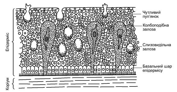
Fig. 4.20. Skin of a teleost fish
The Skeleton of bony fishes consists of endochondral bones, formed by the replacement of Cartilage, and dermal bones, formed in the corium (present in the Skull, Pectoral Girdle, and other structures). Some bones are of mixed origin. Certain PARTS OF THE skeleton may remain cartilaginous.
In coelacanths, lungfishes, and acipenseriforms, the notochord Functions as a support throughout life (Fig. 4.22). Representatives of the remaining taxa of the class are characterized by amphicoelous bony vertebrae (Fig. 4.23). Their neural arches form the neural canal. Ribs are attached to the transverse processes of the trunk vertebrae; the processes of the caudal vertebrae form the hemal canal (see Fig. 4.23). The intervertebral space is filled with remnants of the notochord. The vertebrae are connected to each other by articulating processes located at the Base of the neural spines. In cypriniforms and siluriforms, the first four trunk vertebrae form the Weberian apparatus, which transmits pressure Changes in the swim bladder to the labyrinth of the Inner ear (Fig. 4.24).

Fig. 4.21. Structure of teleost fish scales (A - cosmoid; B - ganoid; C-E - bony; D - ctenoid bony; E - cycloid bony):
1 - cosmine layer; 2 - Cells of the cosmine layer; 3 - cells of the spongy layer; 4 - ganoin layer; 5 - isopedine (bony) layer;
6 - center of the scale; 7 - sclerites of annual growth; 8 - annual ring; 9 - primary radii; 10 - hyalodentine Teeth;
11 - secondary radii (not originating from the center of the scale)

Fig. 4.22. Axial Skeleton of a sturgeon
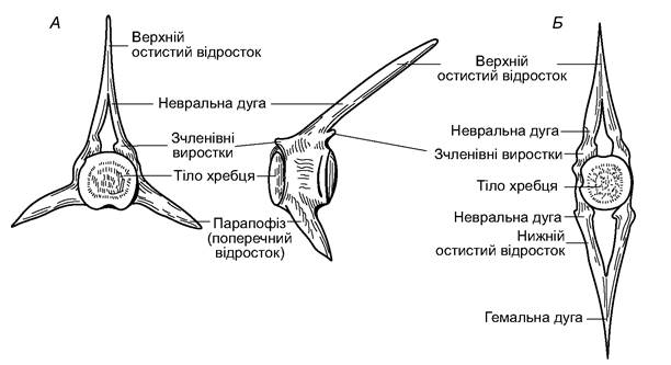
Fig. 4.23. Vertebrae of a bony fish:
A - trunk vertebra; B - caudal vertebra
The skull of bony fishes is divided into the neurocranium and visceral regions.
The neurocranium (axial skull) of acipenseriforms is cartilaginous; ossification of individual fragments is possible only in old fish. Externally, the cartilage is covered by a continuous armor of dermal bones.
The neurocranium of the teleost skull contains over 20 replacing (cartilage) bones and about 10 dermal bones (Fig. 4.25). The occipital bones—basioccipital (basioccipitale), paired exoccipitals (occipitale laterale), and supraoccipital (supraoccipitale)—frame the foramen magnum; five otic bones (otici) on each side, and the sphenoid bones—orbitosphenoid (orbitosphenoideum), basisphenoid (basisphenoideum), and laterosphenoid (laterosphenoideum)—form the lateral walls of the braincase and the Orbit; anteriorly, in the olfactory region, the unpaired mesethmoid (mesethmoideum) and paired ectethmoid (ectoethmoideum) bones are distinguished; the skull roof is formed by the paired nasals (nasale), frontals (frontale), and parietals (parietale); the floor is supported by the unpaired parasphenoid (parasphenoideum) and Vomer (vomer). The orbital cavities are separated by a thin, often incomplete septum. The Brain is located posteriorly. Such a skull is called tropibasic (with a narrow base), in contrast to the platybasic skull (with a wide base, in which the brain lies between the orbits) characteristic of cartilaginous fishes.
There are even more individual bones in the splanchnocranium (visceral skeleton). They form the visceral arches (mandibular, hyoid, five branchial) and opercula. The primary upper jaw consists of the palatine (palatinum), metapterygoid (metapterygoideum), and quadrate (quadratum) bones, formed by the Ossification of the palatoquadrate cartilage; the secondary upper jaw is formed by the dermal ectopterygoid (ectopterygoideum) and endopterygoid (entopterygoideum), Maxilla (maxillare), and premaxilla (praemaxillare) bones; the lower jaw consists of the dentary (dentale), articular (articulare), and angular (angulare) bones. The hyoid arch is formed by paired main bones: the hyomandibula (hyomandibulare), which articulates with the otic region of the neurocranium, and the hyoid (hyoideum), as well as smaller elements—the symplectic (symplecticum) and the ossified interhyal (interhyale). There are often several ossification Zones of the hyoid, resulting in The formation of the epihyal (epihyale), ceratohyal (ceratohyale), two hypohyals (hypohyale), and the unpaired basihyal (basihyale). Long, curved branchiostegal rays of dermal origin (radii branchiostegii) are attached to the ceratohyal and epihyal bones.

Fig. 4.24. Weberian apparatus of the crucian carp: A - general view; B - diagram

Fig. 4.25. Diagram of the skull structure of bony fishes:
1 - basioccipital bone; 2 - exoccipital; 3 - supraoccipital; 4 - otics; 5 - basisphenoid; 6 - pterosphenoid;
7 - orbitosphenoid; 8 - mesethmoid; 9 - ectethmoid; 10 - parietal; 11 - frontal; 12 - nasal; 13 - parasphenoid; 14 - vomer;
15 - palatine; 16 - quadrate; 17 - pterygoids; 18 - premaxilla; 19 - maxilla; 20 - articular; 21 - dentary; 22 - angular;
23 - hyomandibula; 24 - symplectic (ligament); 25 - accessory bone; 26 - hyoid; 27 - basihyal (copula of the hyoid arch); I-V branchial arches
By the method of attachment of the splanchnocranium to the neurocranium, the skull is hyostylic (the exception is lungfishes, which exhibit autostyly).
In most species, teeth are present on the mandibular arch. They can also be found on the vomer, branchial arches, and other structures bordering the Oral Cavity.
In the paired fins of some ancient bony fishes, as in cartilaginous fishes, basal and radial elements are present (Fig. 4.26). Their arrangement can be biserial (radials on both sides of a series of basal elements, as in Neoceratodus and fossil lungfishes) or uniserial (radials on only one side, as in lobe-finned fishes). Other bony fishes lost basals during evolution. Pelvic fins also lack radials. The free web of paired and unpaired fins is formed by lepidotrichia—bony rays of dermal origin. In some fishes, lepidotrichia are modified into strong, sharp spines, which may be associated with a venom gland. Thus, the fin skeleton can perform a protective function.

Fig. 4.26. Pectoral fins of bony fishes:
A - biserial; B, C — uniserial
The embedded part of the skeleton of unpaired fins is formed by cartilaginous or bony rays pointed downward—pterygiophores (Fig. 4.27).

Fig. 4.27. Fragment of the skeleton of an unpaired fin
The pectoral girdle consists of the primary pectoral girdle, formed by the scapula and coracoid (small bones that play The Role of the lost basals), and the secondary girdle (cleithrum, supracleithrum, posttemporale), which is more massive and attached to the neurocranium. The Pelvic Girdle is represented only by an unpaired bony plate.
The caudal fin of bony fish larvae has a symmetrical structure (its axis is formed by the notochord) and is called protocercal (Fig. 4.28). In acipenseriforms, as in cartilaginous fishes, the tip of the tail bends upward, and the ventral lobe expands, forming an asymmetrical, heterocercal fin. In most bony fishes, the ventral lobe is even larger, giving the fin a symmetrical external appearance, but the end of THE Vertebral Column extends into the upper lobe—a homocercal type of structure. In some lungfishes, lobe-finned fishes, and teleosts, the axial skeleton straightened again during ontogeny, and its upper and lower lobes became secondarily symmetrical; this type of structure is called diphycercal. In a homocercal caudal fin, its ventral lobe is supported by hypurals—elongated and flattened hemal spines of the last vertebrae.

Fig. 4.28. Skeleton of the caudal fin
Muscular System. Somatic striated Muscles of bony fishes are segmented. The segments (myomeres) are separated by connective tissue myosepta (Fig. 4.29). There are also differentiated Muscle groups: ocular, epibranchial and hypobranchial, and paired fin muscles. Visceral Muscles surrounding the digestive tract consist of smooth muscle fibers. In the region of the mandibular and branchial arches, smooth muscles are replaced by striated ones.

Fig. 4.29. Internal anatomy of a female perch
Digestive System. The structure of the digestive system in bony fishes is highly variable, reflecting their adaptation to feeding on A wide variety of food. Three regions are distinguished in the digestive tract: the anterior region includes the oral cavity, Pharynx, and Esophagus; the middle region comprises The Stomach, Small Intestine, and digestive glands (The Liver and Pancreas); and the posterior region is represented by the hindgut.
The oral cavity contains glands that secrete mucus, which facilitates swallowing but does not contain Enzymes. The pharynx narrows as it transitions into the esophagus; the stomach is not clearly demarcated externally from the esophagus, and its walls contain glands that produce Hydrochloric acid and Pepsin. Cyprinids, gobies, and certain representatives of other families lack a stomach. In these species, protein breakdown occurs with the help of Trypsin in the small intestine. The enzymes produced by the digestive glands and the intestinal mucosa function effectively in an alkaline environment.
At the beginning of the intestine in bony fishes of various orders, there are pyloric caeca (see Fig. 4.29), which increase the absorptive surface area (ranging from one in loaches to several hundred in salmonids). Phylogenetically ancient systematic groups possess a spiral valve in their intestine. To increase the active surface area in other fishes, the inner surface of the intestine has numerous folds and longitudinal ridges. The length of the digestive tract in predatory fishes is generally shorter than in non-predatory, especially herbivorous, species (0.5-2.0 versus 2.0-15.0 of body length). The intestine terminates at the anus, and only lungfishes possess a cloaca.
The liver is relatively smaller than in cartilaginous fishes (1.5-8.0% of body weight). The pancreas is dispersed as individual lobes within the mesentery walls and the intestinal loops.
The dietary spectrum of bony fishes is extremely broad, encompassing almost all Water/8.html">LIFE FORMS OF aquatic organisms—from unicellular Algae to higher vertebrates—primarily due to the diverse structure of their mouthparts. The Mouth can be armed with teeth, grasping, protrusible, forceps-like, with sharp cutting jaws, or with powerful crushing jaws, among others. In paddlefishes (Polyodontidae), it is so wide that the fish resembles a bizarre barrel with a tail, slowly gliding through the water column and filtering food. Plankton feeders typically have well-developed, numerous gill rakers that form a kind of sieve. Herbivorous fishes often host symbionts (Protozoans, Fungi, and bacteria) in their intestines to help break down Cellulose.
The daily ration of fish, depending on their physiological state, age, and other conditions, can range from 0.2 to 30.0% of their body weight. Predatory fish may go without food for several days or even weeks after a successful hunt. During spawning Migrations, some fishes do not feed at all; in salmon of the genus Oncorhynchus and in freshwater eels, for example, the intestine even undergoes degeneration (Fig. 4.30).
Respiratory system. Gas Exchange in bony fishes involves the gills, the body surface, and the walls of the digestive tract. The gills are the primary respiratory organ. They are located on the first to fourth gill arches. The interbranchial septa are reduced (remnants are present in Acipenseriformes). Rows of gill filaments sit in pairs on the bony arches, fused at their bases. Gill Arteries run along the arches and branch into the filaments, forming a capillary network. The arches are concealed beneath the opercula. In some fish, an accessory pseudobranch may be located on the inner side of the operculum.
The gill filaments have thin transverse folds on their surface—up to 15 per millimeter, which increases their surface area (overall, there are 1-3 cm2 of filament surface area per 1 g of fish body mass). During the passage of water through the gills, the distal ends of the filaments meet, forming a canopy. Blood flows through the capillaries in a countercurrent direction to the water flow. All of this allows the fish to extract 46 to 82% of the dissolved oxygen from the water passing through the gills, remove over 90% of carbon dioxide from the capillary blood, and ensures water-salt exchange and the elimination of metabolites.
The integument also participates in gas exchange (typically accounting for 4 to 30% of the total volume; in loaches and some other species, up to 85%).

Fig. 4.30. Stages of intestinal reduction in chum salmon Oncorhynchus keta during spawning migration
If dissolved oxygen in the water is insufficient, certain fish species can utilize atmospheric oxygen. The simplest case is gulping air at the surface to aerate the water in the oral cavity. This is done by crucian carp, minnows, and other inhabitants of shallow, small water bodies. Loaches, spined loaches, and gudgeons force the air bubble gulped by the mouth into the intestine, where they have a region adapted for gas exchange capable of absorbing up to 50% of the oxygen from the bubble. Paradise fish, gouramis, and other labyrinth fishes possess specialized, folded cavities above the gills connected to the pharynx, where up to 70% of atmospheric oxygen is absorbed, making the gills play a secondary role in respiration. Similar adaptations have been found in snakeheads (Perciformes), swamp eels (Synbranchiformes), and certain species of Siluriformes and Clupeiformes.
The swim bladder, In addition to performing hydrostatic (as well as baroreceptive and acoustic) functions, also participates in gas exchange.
Crossopterygians and lungfishes possess Lungs—paired, cellular sacs whose capillary network is connected to the last pair of gill arteries. The duct connecting them to the esophagus originates from the ventral side of the esophagus and remains functional throughout life. Usually, the lungs serve only as an accessory respiratory organ, but when water bodies dry up, Lepidosiren and Protopterus rely solely on them.
The swim bladder of other bony fishes develops as a dorsal outgrowth of the esophagus; the capillaries surrounding it are connected to the intestinal artery. Bony ganoids, such as the bowfin and gar, obtain up to 60-80% of their required oxygen from it, thanks to the Structural Features of the bladder walls.
Dissolved oxygen in water is typically sufficient to meet the needs of fish. However, during the night in slow-flowing, overgrown, and silted water bodies, fish may experience Hypoxia, especially during algal blooms—a rapid growth of unicellular and colonial algae (in the dark, Photosynthesis does not offset the oxygen consumed by autotrophic respiration). In winter, under ice cover, in addition to oxygen deficiency, hypoxia can be caused by increased concentrations of carbon dioxide, hydrogen sulfide, methane, and other organic decomposition products. Fish are classified according to their sensitivity to water aeration. 'Oxyphilic' species inhabit fast-flowing streams, such as trout, bullhead, common minnow, riffle minnow, etc. Significant oxygen deficits can be tolerated by crucian carp, loach, lake minnow, and Chinese sleeper.
Circulatory system. The differences in the STRUCTURE OF THE circulatory system between cartilaginous and bony fishes are minor. All bony fishes possess a sinus venosus, an atrium, and a ventricle (Fig. 4.31). The conus arteriosus with semilunar Valves is retained in crossopterygians, lungfishes, Acipenseriformes, Lepisosteiformes, and Polypteriformes. In the rest, to prevent the rupture of the initial section of the vascular bed due to cardiac contractions, the bulbus arteriosus is used, which is no longer considered part of The Heart. Only the valves remain from the previous structure (Fig. 4.31, B). Blood pressure in bony fishes is slightly higher: 20-120 mmHg compared to 7-45 mmHg in cartilaginous fishes. There are four pairs of afferent and efferent branchial arteries remaining. After the carotid arteries branch off from the roots of the dorsal aorta, the efferent branchial arteries close to form a characteristic circle. In many species, the renal portal System of the right Kidney is partially reduced, and some blood from the right portal vein flows directly into the right posterior cardinal vein (Fig. 4.32).

Fig. 4.31. Compartments of the fish heart: A - cartilaginous; B - bony
There are many deviations from the general scheme. The most peculiar differences are found in lungfishes, due to The Emergence of the Pulmonary Circulation. A septum is developed in their atrium, which extends as a fold through the ventricle to the beginning of the conus arteriosus. Blood from the pulmonary vein enters the left part of the atrium, while blood from the sinus venosus enters the right. The septum and fold direct the blood so that the more oxygenated blood from the lung primarily enters the anterior afferent branchial arteries, becomes further oxygenated, and goes to the brain, while the more deoxygenated blood passes through the Vessels of the posterior gill arches to other Organs of the body and enters the lung via the pulmonary artery. Lungfishes have five, rather than four, pairs of afferent and efferent branchial vessels.
The blood volume, depending on locomotor activity, can range from 1.1 to 7.3% of body weight. THE RED BLOOD Cell count ranges from 600,000 to 4.1 million per 1 mm3; they are oval and nucleated. Cellular blood elements are formed in the anterior kidney and the Spleen. In some representatives of the notothenioids from the family Chaenichthyidae, the blood contains neither erythrocytes nor Hemoglobin; instead, they have a highly developed capillary network in the skin and fins (up to 45 mm of capillary length per 1 mm2 of surface area).
Excretory system. The excretory system proper of bony fishes is represented by paired mesonephric (trunk) Kidneys, although the skin, gills, digestive tract, and liver are also involved in excreting Protein METABOLISM products and maintaining the physicochemical balance of the body. The Significance of any given organ depends not so much on the level of evolutionary development, but rather on whether the fish inhabits fresh or salt water.
The kidneys are located beneath the spine along the upper wall of the body cavity. The Wolffian ducts function as Ureters, fusing and opening through a separate slit on the urogenital papilla (into the cloaca in lungfishes); a Urinary Bladder is present (see Fig. 4.33). Typically, the kidneys, especially in freshwater fishes, feature well-developed Bowman's capsules with large glomeruli (glomerular kidneys). The end product of nitrogenous waste breakdown in ray-finned fishes is ammonia, whereas in other vertebrates, it is urea or uric acid. During estivation, lungfishes produce less toxic urea, but in their active state, they excrete ammonia.

Fig. 4.32. Diagram of the CIRCULATORY SYSTEM OF bony fishes
Osmoregulation in freshwater fish (hypotonic environment) involves The excretion of a significant amount of water by the kidneys, which constantly enters through the skin, gills, and with food (Fig. 4.33). The salt content in Tissues and blood is replenished through reabsorption in the renal tubules, active uptake by the gills, and intake with food.

Fig. 4.33. Excretory ducts of the Urogenital System of a male pike
Marine fish (hypertonic environment) lose water through the skin, gills, urine, and feces. To replenish water in the body, they drink seawater (40-200 ml per 1 kg of fish body weight per day), excreting excess salts via specialized cells in the gill filaments (Fig. 4.34).

Fig. 4.34. Diagram of osmoregulation in marine and freshwater fishes
Diadromous fish are capable of rapid physiomorphological adaptations to meet the demands of osmoregulation.
The Reproductive System of bony fishes also exhibits certain differences among various representatives. Typically, paired Gonads are attached by the mesentery to the body walls beneath the kidneys. In teleosts, the testicular tubules empty into specially formed sperm ducts, whereas in ganoids and some other ancient groups, as in cartilaginous fishes, they open into the renal tubules of the kidney (specifically, into its middle rather than anterior lobe). The lower section of the sperm duct has an expansion—the Seminal Vesicle. The genital pore is unpaired (as already mentioned, some ancient groups possess a cloaca).
In female lungfishes, as in cartilaginous fishes, the oviducts are elongated Müllerian ducts, which form a funnel at the inner end and open externally into the cloaca. In bony fishes, the Müllerian ducts are reduced, and the connective tissue wall of the gonads extends in a tube-like fashion to the genital pore. Only in some salmonids do mature eggs rupture the gonad wall and enter the body cavity, from where they exit through a short, wide oviduct.
The vast majority of bony fishes are gonochoristic, but there are many Examples of synchronous (Testes and Ovaries mature simultaneously) or sequential (testes mature first, but in older age they degenerate and ovaries develop instead) Hermaphroditism. In most cases, Fertilization is external. Sexual maturity is reached at an age ranging from two months (some cyprinodontiforms) to over 20 years (acipenseriforms). Males usually mature faster and are smaller than females.
Sexual Dimorphism is manifested not only in body size and proportions, but also in fin length and coloration. Males of species characterized by internal fertilization have elongated genital papillae (sculpins), gonopodia (cyprinodontids), etc. Often, sexual dimorphism is apparent only during the breeding season. For example, males of many cyprinid species inhabiting our waters develop "nuptial tubercles" on their heads—small, white, conical structures. However, in some species, these tubercles are also present in females. Hormonal changes in male salmonids are manifested by the elongation and hooking of the jaws.
Reproduction. The fecundity of bony fishes is much higher than that of cartilaginous ones. In very small fish, there are only a few dozen eggs. Mostly, however, the count goes into tens of thousands or millions. The record holder is the ocean sunfish, which lays up to 300 million small (0.7 mm) pelagic (freely floating near the surface) eggs. Yet, less than one percent of them have a chance to develop and survive to one year of age. The fecundity of female pikeperch, pike, and carp is up to 1 million eggs, while that of brown trout is only 200–2,000. Trout eggs are large (up to 5 mm) and incubate from October to April in gravel nests in the middle of mountain stream beds, where they face no threats at this time. However, heavy machinery can destroy these nests if, for example, the stream bed is used for timber transport.
The envelope of fertilized eggs of phytophilous and lithophilous fish remains sticky for some time after spawning, allowing the clutches to be deposited in optimal locations for further development, which increases the success of embryonic development (Fig. 4.35). However, even in such cases, due to the predation of eggs and young by other fish and invertebrates,
survival is only 1–3%. There are many examples of parental care. For example, in many species, the male guards the clutch; by fanning his fins, he improves the oxygen regime. Other species mouthbrood eggs and larvae (tilapias, cardinalfishes), carry them on their bodies (some catfishes, discus fish), or in brood pouches under the integument (pipefishes, seahorses). There are examples of using the digestive tract as an incubator (Arius catfishes). Eggs protected by the body receive oxygen from the capillaries of the parent Organism. Finally, some bony fishes exhibit ovoviviparity (swordtail, eelpout).

Fig. 4.35. Stages of Ontogeny of the bream
Most fish reproduce several times during their lives, meaning they are polycyclic (monocyclic ones include the European eel and Pacific salmon). Fish spawn annually or once every 2–5 years, timing the emergence of fry to the period of maximum development of food organisms. Spawning can be total or fractional (at intervals of 2–4 weeks). European eels, Pacific salmon (genus Oncorhynchus), and spawners of some other species from various orders die after spawning. The carcasses of adult individuals enrich the spawning grounds with nutrients, which contributes to the local improvement of the food supply for the young. For spawning to begin, the eggs and milt must be mature and "ripe." Synchrony of final maturation is ensured by Sex Hormones (specifically, Steroid Hormones), and spawning itself begins in the presence of several factors—spawning cues. These include appropriate water chemistry, Temperature, and aeration, a specific spawning substrate, and The behavior of partners. Phytophilous species lay eggs on plants, psammophilous on sand, and lithophilous on stones. Pelagic fish mostly have floating eggs (specific gravity is reduced due to oil droplets). After a more or less prolonged incubation
(from two to over 200 days), larvae hatch from the eggs, and in their free-living state, The Development of all major Organ Systems is completed, and scales are formed. Only after this does the fry period begin. Subsequently, the fish grows throughout its life without significant changes in structure, except for those associated with sexual maturation. Individual Stages of the Ontogeny of the bream Abramis brama are shown in Fig. 4.35.
The first stages of ontogeny in ovoviviparous species occur within the female's body. After the incubation period is complete, females give birth to larvae or even fry. Such "spawning" is well known to aquarists, as it is characteristic of guppies, swordtails, and other popular aquarium fish of the family Poeciliidae. The eggs, which are generally few (10–2,000), have no direct connection with the maternal organism during incubation. Only in representatives of the family Goodeidae from the mountain streams of Mexico do yolk-poor eggs develop in ribbon-like outgrowths of the ovarian walls—trophotaeniae; often only one embryo develops at a time, and the length of the newborn fry is the same as that of the mother's body.
Spawning in bony fishes is associated with more or less prolonged journeys—migrations. They allow for the rational use of the species' living space, in particular, the stocks of food organisms. In riverine fish, a distinction is made between anadromous (upstream) and catadromous (downstream) migrations. Deep-sea fish may make vertical (ascending) movements for spawning. In addition to spawning migrations, there are also feeding and wintering migrations. The total length of seasonal movements can reach several thousand kilometers. More pronounced migrations are characteristic of schooling fish.
The Nervous System and Sensory Organs are generally similar to their counterparts in cartilaginous fishes. The relative size of the brain is somewhat larger. The Forebrain (Telencephalon) is small; its roof lacks Nerve Cells, or they form only small clusters. In the Diencephalon, the Pineal Gland and Pituitary Gland are clearly visible. The Midbrain (mesencephalon), with its optic lobes, stands out among other sections (Fig. 4.36). The Cerebellum is better developed in active fish, ensuring coordination of movements and controlling the somatic muscular system. The medulla oblongata (myelencephalon) manages reflex actions and controls the Autonomic nervous system. Ten pairs of Cranial Nerves have the same function as in cartilaginous fishes.

Fig. 4.36. Brain of a bony fish: I-XII - cranial nerves
The Spinal Cord (medulla spinalis) performs the same functions as in cartilaginous fishes, but it is under greater control of the brain.
Fish orientation, in the broad sense of the term, occurs through the interaction of six Sensory systems: tactile (Touch), chemoreceptive (Smell and Taste), seismosensory (infrasound perception), auditory combined with gravitational (perception of sound waves, including ultrasound, and balance), visual, and electrosensory (analysis of magnetic and electric fields).
The receptor arcs of the tactile sense have the simplest structure. Sensory cells are arranged in small groups all over the body. On the barbels, at the tips of the fin rays, and at certain other "prominent" points, they form clusters called tactile corpuscles. These corpuscles also contain thermoreceptors with a resolution of 0.5°C.
As with all aquatic animals, information from chemical receptors—taste and smell—is of great importance to fish. The structure of the olfactory sacs ensures a constant inflow of external Water and Its analysis on the sensitive folds of their walls. Taste receptors are usually organized into so-called taste buds and are located on the walls of the oral cavity, on the barbels, and often on the outer body surface. It has been established that fish detect Organic compounds better than inorganic ones. Overall, the ability of fish to recognize chemical signals is comparable to that of dogs. Among known species, the European eel has the keenest chemical sense. Thanks to "chemical memory," fish undergoing large-scale migrations find the correct path through the water column. As for taste receptors, they function better in non-predatory fish than in predators. Predators rely more heavily on the lateral line system.
Actually, the lateral line is not an obligatory element of the seismosensory system; it is merely the most noticeable one. The essential components are the subdermal canals on the HEAD (Fig. 4.37), which open to the outside through short branches. The walls of these canals contain clusters of sensitive ciliated Hair cells innervated by the Vagus nerve (cranial nerve X). The lateral line organs can perceive vibrations with a frequency of up to 500 Hz, meaning they can detect not only infrasound but also the lower range of audible sound. The seismosensory system is particularly important for fish living in turbid water.

Fig. 4.37. Seismosensory system (lateral line organs) of the perch
The Organ of Hearing and Balance is the inner ear. Balance is monitored by the three semicircular canals of the membranous labyrinth and the utricle. Inside, they contain otoliths—"ear stones" made of calcium carbonate, which develop from crystals called otoconia and have a layered structure (Fig. 4.38). The Mechanism of the balance organ relies on the response of the sensitive processes of specialized cells to the inertial Displacement of the otolith from its static position as soon as the fish's body accelerates in any direction. The saccule with a hollow outgrowth—the lagena—is the actual Organ of Hearing. In some bony fishes, the swim bladder also participates in the perception of sound vibrations.
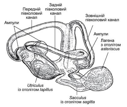
Fig. 4.38. Inner ear labyrinth of the common minnow with otoliths
Its blind outgrowths lie adjacent to the membranous "windows" of the perilymphatic cavity of the labyrinth, as in Perciformes and Gadiformes, or the contact is formed via the modified first four vertebrae (Weberian apparatus), as in Cypriniformes and Siluriformes. In both cases, the swim bladder acts as a resonator. Water conducts sound very well. For fish, the most interesting sounds are jaw-smacking and the rubbing of scales (mechanical or non-specific); also important are acoustic signals specifically generated by individuals of the same species or by neighbors belonging to other taxa. These sounds accompany behavioral acts (courtship, feeding behavior, maintaining school Organization, etc.) and are produced using the swim bladder, stiff fin rays, or articulating bones. The frequency range of the generated sounds is between 20 and 12,000 Hz, almost completely overlapping with the perception range of the hearing organs.
The eye of a bony fish has a spherical lens and a flat cornea. It is capable of perceiving light wavelengths of 380–750 nm. Accommodation is achieved by the falciform process, which adjusts THE POSITION OF the lens relative to the retina. The retina contains rods and cones, providing Color Vision. Adaptation to lighting conditions (retinomotor response), as in other vertebrates, involves pigment cells that elongate in light to shield the rods and, conversely, flatten in the dark to expose the sensitive cells. The choroid of the eye contains silvery guanine granules, which can form a reflective layer (tapetum lucidum) on the inner surface to enhance vision. The quality of Visual Perception depends on the fish's habitat conditions and ensures reliable orientation at close range (up to 10 m). Fish that constantly live in low-light conditions have enlarged, protruding (telescopic) eyes and a well-developed tapetum. In other dwellers of the dark, the eyes may be partially reduced or hidden under the skin (family Amblyopsidae).
The Cytology/practical/76.html">Cornea of the four-eyed fish Anableps tetrophthalmus from Central America, which feeds on flying insects, is divided in two by an epithelial band. This creates the illusion of double eyes. In reality, there is only one lens in each eye; its upper part is flatter, allowing for better vision in the air. Such eyes facilitate the simultaneous monitoring of the situation both in the water and in the air.
Bony fishes are sensitive to changes in electromagnetic fields, which they detect using modified Skin glands. Low field strength prompts the fish to align their bodies along the field lines.
Electromagnetic fields are generated around the bodies of all fish, but they are weak. In schooling fish, a field-merging effect is observed, creating a collective field that significantly influences the coordinated movement of school members. Orientation using electric fields can be even more effective if the fish possess specialized electric organs to generate electromagnetic pulses. If the power of these generators is increased, they can also be used for defense or attack.
Electric organs are most commonly modified striated muscles. Muscle contraction is always accompanied by the release of a small amount of electrical energy. The orderly arrangement of electrogenic elements (cells) makes them similar to capacitors. The fish is capable of regulating the strength and frequency of the electrical discharges. The power of these organs depends on the total volume of the modified Introduction/39.html">Muscles and their level of specialization. For example, the voltage of the field generated by the electric eel (genus Electrophorus) can exceed 700 volts, with pulsed discharges at a frequency of up to 50 per second.
Some fish possess electric organs of a different nature. In particular, in the electric catfish (genus Malapterurus), they are formed by skin glands.
The tendency of fish to move toward the anode under certain electric field parameters is utilized in electrotrawling. The device known to many as an "electrofisher", often used by poachers, operates on a slightly different principle: in addition to the attractive property of the anode, it uses high-voltage pulses with a frequency of about 70 Hz, which cause spasms in the somatic muscular system at close range. The immobilized fish is then collected with an anode net. When used correctly, an electrofisher is an effective research tool. It
causes less harm to the fish than netting when it needs to be weighed, measured, and released unharmed. The spasm passes in a few seconds, and physiological parameters quickly return to normal. However, if the exposure to the electric field lasts longer than necessary, or if its parameters are incorrectly configured, the fish may die.
Last update: 19/08/2026
Editorial and Educational Adaptation: This material has been compiled based on the primary/original source text. The project team performed an editorial review, corrected technical inaccuracies, structured sections, and adapted the content for an educational format.
What was processed:
- elimination of formatting defects (OCR errors, structural breaks, corrupted characters);
- editorial organization of content;
- standardization of terminology in accordance with academic sources;
- verification of factual statements against the original source text.
All mentions of the author, publication year, and origin of the primary text have been preserved in accordance with the source.