Human Anatomy (with the Fundamentals of Dynamic and Sports Morphology) - Ivanitsky, M. F. 2008
The Doctrine of the Nervous System
Central Nervous System
The spinal cord is a part of the Central Nervous System located within the vertebral canal. In the embryo, the length of the spinal cord corresponds to that of the trunk. However, as development proceeds, the growth of the spinal cord lags behind that of the trunk; consequently, by the time of birth, its lower end lies at the level of the 3rd lumbar vertebra, and in adults, at the level of the 1st–2nd lumbar vertebrae.
The spinal cord appears as a cord-like Structure, somewhat flattened in the anteroposterior direction and terminating in the lumbar region as the conus medullaris (see Fig. 104), which is prolonged inferiorly by the filum terminale. The spinal cord is composed of Nerve Cells, nerve fibers, and neuroglia, with the cells forming its Gray matter located internally and the fibers forming the White matter located externally (Fig. 105). The spinal cord consists of 31 segments that share a common structural plan: 8 cervical, 12 thoracic, 5 lumbar, 5 sacral, and 1 coccygeal.
Class="center">
Fig. 105. Scheme of a transverse section of the spinal cord with the pia mater and denticulate ligament:
1 — posterior median sulcus; 2 — posterior septum; 3 — posterior intermediate sulcus; 4 — posterior lateral sulcus and posterior ROOT of spinal n.; 5 — thoracic Column (thoracic Nucleus); 6 — Cells of the lateral horn; 7 — posterolateral and posteromedial nuclei of the anterior horn; 8 — central nucleus; 9 — anterolateral and anteromedial nuclei; 10 — central intermediate gray matter; 11 — white commissure; 12 — anterior spinal a.; 13 — anterior median fissure; 14 — anterior root of spinal n.; 15 — anterior corticospinal (pyramidal) tract; 16 — anterior proper fasciculi; 17 — anterior horn; 18 — lateral proper fasciculi; 19 — lateral horn; 20 — anterior spinocerebellar tract; 21 — lateral corticospinal (pyramidal) tract; 22 — posterior spinocerebellar tract; 23 — denticulate ligament; 24 — pia mater of the spinal cord; 25 — posterior horn; 26 — posterior root; 27 — cuneate fasciculus; 28 — gracile fasciculus (after Rauber)
On each side, the following structures are associated with a spinal cord segment (Fig. 106):
1. Two roots of the Spinal Nerves, anterior and posterior. The anterior root consists of the processes of motor cells in the anterior horns (Fig. 106) and the processes of cells in the lateral horns of the spinal cord; the posterior root consists of the processes of sensory nerve cells located in the intervertebral ganglion.
2. The spinal ganglion, which houses The Cell bodies of the sensory Neurons of the spinal nerves.
3. The spinal nerve, formed by the union of the anterior and posterior roots and therefore containing both motor and sensory fibers. Thus, the spinal nerve is a mixed nerve.
4. The Branches of the spinal nerve: anterior, posterior, meningeal, and communicating.
The anterior branch contains both motor and sensory fibers; in the Thoracic nerves, it continues directly into the intercostal nerves, while in the remaining spinal nerves, it forms plexuses—cervical, brachial, lumbar, sacral, and coccygeal.
The posterior branch, also mixed in terms of its fiber composition, passes posteriorly and participates in the Innervation of the Muscles and Skin of the back.
The communicating branch serves to connect the nerve segment with the ganglia of the Sympathetic trunk belonging to the autonomic system, which lie along THE Vertebral Column (see p. 331).
The meningeal branch runs toward the spinal cord to supply its dura mater. The spinal nerve roots emerging from the inferior part of the spinal cord extend downward to form the cauda equina. In the center of the cauda equina runs the aforementioned terminal filament (filum terminale), which extends downward from the apex of the medullary cone along the vertebral canal.

Fig. 106. Scheme of the formation and branching of a spinal nerve:
1 — segment of the spinal cord; 2 — posterior root; 3 — spinal ganglion; 4 — spinal n.; 5 — posterior branch; 6 — anterior branch; 7, 8 — n. n., innervating the skin; 9 — communicating branch; 10 — meningeal branch; 11 — anterior root (after Rauber)
The spinal cord features two enlargements: the cervical and lumbosacral enlargements (see Fig. 104). The cervical enlargement extends from the level of the 2nd cervical to the 2nd thoracic vertebra, reaching its maximum thickness at the level of the 5th–6th cervical vertebrae; the lumbosacral enlargement begins at the level of the 10th thoracic vertebra and attains its greatest dimensions at the level of the 12th thoracic vertebra. In these regions, the largest spinal nerves arise from the spinal cord to supply the upper and lower limbs.
The structure of the spinal cord is symmetrical: it is divided into two halves, right and left. Its anterior and posterior surfaces bear longitudinally oriented depressions (see Fig. 105). The anterior depression is termed the anterior median fissure, and the posterior one is the posterior median sulcus. The anterior median fissure is considerably deeper and wider. In addition, each half of the spinal cord exhibits lateral sulci, anterior and posterior. The anterior roots emerge through the anterior sulcus, and the posterior roots enter through the posterior sulcus, numbering 31 pairs on each side. According to the position of the sulci, the WHITE MATTER OF the spinal cord is divided into funiculi (cords): anterior, lateral, and posterior. The anterior funiculus lies between the anterior median fissure and the anterior lateral sulcus; the lateral funiculus lies between the anterior and posterior lateral sulci; and the posterior funiculus lies between the posterior lateral and posterior median sulci. The funiculi of the spinal cord's white matter contain ascending or descending bundles of nerve fibers. They conduct impulses from one segment of the spinal cord to another, or from the spinal cord to the Brain and vice versa, thus forming the conduction PATHWAYS OF THE spinal cord. Their course will be described in greater detail on page 302.
A transverse section of the spinal cord reveals that the gray matter has a characteristic shape, commonly compared to the letter H or a butterfly with spread wings (Fig. 105). Throughout the length of the spinal cord, it forms three columns: anterior, posterior, and lateral. Corresponding to these columns, the gray matter is subdivided into the anterior, posterior, and lateral horns of the spinal cord. Furthermore, the anterior and posterior horns are present in all segments of the spinal cord, whereas the lateral horns are found only in the thoracic and upper lumbar segments. The right and left halves of the gray matter are interconnected by the central intermediate (gray) substance. In addition, a white commissure connects the anterior funiculi of the spinal cord and lies anterior to the central intermediate substance. Running through the center of this substance is the central canal, which represents the remnant of the embryonic neural tube lumen.
Within the gray matter of the spinal cord, nerve cells form clusters known as nuclei. The posterior horn contains the proper Nucleus of the posterior horn and the thoracic nucleus. The anterior horn houses six nuclei: posterolateral, posteromedial, anterolateral, anteromedial, central, and retroposterolateral (or retro-posterolateral). These six nuclei contain motor nerve cells that innervate the somatic musculature.
The clusters of nerve cells in the lateral horn, which form the lateral intermediate column throughout the thoracic and upper lumbar segments, constitute the central part of the Autonomic (vegetative) nervous system, specifically its sympathetic division (for details, see p. 330). Analogous sacral parasympathetic nuclei form the central part of the Parasympathetic division of the Autonomic Nervous System.
Brain
The brain is housed within the cranial cavity and has a spheroid shape (see Fig. 104). By mass, it exceeds the spinal cord by 50 times.
The weight of the human brain averages 1,375 g (ranging from 1,000 to 2,200 g). No correlation has been established between brain mass and a person's level of intelligence or talent. Highly gifted individuals can be found among those with both large and small brain masses. For example, the brain of Ivan Turgenev weighed over 2,000 g, whereas Anatole France's brain weighed about 1,000 g.
Based on data concerning the evolutionary METABOLISM/13.html">History of the brain, it is generally divided into five main parts.
2. Hindbrain (metencephalon), which includes the Pons and the Cerebellum.
3. Midbrain (mesencephalon).
4. Diencephalon.
5. Telencephalon.
The telencephalon makes up the bulk of the brain—the cerebrum—while all PARTS OF THE brain excluding the cerebral hemispheres and the cerebellum (i.e., the diencephalon, midbrain, medulla oblongata, and pons) form the Brainstem.
The medulla oblongata lies on the clivus of the Skull base and is a direct continuation of the spinal cord. The conventional boundary between these two parts of the central national nervous system is considered to be the exit point of the rootlets of the right and left first cervical nerves. In shape, the medulla oblongata resembles a truncated cone, slightly flattened in the anteroposterior direction and expanding upward.

Fig. 107. Medulla oblongata and pons (anterior view):
1 — anterior median fissure; 2 — pyramid; 3 — olive; 4 — anterior lateral sulcus; 5 — decussation of the pyramids; 6 — middle cerebellar peduncle; 7 — pons; 8 — Basilar artery; 9 — cerebral peduncle; 10 — optic tract; 11 — posterior perforated substance; 12 — cerebellum. III — oculomotor n.; IV — trochlear n.; V — trigeminal n.; VI — abducens n.; VII — facial n.; VII' — intermediate n.; VIII — vestibulocochlear n.; IX — glossopharyngeal n.; X — vagus n.; XI — accessory n.; XII — hypoglossal n.; C — 1st cervical spinal nerve (after B. N. Tonkov)
On the anterior surface of the medulla oblongata (Fig. 107), directly adjacent to the anterior median fissure on the right and left, are the pyramids of the medulla oblongata, which are formations containing the fibers of the corticospinal tracts. In their lower part, these fibers cross to the opposite side, forming the decussation of the pyramids, which is clearly visible at the bottom of the median sulcus. Lateral to the pyramid lies an elevation known as the olive. The Hypoglossal nerve emerges from the sulcus between the pyramid and the olive. The olive contains the inferior olivary nucleus, the high development of which in humans is associated with bipedalism.
On the posterior surface of the medulla oblongata and pons lies the Rhomboid fossa, which forms the floor of the Fourth ventricle. It is divided by a longitudinal sulcus into two halves—right and left. On each half, alongside the sulcus, runs an elevation featuring a thickened region known as the facial colliculus. It is located where nerve fibers, which later become part of the Facial Nerve, course across The surface of the rhomboid fossa. Along the lateral surface of the rhomboid fossa, running from medial to lateral, are the medullary striae, where the fibers of the Vestibulocochlear nerve pass. In the lower part of the rhomboid fossa are small elevations: the hypoglossal trigone and the vagus trigone.
The first corresponds to the Location OF THE hypoglossal nerve nucleus, and the second to the location of the Vagus nerve nucleus. In its upper part, the rhomboid fossa narrows toward the cerebral aqueduct, while in its lower part it narrows toward the central canal of the spinal cord and is closed posteriorly by a plate of white matter called the obex.
Below the rhomboid fossa on the posterior surface of the medulla oblongata, on both sides of the posterior median sulcus, lie two bundles: the gracile and cuneate fasciculi. These are clusters of nerve fibers that conduct impulses of conscious kinesthetic sense (Muscle and joint sense) to the Cerebral Cortex. These bundles terminate in enlargements known as the gracile and cuneate tubercles, where synaptic Relay of the pathways occurs on neurons.
On the lateral surface of the medulla oblongata is the continuation of the lateral funiculus of the spinal cord, referred to here as the lateral funiculus of the medulla oblongata. Extending upward, the cuneate and lateral funiculi become part of the inferior cerebellar peduncle, which runs from the cerebellum to the medulla oblongata.
The medulla oblongata contains the nuclei of cranial nerve pairs IX–XII (see pp. 368, 371). At the border between the medulla oblongata and the pons lies The Nucleus of the VIII nerve. Nerves whose nuclei are located within the medulla and pons emerge in the region of the medulla oblongata (see Fig. 107). The Abducens nerve emerges between the pyramid and the pons; in the rostrolateral part of the medulla oblongata, posterior to the olive, emerge the facial and vestibulocochlear nerves, and between them is the intermediate nerve, which is usually described together with the facial nerve. Inferior to the vestibulocochlear nerve emerge the glossopharyngeal and vagus nerves, and inferior to them is the Accessory nerve. The hypoglossal nerve emerges between the olive and the pyramid.
The medulla oblongata houses the cardiac and respiratory centers, as well as the vasomotor center. In addition to gray matter, the medulla oblongata contains white matter consisting of nerve fibers that connect the medulla to other parts of the central nervous system or pass through it, linking the spinal cord with higher brain centers and vice versa.
The hindbrain consists of the pons and the cerebellum.
The anterior surface of the pons faces the clivus, which extends along the posterior surface of the body of the Sphenoid bone and the basilar part of the Occipital bone. The pons consists of A large number of nerve fibers running vertically and transversely, forming the white matter of the pons (see Fig. 107). Interspersed among these fibers are numerous clusters of gray matter that make up the pontine nuclei. The pons serves as a structure connecting the cerebellum and medulla oblongata with the cerebral hemispheres. Located within the region of the pons are the nuclei of the trigeminal, abducens, facial, and vestibulocochlear nerves (within the rhomboid fossa). The pons is connected to the cerebellum via the middle cerebellar peduncles. A sulcus runs along the midline of the anterior surface of the pons, corresponding to the course of the basilar artery. The Trigeminal nerve emerges onto the brain surface at the border between the pons and the middle cerebellar peduncle.
The cerebellum (Fig. 108) is the largest part of the hindbrain. It appears as a flattened ellipsoidal body and is divided into two lateral parts, called the cerebellar hemispheres, and a median part connecting these hemispheres, called the vermis.
Anteriorly and inferiorly, the cerebellum connects with the medulla oblongata. Superiorly, the cerebellum lies adjacent to the occipital lobes of the cerebrum, separated from them by a fold of the dura mater known as the tentorium cerebelli.
The cerebellum presents anterior and posterior margins, as well as superior and inferior surfaces.
The cerebellum is connected via the middle, inferior, and superior cerebellar peduncles to adjacent parts of the brain: the pons, the medulla oblongata, and the midbrain (see Figs. 107 and 108).
A midsagittal section through the cerebellum reveals that its central portion consists of white matter, while the periphery is composed of gray matter, known as the cerebellar cortex. The characteristic pattern formed by the white and gray matter on a midsagittal section of the cerebellum is called the arbor vitae (tree of life). Both the vermis and the cerebellar hemispheres feature transverse sulci running from one side to the other, dividing the vermis and hemispheres into distinct lobules.
A horizontal section through the cerebellum shows that nuclei, which are clusters of gray matter, are embedded within the white matter of its hemispheres. The largest of these is the dentate nucleus, which resembles a convoluted lamina.
The primary function of the cerebellum is the reflex coordination of muscle contractions and muscle groups, ensuring both the maintenance of bodily balance in various postures and the execution of complex physical movements, including athletic activities.
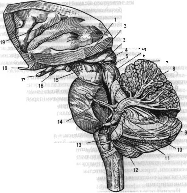
Fig. 108. Brainstem with part of the cerebellum and cerebral hemispheres:
1 - optic tract; 2 - lateral geniculate body; 3 - medial geniculate body; 4 - pulvinar of the thalamus; 5 - Pineal Gland; 6 - inferior colliculus of the midbrain tectum; 7 - cerebellar vermis; 8 - superior cerebellar peduncle; 9 - fibers projecting to the dentate nucleus; 10 - inferior cerebellar peduncle; 11 - vestibulocochlear n.; 12 - facial n.; 13 - olive; 14 - trigeminal n.; 15 - cerebral peduncle; 16 - tuber cinereum; 17 - optic chiasm; 18 - olfactory tract; 19 - insular lobe (after Braus)
The fourth ventricle is situated between the cerebellum dorsally, and the medulla oblongata and pons ventrally. Its boundaries are formed by the following structures: the floor is the rhomboid fossa, the superior-posterior wall is the superior medullary velum, and the inferior-posterior wall is the inferior medullary velum. These medullary vela give the entire cavity of the fourth ventricle a tent-like appearance. Anteriorly and superiorly, the fourth ventricle opens into the cerebral aqueduct, through which it communicates with the Third ventricle; posteriorly, an aperture in the inferior medullary velum connects the ventricular cavity with the subarachnoid space; further caudally, the cavity narrows and directly continues into the central canal of the spinal cord.
The midbrain consists of a superior (posterior) and an inferior (anterior) region.
The superior region comprises the tectum of the midbrain (quadrigeminal plate), which features four colliculi (a superior pair and an inferior pair) (Fig. 109) containing internal clusters of gray matter. Extending laterally from each colliculus are white matter elevations known as the brachia of the colliculi. One brachium runs from the superior colliculus to the lateral geniculate body and the pulvinar of the posterior thalamus, while the other runs from the inferior colliculus to the medial geniculate body. The geniculate bodies, like the posterior thalamus, belong functionally to the diencephalon rather than the midbrain. The superior colliculi serve as subcortical visual centers and act as relay stations for visual impulses. The inferior colliculi are linked to the auditory pathways and function as subcortical auditory centers.
The inferior region of the midbrain is formed by large bundles known as the cerebral peduncles. Anteriorly, they diverge to form the interpeduncular fossa. In cross-section, each cerebral peduncle exhibits a broad lower part, the crus cerebri, and a narrower upper part, the tegmentum.
Between the crus cerebri and the tegmentum lies a distinct layer known as the substantia nigra, its dark coloration resulting from the pigmentation of the local nerve cells. Fibers coursing from the cerebral hemispheres to the medulla oblongata and spinal cord pass through the crus cerebri. The tegmentum contains a prominent mass of gray matter called the red nucleus, which serves as one of the most important motor subcortical centers.
The cavity of the midbrain is a narrow channel known as the cerebral aqueduct, which connects the cavities of the third and fourth ventricles. The aqueduct is approximately 15 mm long and is surrounded by a layer of gray matter. Located ventral to the aqueduct are the nuclei of the oculomotor and trochlear nerves. The oculomotor nucleus itself consists of several subnuclei, including the nucleus of the parasympathetic division of the autonomic nervous system.
The diencephalon is located between the cerebral hemispheres and includes the thalamus and associated structures situated ventral, dorsal, and lateral to it (Hypothalamus, epithalamus, and metathalamus).
The thalamus is the largest STRUCTURE OF THE diencephalon and its principal subcortical sensory relay center. Composed of gray matter and divided into anterior and posterior portions, the thalamus forms the lateral wall of the third ventricle. Thin sheets of white matter subdivide the posterior thalamus into three main nuclear groups: anterior, medial, and ventrolateral.
Posterirolateral to the thalamus lies the metathalamus, a region comprising two geniculate bodies on each side: the lateral and the larger medial geniculate body. The medial geniculate body serves as the subcortical relay center for the Auditory pathway, whereas the lateral geniculate body Functions as that for the visual pathway.
The epithalamus includes the pineal gland (see Fig. 108), which is an endocrine organ (see p. 412).
The hypothalamus comprises the mamillary bodies, the tuber cinereum with the Pituitary Gland, and the optic chiasm.
The mamillary bodies appear as two rounded elevations about 5 mm in diameter, located in the posterior part of the hypothalamus between the tuber cinereum and the posterior perforated substance. Each mamillary body contains a cluster of gray matter organized into two nuclei: medial and lateral. The columns of the fornix terminate within the mamillary bodies.
The tuber cinereum is situated between the mamillary bodies posteriorly and the optic chiasm anteriorly. It consists of a thin layer of gray matter whose cells act as higher autonomic centers influencing thermoregulation and metabolism. The terminal lamina lies adjacent to the optic chiasm anteriorly, positioned within the recess of the longitudinal cerebral fissure between the right and left hemispheres. Laterally and superiorly, the tuber cinereum transitions into the gray matter of the cerebral hemispheres. Viewed from above—that is, from the cavity of the third ventricle—a depression leading to its apex, called the infundibulum, can be observed. Ventrally, the tuber cinereum connects to the pituitary gland.
The pituitary gland (hypophysis) is an unpaired endocrine organ, much like the pineal gland. It rests within the hypophysial fossa (sella turcica) and is covered superiorly by a fold of the dura mater. Only its posterior lobe (neurohypophysis) has a developmental origin directly tied to the brain, whereas the anterior lobe (adenohypophysis) arises as an outpouching of the pharyngeal epithelium. The pituitary gland is directly related to surrounding structures: the optic chiasm anteriorly, the tuber cinereum superiorly, and the mamillary bodies posteriorly.
The optic chiasm is formed by nerve fibers continuing from the optic nerves. These fibers undergo only a partial decussation: medial fibers cross to the opposite side, while lateral fibers continue into the optic tract of the same side.
The optic tracts are two nerve bundles originating from the optic chiasm that course posteriorly and laterally around the cerebral peduncles. They terminate in three distinct brain regions: the pulvinar of the thalamus, the superior colliculi of the midbrain tectum, and the lateral geniculate bodies.
The cavity of the diencephalon is the third ventricle.
The third ventricle is a narrow, slit-like cavity situated in the median plane between the right and left thalami. This cavity communicates with each lateral ventricle of the telencephalon via the interventricular foramen, and posteriorly with the fourth ventricle via the cerebral aqueduct. The cavity of the third ventricle is filled with CEREBROSPINAL FLUID produced by its choroid plexus.
The third ventricle presents two lateral walls, as well as a superior, anterior, inferior, and posterior wall. The lateral walls are formed by the medial surfaces of the thalami. The superior wall features the choroid plexus, which is covered above and below by a delicate layer of Epithelial Tissue. The anterior wall contains the rostral portions of the fornix, known as the columns of the fornix. Anterior to these columns lies the anterior commissure, composed of white matter, and ventral to it is the thin terminal lamina, consisting of gray matter. The inferior wall is formed by the superior surfaces of the cerebral peduncles, the posterior perforated substance located within the floor of the interpeduncular fossa, the mamillary bodies, the optic chiasm, and the tuber cinereum along with its funnel-shaped infundibulum. The posterior wall contains the posterior commissure, beneath which lies the opening leading into the cerebral aqueduct, connecting the cavities of the third and fourth ventricles.
A cluster of small nerve cells in the region of the lateral funiculi of the spinal cord, as well as in the medulla oblongata, midbrain, and diencephalon, is called the reticular formation. The reticular formation plays a crucial functional role by regulating the tone of all the aforementioned central nervous system structures and exerting an activating influence on the cerebral cortex. It is through the reticular formation that many pharmacological agents—both those that stimulate and those that inhibit activity—exert their effects on the body.
The telencephalon consists of the right and left cerebral hemispheres, which are interconnected by the corpus callosum and the fornix. Each hemisphere comprises the cerebral cortex (Pallium) and the rhinencephalon (olfactory brain). The basal nuclei and Lateral ventricles also belong to the hemispheres.
The pallium of the cerebral hemispheres features three surfaces: superolateral, medial, and inferior (Figs. 109 and 110). It is customary to distinguish specific terminal regions known as poles. These include the frontal pole—the most anteriorly projecting part of the hemispheres; the occipital pole, directed posteriorly; and, finally, the temporal pole—the most anteriorly projecting part of the temporal lobe.
A deep longitudinal fissure of the cerebrum lies between the right and left hemispheres.
On the surface of the hemispheres, the cortex forms numerous sulci, between which lie the gyri. The largest of these are the central sulcus and the lateral sulcus. Their positions serve as landmarks for subdividing the cerebral hemispheres into lobes.
Each hemisphere has four lobes: the frontal lobe, located anterior to the central sulcus; the parietal lobe, situated posterior to the central sulcus and superior to the lateral sulcus; the temporal lobe, positioned inferior to the lateral sulcus; and the occipital lobe. The boundary between the temporal and occipital lobes is conventionally considered to be a small indentation located on the Inferior surface of the brain. The boundary between the parietal and occipital lobes is the parieto-occipital sulcus, located on the Medial surface of the hemisphere, along with its imaginary continuation across the superolateral surface.

Fig. 109. Diagram of the arrangement of sulci (A) and gyri (B) on the Superolateral surface of the left cerebral hemisphere:
1 - lateral sulcus; 2 - central sulcus; 3 - superior precentral sulcus; 4 - inferior precentral sulcus (3 and 4 frequently merge into a single sulcus); 5 - superior frontal sulcus; 6 - inferior frontal sulcus; 7 - postcentral sulcus; 8 - intraparietal sulcus; 9 - superior temporal sulcus; 10 - inferior temporal sulcus; 11 - occipital sulci;
1 - precentral gyrus; 2 - superior frontal gyrus; 3 - middle frontal gyrus; 4 - inferior frontal gyrus; 5 - postcentral gyrus; 6 - superior parietal lobule; 7 - supramarginal gyrus; 8 - angular gyrus (7, 8 - inferior parietal lobule); 9 - superior temporal gyrus; 10 - middle temporal gyrus; 11 - inferior temporal gyrus (after V. N. Tonkov)
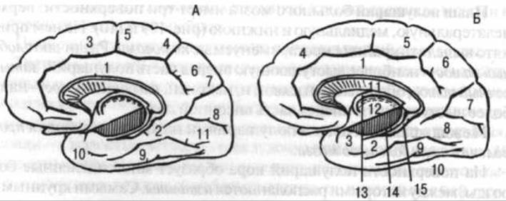
Fig. 110. Diagram of the arrangement of sulci (A) and gyri (B) on the medial surface of the right cerebral hemisphere.
The brainstem has been removed, as has the posteroinferior part of the thalamus via a deep section (its plane indicated by vertical hatching):
1 - sulcus of corpus callosum; 2 - hippocampal sulcus; 3 - peduncular sulcus; 4 - marginal ramus of cingulate sulcus; 5 - paracentral sulcus; 6 - subparietal sulcus; 7 - parieto-occipital sulcus; 8 - calcarine sulcus; 9 - collateral sulcus; 10 - rhinal sulcus; 11 - occipitotemporal sulcus; 1 - cingulate gyrus; 2 - parahippocampal gyrus; 3 - uncus; 4 - medial frontal gyrus; 5 - paracentral lobule; 6 - precuneus; 7 - cuneus; 8 - medial occipitotemporal gyrus; 9 - lateral occipitotemporal gyrus; 10 - inferior temporal gyrus; 11 - corpus callosum; 12 - thalamus; 13 - fornix; 14 - fimbria of hippocampus; 15 - dentate gyrus (after V. N. Tonkov)
The frontal lobe (see Fig. 109) exhibits the following sulci: precentral, superior frontal, and inferior frontal, between which lie the precentral, superior frontal, middle frontal, and inferior frontal gyri. Additionally, on the inferior surface of the frontal lobe, there is the olfactory sulcus, which houses the olfactory tract and olfactory bulb belonging to the rhinencephalon. This sulcus bounds the gyrus rectus (straight gyrus) laterally. The inferior surface of the frontal lobe also features orbital sulci that somewhat resemble the letter "H", with correspondingly named gyri situated between them.
The parietal lobe features the postcentral and intraparietal sulci, which delimit the postcentral gyrus, as well as the superior and inferior parietal lobules.
The temporal lobe has two temporal sulci—superior and inferior—running in an approximately anteroposterior direction. Furthermore, on the inferior surface of the temporal lobe lie the occipitotemporal and collateral sulci (see Fig. 109). Between them are located the superior, middle, and inferior temporal gyri, as well as the lateral occipitotemporal gyrus. On this same inferior surface of the temporal lobe lies the parahippocampal gyrus, situated medially to the lateral occipitotemporal gyrus, between the collateral sulcus and the hippocampal sulcus. Anteriorly, this gyrus presents a thickening known as the uncus, and posteriorly it transitions into the medial occipitotemporal gyrus.
The occipital lobe features several relatively small transverse and longitudinal occipital sulci and rather irregularly shaped gyri.
On the medial surface of the cerebral hemisphere are the callosal sulcus, located directly above the corpus callosum, and the cingulate sulcus, running approximately parallel to it. These sulci traverse all lobes of the brain (see Fig. 110). The cingulate gyrus lies between these two sulci. The cingulate, medial occipitotemporal, and parahippocampal gyri together form the limbic gyrus (fornicated gyrus).
In the posterior region of the medial surface of the hemispheres, two sulci are distinguished: the calcarine sulcus and the parieto-occipital sulcus. Between them lies a region of the occipital lobe called the cuneus. The area located anterior to it in the parietal lobe is termed the precuneus. Anterior to the precuneus, above the cingulate sulcus, lies the so-called paracentral lobule. Meanwhile, the large anterior portion of the medial surface of the hemispheres above this sulcus is occupied by the medial frontal gyrus.
At the floor of the lateral sulcus of the cerebral hemispheres lies the insular lobe, or insula (Fig. 111). This is a rudimentary fifth lobe of the brain, representing a direct continuation of the cerebral cortex.
As previously mentioned, in addition to the pallium, the hemisphere incorporates the rhinencephalon. Aside from the olfactory bulb and olfactory tract, this includes the olfactory trigone and the anterior perforated substance located posterior to the tract. These four structures constitute the peripheral part of the rhinencephalon. Its central part comprises the limbic gyrus, the hippocampus located in the inferior horn of the lateral ventricle, and several other structures.
The cerebral hemispheres feature gray matter on their outer surface—a cluster of nerve cells and their processes that make up the cerebral cortex—and white matter internally, consisting of bundles of nerve cell processes along with the aforementioned basal nuclei and ventricles.
The most complex part of the entire nervous system is the cortex. It receives impulses both from the external environment and from all Organs of the body. The cortex serves as the anatomical basis for higher nervous (mental) activity and regulates all bodily functions.
The complexity of the microscopic structure of the cerebral cortex—its cytoarchitecture—is evidenced simply by the sheer number of cortical nerve cells, which is estimated in the billions. The thickness of the cortex reaches up to 3 mm.
Six successively arranged layers can be distinguished in the cerebral cortex (Fig. 112).
The molecular layer is the most superficial layer. It contains a small number of small nerve cells and consists mainly of glial fibers (trophic interstitial tissue) and terminal branches of protoplasmic processes from deeper-lying cells.
The external granular layer contains a large number of rounded cells and small pyramidal cells ranging from 4 to 10 µm in diameter.
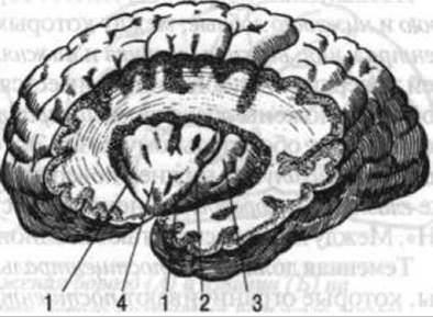
Fig. 111. Insular lobe of the brain:
1 — circular sulcus; 2 — central sulcus; 3 — long gyrus of the insula; 4 — short gyri of the insula

Fig. 112. Cellular and fibrous structure of the cerebral cortex (diagram).
Roman numerals indicate the layers of the cellular structure of the cortex (cytoarchitectonics), and Arabic numerals indicate the fibrous structure (myeloarchitectonics):
I - molecular layer; II - external granular layer; III - external pyramidal layer; IV - internal granular layer; V - internal pyramidal layer; VI - multiform layer;
1 - tangential nerve fibers; 2 - layer with a sparse number of fibers; 3 - suprastriate fiber layer; 4 - outer fiber layer; 5 - interstriate layer and inner stripe of fibers; 6 — substriate layer
The external pyramidal layer consists of cells 10–20 µm in diameter.
The internal granular layer is absent in some areas of the cortex. It contains small rounded and angular cells of stellate or pyramidal shape. Their size is similar to that of the cells In the second layer.
The internal pyramidal layer comprises cells ranging from 15 to 40 µm in diameter. Their processes participate in The formation of the corticospinal (pyramidal) tracts.
The cell diameter ranges from 10 to 30 µm.
Like the cells, nerve fibers in the cerebral cortex are arranged in layers, mainly running parallel or perpendicular to the cortical surface. The structure of the cerebral cortex varies across its different regions. On this basis, it is customary to divide the entire cortex into distinct areas. Among the Structural Features of the cortex, the following should be noted: in the region of the precentral gyrus, it is characterized by the presence of large and sparsely distributed giant cells, but lacks the fourth layer; in the region of the occipital pole of the hemispheres, the cortex features a dense arrangement of nerve cells, with the fourth layer being well developed and containing a large number of stellate neurons; the internal granular layer performs primarily a receptive function, whereas the upper cortical layers carry out associative functions, and the lower layers, the fifth and sixth, perform effector functions.
According to the teachings of I. P. Pavlov, the entire cortex functionally consists of the cortical ends of analyzers. Structurally, an analyzer comprises a structure that perceives stimuli at the periphery of the body (Sense Organs, sensory nerve endings), a pathway that conducts these stimuli from the periphery to the center, and a central department. Within the central department, the cortical end of the analyzer is of particular importance: it not only perceives incoming stimuli, not only breaks them down and "analyzes" them, but also links and synthesizes them; it also participates in the integrative and transmission Functions of the cortex, which consist in establishing temporary connections between individual cortical areas and between the central departments of analyzers, and in transmitting impulses to the nerve cells of underlying structures of the BRAIN AND SPINAL cord. From these structures, effector impulses travel to a specific "working organ" (muscle, gland). Let us examine the location of the central departments of certain analyzers within the cortex.
The motor analyzer is located in the precentral gyrus. This cortical area receives stimuli (proprioceptive, kinesthetic) arising mainly in muscle tendons, joints, ligaments, and partly in the skin and skeletal musculature. The motor analyzer ensures the formation of motor conditioned Reflexes in response to various sensory stimuli (pain, thermal, visual, auditory, etc.).
The general sensory analyzer (for pain, Temperature, and Touch) is located in the postcentral gyrus.

Fig. 113. Cerebral hemispheres at various levels of horizontal section (right — below the level of the floor of the lateral ventricle; left — above the floor of the lateral ventricle): the fourth ventricle is opened from above, the rhomboid fossa is visible: 1 — HEAD of the caudate nucleus; 2 — putamen; 3 — cortex of the insular lobe; 4 — globus pallidus; 5 — claustrum; 6 — tail (of the caudate nucleus); 7 — nucleus of the medial geniculate body; 8 — inferior horn of the lateral ventricle; 9 — superior cerebellar peduncle; 10 — middle cerebellar peduncle; 11 — inferior cerebellar peduncle; 12 — medullary striae; 13 — trigone of the hypoglossal nerve; 14 — trigone of the vagus nerve; 15 — tubercle of the gracile nucleus; 16 — cerebellum; 17 — superior medullary velum; 18 — Trochlear nerve; 19 — thalamus; 20 — red nucleus; 21 — stria terminalis of the thalamus; 22 — one of the hypothalamic nuclei; 23 — caudate nucleus (body); 24 — cortex of the insular lobe; 25 — cavity of the septum pellucidum; 26 — anterior horn of the lateral ventricle (after G. F. Ivanov)
The Auditory Analyzer is located in the middle part of the superior temporal gyrus, primarily on its surface facing the insular lobe.
The olfactory analyzer is situated in the region of the uncus, i.e., at the anterior end of the parahippocampal gyrus of the temporal lobe of the brain. The gustatory analyzer is located in the same area.
The visual analyzer is located along the margins of the calcarine sulcus, i.e., in the occipital lobe.
The motor analyzer for complexly coordinated movements (praxis) is located in the left inferior parietal lobule in right-handed individuals and in the right one in left-handed individuals.
The tactile object recognition analyzer (stereognosis) is located in the superior parietal lobule of the right and left hemispheres.
The motor analyzer of written speech is situated in the posterior part of the middle frontal gyrus and serves as the analyzer for the fine motor skills involved in writing not only individual letters but also various conventional symbols.
The motor analyzer of speech movements is located in the posterior part of the inferior frontal gyrus.
The auditory speech analyzer, much like the general auditory analyzer, is located in the posterior part of the superior temporal gyrus.
Several other analyzers are also distinguished. Among the cortical Regions of the human brain's analyzers, those associated with speech capabilities play a significant role. Specifically, these include the motor and auditory speech analyzers as well as the motor analyzer of written speech.
On a horizontal section through the cerebral hemispheres, made closer to the base, clusters of gray matter can be seen located near the medial region of each hemisphere. These clusters are called basal nuclei (Fig. 113). The largest nucleus is the corpus striatum, which is further subdivided into the caudate nucleus and the lentiform nucleus. The former features an expansion at its anterior end known as the head, and a tail at its posterior end; the latter is divided into a lateral part called the putamen, and a medial part, which in turn is divided into two segments of the globus pallidus.
Between the thalamus and the head of the caudate nucleus medially, and the lentiform nucleus laterally, lies a distinct layer of white matter known as the internal capsule. This is where the major Neural Pathways connecting the cerebral cortex to the brainstem and spinal cord pass through.
Laterally from the lentiform nucleus, between it and the cortex, lies a very narrow layer of gray matter, which is also a basal nucleus and is called the claustrum. The layer of white matter situated between the lentiform nucleus and the claustrum is termed the external capsule.
Lateral ventricles (Fig. 114). The lateral ventricles are clefts representing the residual cavity of the telencephalon. Each lateral ventricle in both the right and left hemispheres comprises a central part and three horns: anterior, posterior, and inferior.
The central part is located posterior to the interventricular foramen (which connects the lateral ventricle to the third ventricle) and anterior to the point where the lateral ventricle diverges into the posterior and inferior horns. Within the central part of the lateral ventricle, one can observe the continuation of the fornix, which begins as two pillars anterior to the interventricular foramen, extends posteriorly into the body and columns of the fornix, and then continues downward into the inferior horn of the lateral ventricle as the fimbria of the hippocampus (see Fig. 110). Within the central part of the lateral ventricle lies the choroid plexus, which is a direct continuation of the choroid plexus of the third ventricle. This plexus is covered by a thin layer of epithelial tissue.
The anterior horn of the lateral ventricle forms an expansion within the frontal lobe. The head of the caudate nucleus projects into the cavity of the anterior horn, forming its lateral and partially inferior wall. The medial wall of the anterior horn is formed by the septum pellucidum, located between the corpus callosum and the fornix, which encloses the cavity of the septum pellucidum.
The posterior horn of the lateral ventricle extends into the occipital lobe. Its medial surface features elevations, the most prominent of which is the calcar avis, corresponding to the calcarine sulcus on the medial surface of the cerebral hemisphere.
The inferior horn of the lateral ventricle extends into the temporal lobe. It features an elevation running along its medial wall known as the hippocampus, which terminates anteriorly and inferiorly in the pes hippocampi. The cavity of the lateral ventricle is filled with cerebrospinal fluid produced by the choroid plexus and, in part, by the ependymal cells lining the ventricular cavity.
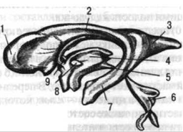
Fig. 114. Cast of the brain ventricular cavities:
1 — anterior horn of the lateral ventricle; 2 — central part; 3 — posterior horn of the lateral ventricle; 4 — third ventricle; 5 — cerebral aqueduct (aqueduct of midbrain); 6 — fourth ventricle; 7 — inferior horn of the lateral ventricle; 8 — interthalamic adhesion; 9 — interventricular foramen
The corpus callosum (see Fig. 110) is the largest and most prominent commissure connecting the cerebral hemispheres. On a medial section, it appears as an anteroposteriorly elongated structure, 5–7 cm long and 1.5 cm wide. The posterior part of the corpus callosum features a thickening (splenium). Its anterior region forms a bend, or genu, below which lies the rostrum, continuing downward and backward as a thin sheet of white matter toward the optic chiasm.
The surface of the corpus callosum exhibits longitudinal and transverse striations. The transverse striations are caused by the transverse orientation of nerve fibers connecting the right and left hemispheres. These fibers diverge laterally to form the corona radiata of the corpus callosum. The fibers of the corona radiata radiate in various directions toward the cerebral cortex. Specifically, the fibers from the anterior part of the corpus callosum run toward the frontal lobes, while those from the posterior part run toward the occipital lobes. On horizontal sections, these fibers converge to form structures known respectively as the forceps anterior (minor) and forceps posterior (major).
On its inferior surface, the corpus callosum is fused with the fornix and the septum pellucidum. The fornix, much like the corpus callosum, is composed of white matter, i.e., nerve fibers.
The function of the fornix fibers is to connect the diencephalon with the temporal lobe.
Pathways of the Central Nervous System
During the execution of all movements, including athletic ones (as well as during their acquisition), the central nervous system plays a leading role. The motor apparatus, while under its control, in turn exerts an influence upon it. Learning any Physical Exercise and perfecting its execution is, above all, training for The Nervous System. Only through the nervous system is that high degree of movement coordination achieved. The anatomical substrate of coordination consists of billions of nerve cells in the central nervous system and their processes, which together form the Pathways of the brain and spinal cord, as well as the peripheral nerves.
The pathways of the central nervous system are represented by nerve fibers—that is, neuronal processes interconnecting discrete clusters of cell bodies (nuclei, centers). They make up the bulk of the cerebral white matter, which is located deep within the cerebral hemispheres, in certain regions of the brainstem, and in the funiculi of the spinal cord. By transmitting nerve impulses from one part of the central nervous system to another, these pathways establish functional connections between them. Depending on the location and Functional Characteristics of the nerve fibers within the cerebral hemispheres, pathways are classified into association, commissural, and Projection Pathways.
Association Pathways of the brain interconnect different cortical areas within the same hemisphere. They are divided into short and long pathways. Short association pathways connect cortical neurons of adjacent gyri, whereas long ones link more distant cortical areas (for example, gyri in different lobes of the hemispheres). In the spinal cord, intrinsic spinal tracts (fasciculi proprii) serve as association pathways, connecting neurons of superior and inferior segments.
Commissural pathways interconnect symmetrical regions of the brain and also contribute to the brain commissures. The largest portion of commissural fibers forms the corpus callosum, which is the largest cerebral commissure (see Fig. 110). The anterior fibers of the corpus callosum link the frontal lobes of the hemispheres, the middle fibers link the parietal and temporal lobes, and the posterior fibers link the occipital lobes. For the cerebellar hemispheres, the transverse fibers of the pons serve a similar commissural function.
Projection pathways connect the cerebral cortex with the underlying structures of the brain (the brainstem) and the spinal cord, and through them, with various organs of the body. They are divided into short and long projection pathways. Short projection pathways connect the cerebral cortex with the corpus striatum, thalamus, superior colliculi of the midbrain, cerebral peduncles, cerebellum, medulla oblongata, and Sensory Organs. Long projection pathways connect the cerebral cortex with the spinal cord and, via the cord, with all bodily organs. Both short and long projection pathways include sensory (afferent) and motor (efferent) pathways.
Short sensory projection pathways include the visual, auditory, vestibular, olfactory, and gustatory pathways. All of these serve as conduction pathways for their respective analyzers, transmitting neural impulses of Vision, Hearing, Olfaction, gustation, and statokinetic sense to the corresponding subcortical and cortical centers. They are discussed in detail in the section on sensory organs (see p. 334).
Short motor projection pathways comprise the corticonuclear (corticobulbar) and corticocerebellar pathways. The corticonuclear pathways connect the pyramidal cells of the motor cortex (precentral gyrus) with the motor nuclei of the Cranial Nerves (pairs III, IV, V, VI, VII, IX, X, XI, and XII). These pathways transmit conscious motor commands to the extraocular muscles, masticatory and facial muscles, Muscles of the Larynx, Pharynx, and Tongue, as well as certain Neck Muscles (hyoid, trapezius, and sternocleidomastoid muscles). The corticocerebellar pathway connects the cerebral cortex to the cerebellum. It courses through the pontine nuclei, which divide it into the corticopontine and pontocerebellar pathways.
Long sensory projection pathways include the lateral spinothalamic tract, the fasciculus gracilis and fasciculus cuneatus, the posterior spinocerebellar tract, and the anterior spinocerebellar tract.
Long motor projection pathways include the lateral corticospinal (pyramidal) tract, the anterior corticospinal (pyramidal) tract, and the rubrospinal tract.
The lateral spinothalamic tract (Fig. 115) is the pathway for exteroceptive sensitivity. It conducts pain and temperature impulses to the cortical center of general sensation. Its receptors are located in the skin, and the cell bodies of the first-order neurons lie in the dorsal root ganglia. Their peripheral processes conduct impulses from Skin Receptors to the cell bodies in the dorsal root ganglia, and from there, via central processes within the dorsal roots, to the neurons of the posterior horn proper (substantia gelatinosa/proper nucleus) of the spinal cord, which serve as the second-order neurons of this pathway.
The axons of the second-order neurons cross to the contralateral lateral funiculus of the spinal cord, where they converge to form the lateral spinothalamic tract. Ascending within the lateral funiculus, this bundle of second-order axons passes through the medulla oblongata, pons, and midbrain as part of the medial lemniscus, ultimately reaching the thalamus, where it synapses with the cell bodies of the third-order neurons. The processes of the latter travel through the internal capsule to the cortex of the postcentral gyrus in the parietal lobe of the hemisphere—that is, to the cortical (central) end of the pain and temperature analyzer.
The fasciculus gracilis and fasciculus cuneatus are the conduction pathways for conscious muscle-joint (proprioceptive) sense and tactile sensation. The cell bodies of the first-order neurons reside in the spinal ganglion (see Fig. 115). Their peripheral processes originate as receptors located in organs of movement—muscles, tendons, ligaments, and joints (proprioceptors)—and in the skin (exteroceptors), while their central processes enter the spinal cord via the dorsal roots into the dorsal funiculi, forming the fasciculus gracilis and fasciculus cuneatus. Within these tracts, the neurites of the first-order neurons ascend to the medulla oblongata, where they synapse with cells in the gracile and cuneate nuclei. The axons of these cells, acting as the second-order neurons of the pathway, ascend, decussate in the medulla oblongata, course through the brainstem (pons, midbrain) as part of the medial lemniscus, and terminate in the thalamus. The axons of the thalamic neurons (third-order neurons of the pathway) deliver proprioceptive and tactile impulses to the cortex of the postcentral gyrus in the parietal lobe, the center for general sensation.
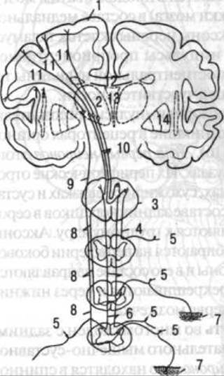
Fig. 115. Sensory projection pathways (exteroceptive):
1 - cerebral cortex; 2 - thalamus; 3 - medulla oblongata; 4 - cuneate fasciculus; 5 - sensory neuron of the spinal ganglion; 6 - gracile fasciculus; 7 - skin; 8 - exteroceptive pathways in the lateral funiculi of the spinal white matter; 9 - spinothalamic tract (lateral lemniscus); 10 - spinothalamic tract (medial lemniscus); 11 - thalamocortical fibers; 12 - corpus callosum; 13 - third ventricle; 14 - lentiform nucleus

Fig. 116. Sensory projection pathways (proprioceptive):
1 — cerebral cortex; 2 — cerebellum; 3 — posterior spinocerebellar tract; 4 — central process of the sensory neuron; 5 — spinal ganglion; 6 — peripheral process of the sensory neuron; 7 — muscle; 8 — anterior spinocerebellar tract; 9 — spinal cord; 10 — medulla oblongata
The posterior spinocerebellar tract conducts impulses of unconscious muscle-joint sense, originating in the movement receptors, to the cerebellar cortex (Fig. 116). The cell bodies of the first-order neurons of this pathway are located in the spinal ganglia. Their peripheral processes begin as receptors in muscles, tendons, ligaments, and joints, whereas their central processes enter the gray matter of the spinal cord via the dorsal roots, heading toward the thoracic nucleus (nucleus dorsalis/Clark's column). The axons of these cells (second-order neurons) gather at the periphery of the ipsilateral lateral funiculus of the spinal cord and ascend toward the medulla oblongata, where they decussate and pass through the inferior cerebellar peduncles to reach the cortex of the cerebellar vermis.
The anterior spinocerebellar tract is largely similar to the posterior one. It also transmits impulses of unconscious muscle-joint sense to the cerebellum. The cell bodies of its first-order neurons are situated in the spinal ganglia. Their central processes enter the gray matter of the spinal cord via the dorsal roots, synapsing with cells of the intermediate medial nucleus, which function as the second-order neurons. The axons of the second-order neurons also concentrate at the periphery of the lateral funiculus, but on the contralateral side; ascending within these funiculi, they terminate upon the cells of the cerebellar vermis, entering it through the superior cerebellar peduncles (see Fig. 116).
Proprioceptive pathways, which carry impulses of muscle-joint sense and touch, are of paramount importance in athletic activity. They enable athletes to orient themselves in space, perceive their posture and movements, and receive a continuous stream of feedback regarding the state of The Musculoskeletal System. Highly developed proprioceptive sensitivity protects athletes from injury and allows them to execute extremely rapid and precise differentiated movements (as in boxing, fencing, and sambo). The fasciculus gracilis and fasciculus cuneatus, which transmit muscle-joint and tactile sensations, provide a highly refined proprioceptive and tactile awareness. This allows athletes not only to feel their own body but also to sense the opponent's position, sometimes through mere contact with their hands or even just the opponent's uniform.

Fig. 117. Course of the Pyramidal Tracts and the corticonuclear pathways of the trigeminal and facial nerves:
1 - corpus callosum; 2 - caudate nucleus; 3 - thalamus; 4 - fibers of the corticospinal (pyramidal) tracts; 5 - corticonuclear fibers of the facial n.; 6 - corticonuclear fibers of the trigeminal n.; 7 - lentiform nucleus; 8 - pons; 9 - motor nucleus of the trigeminal n.; 10 - trigeminal ganglion; 11 - motor component of the III branch of the trigeminal n.; 12 - nucleus of the facial n.; 13 - facial n.; 14 - lateral corticospinal tract; 15 - anterior corticospinal tract; 16 - anterior root (after Rauber)
The lateral corticospinal (pyramidal) tract transmits voluntary motor impulses from the cerebral cortex through the spinal cord to the Muscles of the Trunk and limbs. The primary neurons of this pathway are the pyramidal cells of the precentral gyrus in the cerebral cortex (Fig. 117). The axons of these cells pass through the internal capsule, the crus cerebri, the pons, and enter the pyramids of the medulla oblongata. Within the pyramids, the majority of the fibers cross to the opposite side (forming the pyramidal decussation), after which they descend within the lateral funiculi of the spinal cord. The fibers of the lateral corticospinal tract synapse with the motor neurons of the anterior horns in each spinal segment, which serve as the second-order neurons. The axons of these anterior horn motor cells travel within the ventral roots of the Spinal nerves and their branches, delivering voluntary motor impulses to the skeletal muscles.
The anterior corticospinal (pyramidal) tract is largely analogous to the preceding one. It also conducts voluntary motor commands from the cerebral cortex via the spinal cord to the skeletal muscles of the trunk and limbs. Up to the medulla oblongata, the fibers of the lateral and anterior corticospinal tracts travel together. The uncrossed bundle of fibers descends into the anterior funiculi of the spinal cord, where it forms the anterior corticospinal tract (see Fig. 117). Its fibers likewise synapse with the motor neurons of the spinal gray matter (both ipsilateral and contralateral). The peripheral processes of the second-order neurons travel via the anterior roots of the spinal nerves to the skeletal muscles, terminating in effector nerve endings known as motor endplates.
As they descend through the spinal cord, the pyramidal tracts progressively decrease in thickness because their fibers terminate segmentally upon the motor neurons of the anterior horns.
The pyramidal system, which typically encompasses the cortical motor center and the pyramidal tracts, plays a crucial role in the execution of voluntary movements. It fulfills three primary functions: 1) it dispatches conscious starting impulses—movement commands—to the motor neurons of the spinal cord; 2) it facilitates the transmission of nerve impulses through the interneurons of the spinal cord; and 3) it exerts regulatory control over the stream of sensory feedback.
The extrapyramidal system is a phylogenetically older structure than the pyramidal system. It includes subcortical motor centers (the corpus striatum, claustrum, amygdala, and red nucleus) along with the rubrospinal tract. In vertebrate animals with an undeveloped cortex, this system plays a leading role in governing motor functions. In humans, owing to the high degree of Development of the cerebral cortex, the pyramidal pathways—together with the cortical motor center—have undergone significant differentiation to form the pyramidal system. Nevertheless, the extrapyramidal system remains vital in humans for executing motor acts: it modulates the activity level of spinal motor neurons and the tonus of corresponding skeletal muscles, and it plays a major role in organizing postural-tonic bodily reactions.
The rubrospinal tract is a motor pathway of the extrapyramidal system. It connects subcortical motor centers (basal nuclei of the hemispheres, red nuclei) and the cerebellum with the motor neurons of the spinal cord, and through them, with skeletal muscles. The cell bodies of the first neurons of this pathway are located in the red nuclei of the cerebral peduncles. Their axons decussate and descend in the lateral funiculi of the white matter of the spinal cord, where they form segmental synaptic connections with the motor neurons of the anterior horns of the gray matter, which are the second neurons of this pathway. The axons of the second neurons, as part of the anterior roots, spinal nerves, and their branches, transmit impulses from the extrapyramidal system and cerebellum to skeletal muscles. Thus, each motor cell in the anterior horns of the spinal gray matter simultaneously receives impulses via the fibers of both the pyramidal (lateral and anterior corticospinal) and extrapyramidal (rubrospinal) pathways.
In Conclusion, it should be emphasized that the conduction pathways, along with the cerebral cortex, nuclei, and subcortical centers of the brain and spinal cord, serve as the material substrate for complex innate (unconditioned) and acquired (conditioned) reflexes, which ensure the normal functioning of the nervous system as a whole. The centers of certain motor pathways (such as the rubrospinal tract) are connected to the corpus striatum, a subcortical motor center. In turn, the corpus striatum connects not only with the red nucleus and other clusters of nerve cells (such as the thalamus), but also with the cerebral cortex.
Fig. 118. Dura mater of the brain and venous sinuses:
1 — falx cerebri; 2 — tentorium cerebelli; 3 — superior sagittal sinus; 4 — inferior sagittal sinus; 5 — transverse sinus; 6 — superior petrosal sinus; 7 — cavernous sinus; 8 — straight sinus (after V.N. Tonkov)
The brain and spinal cord are enclosed by three membranes: the outer dura mater, the middle arachnoid mater, and the inner pia mater. The arachnoid and pia maters are collectively referred to as the leptomeninges (or soft meninges).
The dura mater of the brain (Fig. 118) serves simultaneously as the internal periosteum of the cranial bones and the outer Sheath of the brain. It is tightly fused with the cranial bones only at the Base of the skull, whereas in the area of the cranial vault, it is loosely connected to them.
The cranial dura mater is composed of dense regular Connective Tissue lined on the inside by flattened cells and kept slightly moist. It forms several folds that extend into the longitudinal fissure between the cerebral hemispheres and the transverse fissure between the cerebellum and the occipital lobes of the cerebrum. Additionally, it sends out prolongations along the nerves exiting the cranial cavity, as well as a fold known as the diaphragma sellae. The most important folds of the cranial dura mater are described below.
Falx cerebri. It lies in the sagittal plane between the right and left hemispheres, attaching anteriorly to the crista galli of the Ethmoid bone and the margins of the sulcus for the superior sagittal sinus—which runs along the internal surface of the cranial roof bones in the midline—and continuing posteriorly into the tentorium cerebelli. Inferiorly, the falx cerebri extends as a small fold located between the right and left cerebellar hemispheres posteriorly, known as the falx cerebelli.
Tentorium cerebelli. This fold is situated between the occipital lobes of the cerebral hemispheres and the superior surface of the cerebellum. It attaches posteriorly to the margins of the transverse sinus sulcus on the occipital bone, and laterally, partly anteriorly, to the petrous part of the Temporal bone. It creates a compartment in the posterior cranial fossa that houses the cerebellum.
Diaphragma sellae. This is a fold of the cranial dura mater that extends from the dorsum sellae and the bases of the lesser wings of the sphenoid bone. In the center of the diaphragma sellae is an aperture through which passes the infundibulum (stalk), a continuation of the tuber cinereum. The pituitary gland (hypophysis) is attached to this stalk.
In certain regions of the cranial cavity (most frequently along the paths of the sagittal, transverse, and sigmoid sulci), the cranial dura mater splits into two layers to form channels known as venous sinuses, which drain Blood from the brain (see Fig. 118).
The most prominent venous sinuses of the dura mater include the superior and inferior sagittal, straight, transverse, sigmoid, and cavernous sinuses.
The arachnoid mater is a very delicate membrane. It covers the brain on all sides but does not extend into the sulci and fissures. The arachnoid mater is avascular (lacks Blood Vessels). A narrow gap, the subdural space, lies between the dura and arachnoid maters.
The pia mater adheres closely to the entire surface of the brain, dipping into all its sulci and fissures. Small prolongations extend from the pia mater to penetrate the substance of the brain. Accompanying the pia mater into the brain are blood vessels—primarily branches of the arterial circle located at the base of the brain (anterior, middle, and posterior cerebral Arteries; see p. 254).
Between the arachnoid and pia maters lies the subarachnoid space, which is filled with clear, serous cerebrospinal fluid (CSF). CSF also fills the ventricular cavities. This fluid acts as a medium through which brain cells receive nutrients and discharge their Metabolic waste products. In certain areas, accumulations of cerebrospinal fluid are notably large, forming the so-called subarachnoid cisterns. These include the cistern of the lateral cerebral fossa (located over the lateral sulcus of the brain), the interpeduncular cistern (between the cerebral peduncles), the chiasmatic cistern (situated around the optic chiasm), and the cerebellomedullary cistern (located inferior to the cerebellum, between it and the medulla oblongata).
Cerebrospinal fluid within the ventricular cavities is produced by the choroid plexuses. It flows from the lateral ventricles into the third ventricle, then into the fourth, from which it enters the subarachnoid space—specifically, the cerebellomedullary cistern. From there, the fluid flows into the interpeduncular cistern, then to the chiasmatic cistern, and further into the cistern of the lateral fossa.
In addition to cerebrospinal fluid, tissue fluid containing dissolved metabolic waste products from Nervous Tissue drains from the brain parenchyma into the subarachnoid space. This fluid flows through perivascular adventitial spaces, which compensate for the "deficit" in brain tissue clearance caused by the absence of true lymphatic channels in the central nervous system. The clefts of the endoneurium and perineurium surrounding nerve trunks also participate in the drainage of tissue fluid and CSF. The primary outflow of fluid from the subarachnoid space occurs into the venous sinuses of the dura mater. Here, the fluid enters the arachnoid granulations—projections of the arachnoid mater that pierce the dura mater and protrude into the lumen of the venous sinuses. The granulations are bathed in venous blood, though they have no direct opening into the sinus lumen. However, when the pressure in the subarachnoid space exceeds that in the venous sinus, conditions are established for the diffusion of cerebrospinal fluid from the granulations into the blood of the Dural Venous Sinuses.
Besides the aforementioned subdural and subarachnoid spaces, There are also small slit-like intervals: between the dura mater and the cranial bones, as well as between the pia mater and the brain substance.
The spinal cord is enveloped by the same meninges as the brain; however, the spinal meninges exhibit certain topographical and structural specificities. The spinal dura mater, particularly in its inferior region, splits into internal and external layers, creating a well-defined epidural space between them. This space is filled with adipose tissue and contains internal vertebral venous plexuses. The subarachnoid space is much more extensively developed in the spinal region than in the cranial region and contains a significant volume of cerebrospinal fluid.
Between the lateral surface of the spinal cord and its dura mater lie the denticulate ligaments, situated in the frontal plane. Alongside these are much less distinct posterior ligaments. Together, they constitute the stabilizing fixation apparatus of the spinal cord.
Last update: 08/08/2026
Editorial and Educational Adaptation: This material has been compiled based on the primary/original source text. The project team performed an editorial review, corrected technical inaccuracies, structured sections, and adapted the content for an educational format.
What was processed:
- elimination of formatting defects (OCR errors, structural breaks, corrupted characters);
- editorial organization of content;
- standardization of terminology in accordance with academic sources;
- verification of factual statements against the original source text.
All mentions of the author, publication year, and origin of the primary text have been preserved in accordance with the source.