Human Anatomy (with the Fundamentals of Dynamic and Sports Morphology) - Ivanitsky, M. F. 2008
The Doctrine of the Nervous System
Peripheral Nervous System
Cranial Nerves (Brain nerves)
There are twelve pairs of cranial nerves, designated from I to XII (Fig. 119). Most of them are connected with the rhombencephalon (V–XII), two with the Telencephalon (I), and two with the mesencephalon (III–IV).
Class="center">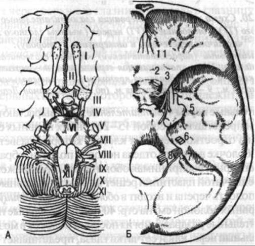
Fig. 119. Sites of exit from the brain (A) and from the cranial cavity (B) of the cranial nerves:
I – olfactory n.; II – optic n.; III – oculomotor n.; IV – trochlear n.; V – trigeminal n.; VI – abducens n.; VII – facial n.; VIII – vestibulocochlear n.; IX – glossopharyngeal n.; X – vagus n.; XI – accessory n.; XII – hypoglossal n.; 1 – cribriform plate (I); 2 – optic canal (II); 3 – superior orbital fissure (III, IV, ophthalmic nerve from V); 4 – foramen rotundum (maxillary n. from V); 5 – foramen ovale (mandibular n. from V); 6 – internal acoustic meatus (VII and VIII); 7 – jugular foramen (IX, X, XI); 8 – hypoglossal canal (XII) (after Lepp and Kogerman-Lepp)

Fig. 120. Diagram of the pathway and innervation Zones of the oculomotor (III), trochlear (IV), and abducens (VI) nerves (the extraocular Muscles are partially sectioned and reflected):
1 — oculomotor (parasympathetic) ROOT of the oculomotor n.; 2 — ciliary ganglion; 3 — levator palpebrae superioris m.; 4 — superior rectus m.; 5 — superior oblique m.; 6 — medial rectus m.; 7 — lateral rectus m.; 8 — inferior oblique m.; 9 — inferior rectus m. (after Latt and Kogerman-Lepp)
I. The olfactory nerves are sensory. They originate from the telencephalon as 15–20 fine nerve trunks formed by the processes of Nerve Cells that branch out within the mucous membrane of the upper Nasal cavity, forming specialized receptors to perceive olfactory stimuli. Passing through the foramina of the cribriform plate of the Ethmoid bone, these nerve trunks enter the cranial cavity and reach the olfactory bulb (for the further course of the nerve fibers, see p. 403). The olfactory nerves transmit olfactory impulses from the nasal cavity to the brain.
II. The Optic nerve, much like the retina of the eye, represents an outgrowth from the wall of the Diencephalon. This nerve is sensory in function. It conducts centripetal nerve impulses generated by light and color stimuli. Accompanied by the ophthalmic artery, the nerve extends from the Eyeball and enters the cranial cavity through the optic canal of the Sphenoid bone. On the superior surface of the body of the sphenoid bone, it contributes to The formation of the optic chiasm (for the further pathway of the visual analyzer pathways, see p. 396).
III. The Oculomotor nerve (its nuclei are located in the floor of the cerebral aqueduct) emerges on the Medial surface of the cerebral peduncles and runs within the lateral wall of the cavernous sinus. Leaving the cranial cavity through the superior orbital fissure, the nerve enters the Orbit, where it divides into superior and inferior branches. It innervates (Fig. 120) the superior, inferior, and medial rectus Muscles of the eye, as well as the inferior oblique Muscle and the levator palpebrae superioris muscle. The oculomotor nerve is the largest of the nerves responsible for moving the eyeball. It is a mixed nerve: In addition to motor fibers, it contains autonomic (preganglionic) fibers directed to the ciliary ganglion (Fig. 120), providing parasympathetic innervation to the smooth muscles located inside the eyeball, particularly the sphincter pupillae muscle.
IV. The Trochlear nerve is a motor nerve and the slenderest of all cranial nerves. It emerges from the superior medullary velum, curves around the lateral aspect of the cerebral peduncle, travels within the lateral wall of the cavernous sinus, and passes through the superior orbital fissure into the orbit. The trochlear nerve innervates the superior oblique muscle of the eye (see Fig. 120).
V. The Trigeminal nerve (Fig. 121) is the largest of the cranial nerves. It is a mixed nerve that emerges from the Pons by two roots: a larger sensory root and a smaller motor root. The sensory root forms the large trigeminal ganglion, which consists of sensory nerve cells and is underlain by the motor root. This ganglion lies on the superior surface of the petrous part of the Temporal bone, in a depression near its apex. Three main nerves arise from the trigeminal ganglion: the ophthalmic, maxillary, and mandibular nerves.
The ophthalmic nerve (see Fig. 121) is sensory. It passes through the superior orbital fissure into the orbit, where it branches into the lacrimal, frontal, and nasociliary nerves.
The lacrimal nerve runs toward the lacrimal gland, supplying it with sensory fibers. Secretory fibers reach this gland via a roundabout pathway: they branch off from the Facial Nerve, pass through the pterygopalatine ganglion (see p. 366), and join the lacrimal nerve only near the lacrimal gland.
The frontal nerve extends toward the upper eyelid and the forehead, where it supplies the Skin.
The nasociliary nerve gives off branches to the ciliary ganglion (see p. 366) and to the mucous membrane of the nasal cavity (ethmoidal nerves).
The maxillary nerve (see Fig. 121) exits the cranial cavity through the foramen rotundum, traverses the upper part of the pterygopalatine fossa, and continues into the orbit as the infraorbital nerve. After passing through the infraorbital canal and the infraorbital foramen of the Maxilla, it branches out in the skin of the middle region of the face. This nerve is sensory.
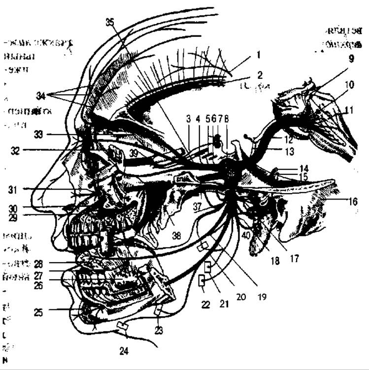
Fig. 127. Diagram of the pathway and innervation zones of the trigeminal nerve:
1 — cutaneous n.; 2 — temporal m.; 3 — sympathetic n.; 4 — oculomotor n.; 5 — abducens n.; 6 — trochlear n.; 7 — internal carotid a.; 8 — trigeminal ganglion; 9 — accessory n.; 10 — trigeminal n.; 11 — floor of the Fourth ventricle; 12 — sensory root of trigeminal n.; 13 — motor root of trigeminal n.; 14 — facial n.; 15 — vestibulocochlear n.; 16 — temporal bone; 17 — otic ganglion; 18, 22 — masseteric n.; 19 — lingual n.; 20 — inferior alveolar n.; 21 — pterygoid n.; 23 — mylohyoid n.; 24 — branch to anterior belly of digastric m.; 25 — Mandible; 26 — submandibular gland; 27 — submandibular ganglion; 28 — sublingual gland; 29 — posterior superior alveolar branches; 30 — anterior superior alveolar branches; 31 — infraorbital n.; 32 — lacrimal gland; 33 — nasociliary n.; 34 — branches of frontal n.; 35 — ophthalmic n.; 36 — mandibular n.; 37 — maxillary n.; 38 — pterygopalatine ganglion; 39 — ciliary ganglion; 40 — parotid gland (after Testut)
The maxillary nerve supplies superior alveolar nerves to the Teeth of the upper jaw and gives off branches to the pterygopalatine ganglion.
The mandibular nerve (see Fig. 121) exits the cranial cavity through the foramen ovale. It is a mixed nerve containing both sensory and motor fibers. The former are incorporated into the branches running from the mandibular nerve to the Tongue, the teeth of the lower jaw, and the skin of the lower facial region, whereas the latter enter the nerves supplying the masticatory muscles.
The sensory Branches of the mandibular nerve include the following: auriculotemporal, buccal, inferior alveolar, and lingual nerves.
The auriculotemporal nerve passes through the parotid gland, runs upward anterior to the external acoustic meatus, and participates in the Innervation of the skin of the temporal region.
The buccal nerve runs toward the buccinator muscle, though it does not innervate it; instead, it passes through the muscle and branches out within the buccal mucosa as sensory fibers.
The inferior alveolar nerve enters the mandibular canal, innervates all the mandibular teeth, and continues as the mental nerve, which emerges through the mental foramen to supply the skin of the anteroinferior region of the face.
The lingual nerve extends toward the mucous membrane of the tongue. It is joined by the chorda tympani, a branch of the facial nerve that carries autonomic secretory fibers to the submandibular and sublingual glands (see p. 240).
The motor branches of the mandibular nerve include the following: the masseteric, temporal, pterygoid, and geniohyoid nerves.
The masseteric nerve innervates the muscle of the same name.
The temporal nerves run to the temporalis muscle, which they innervate.
The pterygoid nerves innervate the pterygoid muscles (both medial and lateral).
The geniohyoid nerve runs to the muscle of the same name and to the anterior belly of the digastric muscle.
Closely associated with the trigeminal nerve are four ganglia: the ciliary, pterygopalatine, otic, and submandibular ganglia. Each of these contains nerve cells and, in addition to sensory fibers from the trigeminal nerve, receives sympathetic and parasympathetic fibers (see "Autonomic Nervous system", p. 328).
The ciliary ganglion (see Figs. 120–121) lies on the outer surface of the optic nerve. It receives branches from the ophthalmic nerve (sensory), the oculomotor nerve (parasympathetic, preganglionic), and the nerve plexus of the ophthalmic artery (sympathetic, postganglionic). In turn, it gives rise to branches that supply the smooth musculature within the eyeball.
The pterygopalatine ganglion (see Fig. 121) is located in the fossa of the same name. It receives branches from the maxillary nerve (sensory), the internal carotid plexus (sympathetic), and the facial nerve (parasympathetic). Branches extend from the pterygopalatine ganglion into the nasal and oral cavities and the orbit, including secretory fibers to the lacrimal gland.
The otic ganglion (see Fig. 121) is connected to the third branch of the trigeminal nerve. This ganglion receives sensory, parasympathetic, and sympathetic fibers: the first originate from the mandibular nerve, the second from the Glossopharyngeal nerve, and the third from the plexus of the maxillary artery. The otic ganglion lies just inferior to the foramen ovale, providing autonomic innervation to the parotid gland.
The submandibular ganglion receives branches from the lingual nerve (sensory), the chorda tympani (parasympathetic), and the plexus of the maxillary artery (sympathetic). This ganglion sends branches to the sublingual and submandibular glands.
Thus, all ganglia associated with the trigeminal nerve contain fibers of threefold origin: somatic (sensory) fibers originating from the trigeminal nerve; sympathetic fibers from the sympathetic plexus accompanying one of the nearby Blood Vessels; and parasympathetic fibers from one of the cranial nerves carrying parasympathetic components (the oculomotor, facial, or glossopharyngeal nerve). Branches from these ganglia extend to smooth muscle (via the ciliary ganglion), to mucous membranes (via the pterygopalatine ganglion), and to glands (via the pterygopalatine, otic, and submandibular ganglia). These branches transmit secretory impulses and also serve a trophic function, helping to regulate blood flow and nutritional processes in the innervated Organs.
Consequently, the innervation area of the trigeminal nerve is very extensive. It encompasses the entire skin of the face, the mucous membranes of the nasal and oral cavities, the Muscles of Mastication, the maxillary and mandibular teeth, PARTS OF THE soft palate, the mucous membrane of the tongue, and the Connective Tissue membrane of the eye.
VI. The Abducens nerve (see Fig. 120) is a motor nerve. It emerges between the pons and the pyramid of the Medulla Oblongata, runs within the lateral wall of the cavernous sinus, passes through the superior orbital fissure, and terminates in the lateral rectus muscle of the eyeball, which it innervates.
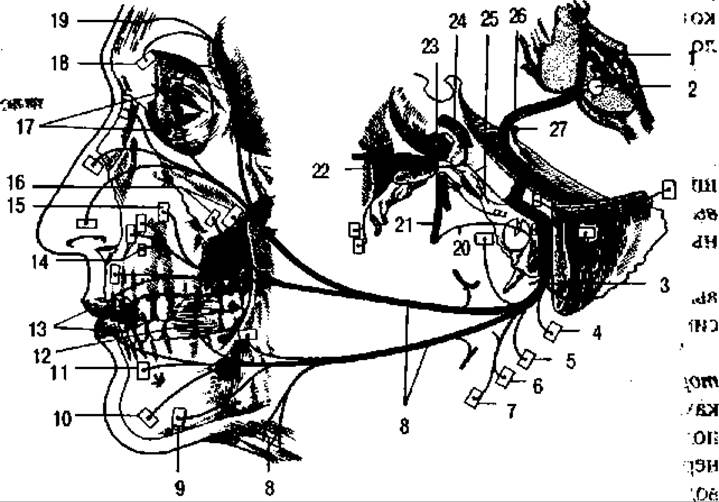
Fig. 122. Diagram of the course and innervation zones of the facial nerve:
1 - floor of the fourth ventricle; 2 - Nucleus of the facial (intermediate) n.; 3 - stylomastoid foramen; 4 - digastric branch; 5 - stylohyoid branch; 6 - branch to the stylohyoid m.; 7 - branch to the glossopharyngeal part of the superior constrictor of the Pharynx; 8 - branches to the Facial Muscles of expression and the platysma; 9 - branch to the depressor anguli oris m.; 10 - branch to the mentalis m.; 11 - branch to the depressor labii inferioris m.; 12 - branch to the buccinator m.; 13 - orbicularis oris m.; 14 - branches to the levator labii superioris and levator labii superioris alaeque nasi m.; 15 - branch to the levator anguli oris m.; 16 - branches to the zygomaticus minor and major m.m. (zygomatic); 17 - orbicularis oculi m.; 18 - branch to the corrugator supercilii m.; 19 - branch to the frontal belly of the occipitofrontalis m.; 20 - chorda tympani; 21 - lingual n.; 22 - pterygopalatine ganglion; 23 - trigeminal ganglion; 24 - internal carotid a.; 25 - greater petrosal a.; 26 - facial n.; 27 - vestibulocochlear n. (after Testut)
VII. The facial nerve is a mixed nerve (Fig. 122). It emerges on the lateral surface of the medulla oblongata, passes through the internal acoustic meatus into the facial canal, and exits the cranial base via the stylomastoid foramen. This nerve innervates all the Muscles of facial expression. It pierces the parotid gland, giving off several branches that form a plexus and course superiorly, anteriorly, and inferiorly toward the facial muscles and the posterior belly of the digastric muscle. While traversing the facial canal within the petrous part of the temporal bone, the facial nerve forms the geniculate ganglion, from which the greater petrosal nerve arises—carrying fibers to the pterygopalatine ganglion (and through it to the lacrimal gland)—along with nerve fibers that continue into the chorda tympani.
The chorda tympani traverses the tympanic cavity and lies directly against the medial surface of the upper part of the tympanic membrane. This nerve contains secretory fibers directed to the submandibular and sublingual glands, as well as taste fibers coming from the lingual mucosa. The fibers of the chorda tympani also become incorporated into the lingual nerve.
Passing between the facial nerve and the adjacent Vestibulocochlear nerve is the slender intermediate nerve, which is predominantly parasympathetic and is considered a functional component of the facial nerve.
The intermediate nerve is a mixed nerve. It contains secretory fibers that innervate the lacrimal gland (via the greater petrosal nerve and pterygopalatine ganglion) and the submandibular and sublingual Salivary Glands (via the chorda tympani, lingual nerve, and submandibular ganglion). Its sensory fibers transmit impulses generated by taste stimuli in certain papillae of the tongue.
VIII. The vestibulocochlear nerve (see Fig. 119) is a sensory nerve. It emerges from the medulla oblongata alongside the facial nerve, slightly inferior to it, passes through the internal acoustic opening and the internal acoustic meatus, and enters the internal ear, where it divides into two parts: the vestibular part and the cochlear part.
The vestibular part innervates the semicircular canals and the vestibule. It is formed by the central processes of nerve cells that constitute the vestibular ganglion. This portion of the nerve is unrelated to the perception of auditory stimuli; instead, it transmits static equilibrium impulses that enable spatial orientation.
The cochlear division Functions specifically as the auditory nerve, as it innervates the spiral organ (see Fig. 139) located within the cochlea, which transduces stimuli generated by sound waves. This nerve component features a spiral ganglion extending along the cochlear turns, containing nerve cells whose peripheral processes extend to the spiral organ, while their central processes form the cochlear part of the vestibulocochlear nerve.
IX. The glossopharyngeal nerve (Fig. 123) is a mixed nerve. It emerges from the medulla oblongata, leaves the Skull through the jugular foramen, and courses anteriorly and inferiorly along the Muscles Attached to the styloid process, branching out within the pharynx and the mucous membrane of the root of the tongue, palatine Tonsils, and arches. This nerve gives rise to a sinus branch, which transmits sensory impulses from the carotid body, and forms two ganglia—the superior and inferior—resembling intervertebral ganglia. These consist of sensory Neurons. The efferent (centrifugal) fibers of the glossopharyngeal nerve pass these ganglionic cells without synapsing.
The glossopharyngeal nerve participates in the Formation of the pharyngeal plexus and innervates both the muscles and the mucous membrane of the pharynx. Along its pathway, it gives off the tympanic nerve, which traverses the tympanic cavity and continues as the lesser petrosal nerve toward the otic ganglion. This nerve delivers secretory fibers that subsequently distribute to the parotid gland. Thus, the glossopharyngeal nerve contains both somatic and autonomic (parasympathetic) fibers.
X. The Vagus nerve (Fig. 124) emerges from the medulla oblongata and exits the skull via the jugular foramen. It is a mixed nerve containing parasympathetic and sensory fibers, alongside a Complement of fibers innervating the striated muscles of the Larynx. The vagus nerve forms superior and inferior ganglia composed of sensory neurons, with the inferior ganglion being notably large. Efferent fibers course past the sensory neurons within these ganglia without synapsing. Upon exiting the skull, the vagus nerve communicates with the glossopharyngeal nerve and the Sympathetic trunk. It descends through the cervical region and thoracic cavity to terminate in the Abdominal cavity, being accordingly divided into cervical, thoracic, and abdominal parts.
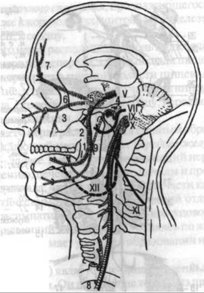
Fig. 123. Diagram of the branching pattern of the trigeminal (V), facial (VII), glossopharyngeal (IX), vagus (X), accessory (XI), and hypoglossal (XII) cranial nerves:
1 — lingual n., carrying parasympathetic secretory fibers from the chorda tympani; 2 — mandibular n.; 3 — maxillary n.; 4 — geniculate ganglion of the facial n. with the emerging greater petrosal n., containing parasympathetic secretory fibers for the lacrimal gland, nasal mucosa, and palate; 5 — trigeminal ganglion; 6 — ophthalmic n.; 7 — frontal n.; 8 — Water/144.html">Origin of the left recurrent laryngeal n. from the vagus n.; 9 — chorda tympani (after Lepp and Kogermann-Lepp)
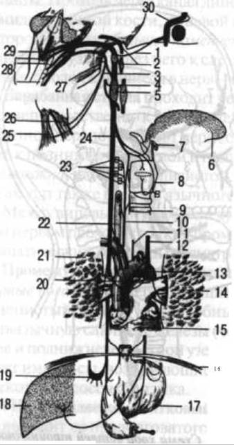
Fig. 124. Diagram of the course and innervation zones of the vagus and accessory nerves: 1 — communication between the vagus n. and the facial n.; 2 — communication between the vagus n. and the glossopharyngeal n.; 3 — accessory n.; 4 — communication between the vagus n. and the hypoglossal n.; 5 — communication between the vagus n. and the sympathetic trunk; 6 — tongue; 7 — Hyoid bone; 8 — larynx; 9 — Trachea; 10 — right recurrent laryngeal n.; 11 — left recurrent laryngeal n.; 12 — left vagus n.; 13 — aorta; 14 — left lung; 15 — Heart; 16 — Diaphragm; 17 — Stomach; 18 — Liver; 19 — celiac ganglion; 20 — cardiac ganglion; 21 — right lung; 22 — Esophagus; 23 — inferior laryngeal n.; 24 — superior laryngeal n.; 25 — trapezius m.; 26 — sternocleidomastoid m.; 27 — accessory n.; 28, 29 — nuclei of the vagus and accessory n.n.; 13 — facial n. (after Testut)
The cervical part of the vagus nerve runs alongside the common carotid artery and supplies branches to the cervical organs. The superior laryngeal nerve pierces the thyrohyoid membrane and distributes to the laryngeal mucosa. Its external branch innervates the cricothyroid muscle. All other laryngeal muscles are innervated by the inferior laryngeal nerve, which is the continuation of the recurrent laryngeal nerve. The latter branches off from the vagus nerve within the thoracic cavity, looping around the aortic arch on the left and the Subclavian Artery on the right. It ascends in the tracheoesophageal groove and supplies both structures.
In the cervical region, the vagus nerve gives off branches to the pharynx—contributing to the pharyngeal plexus, which continues as the esophageal plexus—as well as to the root of the tongue, Thyroid Gland, Thymus, and finally, The Heart.
The thoracic part of the vagus nerve lies alongside the esophagus, with the left vagus positioned anteriorly and the right vagus posteriorly. Within the thoracic cavity, the vagus nerve innervates the esophagus by forming the esophageal plexus, and also participates in the innervation of the Bronchi, Lungs, and heart. Its cardiac branches transmit impulses that slow the heart rate.
The abdominal part of the vagus nerve is situated primarily on The Stomach. The left vagus forms the anterior gastric plexus, while the right forms the posterior. Additionally, the vagus nerve sends branches to the celiac plexus, liver, Spleen, and Kidneys, and continues along the intestinal tract, serving as the primary parasympathetic nerve for the greater part of the gut tube. Only the lower section of the Large Intestine receives parasympathetic fibers from the sacral segment of the Spinal Cord. Distributing throughout the gastrointestinal tract, the vagus nerve participates in forming the intrinsic nerve plexuses of its wall.
XI. The Accessory nerve (see Fig. 123) functions as both a cranial and a spinal nerve, arising from both the medulla oblongata and the spinal cord. Its spinal roots enter the cranial cavity through the foramen magnum and exit again alongside the rest of the nerve via the jugular foramen. The accessory nerve is a motor nerve that, upon exiting the skull in the cervical region, courses posteriorly and inferiorly to innervate the sternocleidomastoid and trapezius muscles.
XII. The Hypoglossal nerve (Fig. 123) is a motor nerve. It emerges from the brain between the olive and the pyramid of the medulla oblongata, traverses the hypoglossal canal of the Occipital bone, and runs anteriorly and inferiorly in the neck to innervate the muscles of the tongue and the anterior Neck Muscles. On the anterior surface of the Internal jugular vein, the hypoglossal nerve anastomoses with the Anterior branches of the Cervical plexus. This loop-like anastomosis is termed the ansa cervicalis, from which branches depart to supply the anterior neck muscles.
The formation and general branching pattern of the spinal nerves were examined in the context of the spinal cord (see p. 284). The posterior branches of these nerves are mixed, containing motor fibers directed to the muscles and sensory fibers to the skin of the back. An exception is the posterior branch of the 1st cervical spinal nerve—the motor suboccipital nerve, which innervates the suboccipital muscles.
The anterior branches of the spinal nerves form plexuses: cervical, brachial, lumbar, sacral, and coccygeal.
The cervical plexus (Fig. 125) is formed by the anterior branches of the upper four cervical nerves. While the majority of its branches supply the skin, it also provides branches to striated muscles.
The cervical plexus communicates with the accessory, hypoglossal, and 5th cervical nerves, as well as with the sympathetic nervous system.
The cutaneous (sensory) NERVES OF THE cervical plexus include:
— the greater auricular nerve, which curves around the sternocleidomastoid muscle and ascends toward the auricle, participating in the innervation of the skin in that region;
— the lesser occipital nerve, which innervates the skin of the posterosuperior region of the neck;
— the transverse cervical nerve, which courses anteriorly across the superficial surface of the sternocleidomastoid muscle to innervate the skin of this area.
Supraclavicular nerves, which supply the skin of the corresponding region of the neck and the upper Thorax.
The branches of the cervical plexus include the mixed phrenic nerve. Descending along the anterior surface of the anterior scalene muscle, it enters the thoracic cavity, runs between the mediastinal Pleura and the Pericardium, supplying sensory branches to them, and upon reaching the diaphragm, innervates it as well. The unusual course of this nerve—running downward for a considerable distance from its point of origin from the cervical plexus—is explained by the fact that the diaphragm originates embryonically in the cervical region and subsequently descends inferiorly; the nerve grows downward following the descent of the diaphragm.

Fig. 125. Cutaneous branches of the right cervical plexus:
1 — posterior auricular m.; 2 — branch of facial n.; 3 — parotid gland; 4 — branches of facial n.; 5 — cervical branch of facial n.; 6 — transverse cervical n.; 7 — external jugular v.; 8 — platysma m.; 9 — omohyoid m. (inferior belly); 10 — supraclavicular nn.; 11 — trapezius m.; 12 — middle scalene m.; 13 — accessory n.; 14 — sternocleidomastoid m.; 15 — levator scapulae m.; 16 — splenius capitis m.; 17 — great auricular n.; 18 — occipital nn. (after Spalteholz)
The motor nerves of the cervical plexus supply the deep and anterior muscles of the neck that attach to the hyoid bone.
Brachial Plexus. The formation of the brachial plexus involves the anterior rami of the four lower cervical nerves and the first thoracic nerve (from the 5th to the 8th inclusively), anastomosing with other cervical and upper Thoracic nerves (Fig. 126). This plexus, like others, contains sensory and motor fibers, which are also joined by sympathetic fibers.
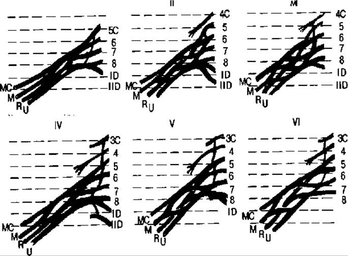
Fig. 126. Types of formation (I-VI) of the brachial plexus in humans:
3, 4, 5, 6, 7, 8, C, I, D - numbers of cervical (C) and thoracic (D) spinal nerves entering the plexus; MC - musculocutaneous n.; R - radial n.; U - ulnar n.; M - median n.; I-VI - types of plexus (after Loth)
The brachial plexus passes together with the subclavian artery through the interscalene triangle between the anterior and middle scalene muscles, directly above the 1st rib. According to its position relative to the clavicle, it is divided into supraclavicular and infraclavicular parts. The Short Branches of the brachial plexus include:
— dorsal scapular nerve, which innervates the scapular muscles (rhomboids, levator scapulae);
— long thoracic nerve, which innervates the serratus anterior muscle;
— suprascapular nerve, running to the supraspinatus and infraspinatus muscles and to the shoulder joint;
— thoracodorsal nerve, running inferiorly and slightly posteriorly, innervating the latissimus dorsi muscle;
— subscapular nerve, innervating the subscapularis and teres major muscles;
— medial and lateral pectoral nerves, running anteriorly and innervating the pectoralis Major and minor muscles and the skin of the chest.
The most massive of the short branches of the brachial plexus is the axillary nerve, which arises from the infraclavicular part of the brachial plexus. Emerging from the axilla through the quadrangular space bounded by the subscapularis muscle, humerus, teres major muscle, and long HEAD of the triceps brachii, this nerve wraps posteriorly around the humerus, passes beneath the deltoid muscle, and innervates this muscle along with the teres minor muscle.
The brachial plexus gives rise to three nerve cords: lateral, medial, and posterior. From these arise the nerves of the free upper limb, among which the largest and longest are the following.
Median nerve (Fig. 127, 128). It is formed by the medial and lateral cords of the brachial plexus and runs alongside the brachial artery. Passing into the forearm, this nerve pierces the pronator teres muscle and participates in the innervation of most muscles located on the anterior surface of the forearm: the pronator teres and pronator quadratus, flexor carpi radialis, flexor digitorum superficialis and partly flexor digitorum profundus, as well as the flexor pollicis longus and palmaris longus. Passing through the carpal tunnel, the median nerve enters the hand and innervates the skin and partly the muscles located on the thumb side. Specifically, it innervates "three and a half digits" (I, II, III, and the radial side of IV), while the remaining "one and a half digits" (V and the ulnar side of IV) are innervated by the ulnar nerve (Fig. 128).
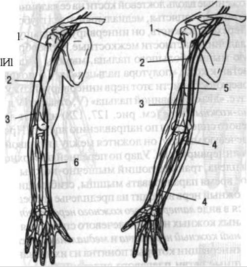
Fig. 127. Nerves of the upper limb:
1 — axillary n.; 2 — radial n.; 3 — ulnar n.; 4 — median n.; 5 — musculocutaneous n.; 6 — deep branch of radial n.
Radial nerve (see Fig. 127, 128). It is formed from the posterior cord of the brachial plexus and extends to the posterior surface of the humerus. In the arm, this nerve innervates the triceps brachii muscle. Passing into the forearm, it divides into two branches—superficial and deep. The first, which is thinner, descends distally along the radial artery and passes to the dorsal surface of the hand, where it innervates the skin of "two and a half digits" (I and partly II and III).
The second (primarily motor) branch courses posteriorly, innervating the extensor muscles of the wrist and fingers, as well as the supinator and anconeus muscles.
Ulnar nerve (see Fig. 127, 128) arises from the medial cord of the brachial plexus and descends along the medial aspect of the arm, winding posteriorly around the medial epicondyle of the humerus, and then runs through the ulnar sulcus, where it lies directly against the bone. Because the overlying layer of soft tissue here is thin, the nerve is easily vulnerable to compression. In such cases, it is squeezed between the bone and the soft Tissues. In the forearm, the ulnar nerve innervates the muscles located along the ulna on its palmar surface (flexor carpi ulnaris, medial part of the flexor digitorum profundus). Passing into the hand, it innervates most of the Deep Muscles of the palm, specifically the interossei, lumbricals (not all), adductor pollicis, muscles of the fifth digit, and in addition, gives off branches to the skin of "one and a half digits" (the Vth and half of the IVth). On the dorsal surface of the hand, this nerve innervates the skin of its ulnar half, i.e., "two and a half digits" (V, partly IV and III).
The musculocutaneous nerve (see Fig. 127, 128) extends anteriorly from the lateral cord of the brachial plexus. Piercing the coracobrachialis muscle, it runs between the biceps brachii and brachialis muscles, innervating them. A blow to the anterior surface of the biceps brachii that injures the musculocutaneous nerve can temporarily paralyze the forearm flexors. The musculocutaneous nerve emerges anteriorly and laterally on the forearm, continuing as the lateral cutaneous nerve of the forearm.
Among the other cutaneous nerves of the brachial plexus, the medial cutaneous nerve of the arm and the medial cutaneous nerve of the forearm are worthy of note; their innervation zones are apparent from their names.
Thus, the major branches of the brachial plexus are distributed across the free upper limb in the following manner.
On the arm, the forearm flexors are innervated by the musculocutaneous nerve, the forearm extensors by the radial nerve, the wrist and finger flexors primarily by the median nerve and partly by the ulnar nerve, the wrist and finger extensors by the radial nerve, and the flexor carpi ulnaris and partly the deep digital flexor by the ulnar nerve.
As previously mentioned, the anterior rami of the thoracic nerves do not form plexuses (see p. 331). They continue as the intercostal nerves and, running through the intercostal spaces, supply the intercostal muscles, as well as the skin and pleura. These nerves reach the body's midline, cross it, and extend slightly to the opposite side. Thus, the terminals of the right and left intercostal nerves overlap in the midline region. The lower six intercostal nerves, emerging into the abdominal region, play a crucial role in innervating the muscles of the anterior abdominal wall (the external and internal oblique muscles, transversus abdominis, rectus abdominis, and pyramidalis) and the abdominal skin. Furthermore, these nerves participate in the innervation of the diaphragm. The dorsal rami of the thoracic nerves innervate the skin, as well as the short and long muscles of the back. Communicating branches exist between the intercostal nerves and the cutaneous nerves of the upper limb.

Fig. 128. Innervation of the hand.
The Lumbar plexus (Fig. 129) is located posterior to the psoas major muscle and partly pierces it. It is formed by the anterior rami of the upper three or four lumbar spinal nerves (most commonly three and a half, as the fourth nerve contributes to both the lumbar and sacral plexuses). In addition, the lower intercostal nerve takes part in its formation. The lumbar plexus gives rise to numerous branches: short branches supply the adjacent muscles (psoas major and minor, quadratus lumborum, iliacus, and partly the muscles of the abdominal wall), while long branches extend to the free lower limb, innervating its skin and muscles.
The main branches of the lumbar plexus are as follows.
The iliohypogastric nerve, together with the ilioinguinal nerve, runs anteriorly, inferiorly, and laterally. They participate in the innervation of the skin and muscles of the abdominal wall (the internal oblique and transversus abdominis muscles). The ilioinguinal nerve also supplies the skin of the anterior thigh and external genitalia.
The genitofemoral nerve descends along the anterior surface of the psoas major muscle and proceeds toward the external genitalia and the skin of the thigh.
The femoral nerve (see Fig. 129) emerges onto the thigh beneath the inguinal ligament alongside the iliopsoas muscle. It gives off numerous cutaneous and muscular branches, innervating the muscles of the anterior thigh, notably the quadriceps femoris, as well as the skin of the anterior and medial thigh. Its longest cutaneous nerve is the saphenous nerve. Initially running alongside the femoral artery, it then passes to the medial surface of the leg, descends along it, crosses to the medial aspect of the FOOT, and reaches the big toe. This nerve is sensory in function.
The lateral cutaneous nerve of the thigh (see Fig. 129) passes beneath the inguinal ligament, innervating the skin of the lateral thigh.
The obturator nerve (Fig. 129) runs along the lateral wall of the lesser pelvis toward the obturator canal; after passing through it, it branches among the adductor muscles of the thigh and innervates them. It also contains sensory fibers that supply a small patch of skin on the medial surface of the thigh just above the knee joint.
The Sacral Plexus (Fig. 130, 131) is formed by the anterior rami of the lower one and a half lumbar and upper three and a half sacral nerves and lies on the anterior surface of the sacrum. Its branches innervate the gluteal region muscles, as well as a significant portion of the skin and Muscles of the Free Lower Limb.
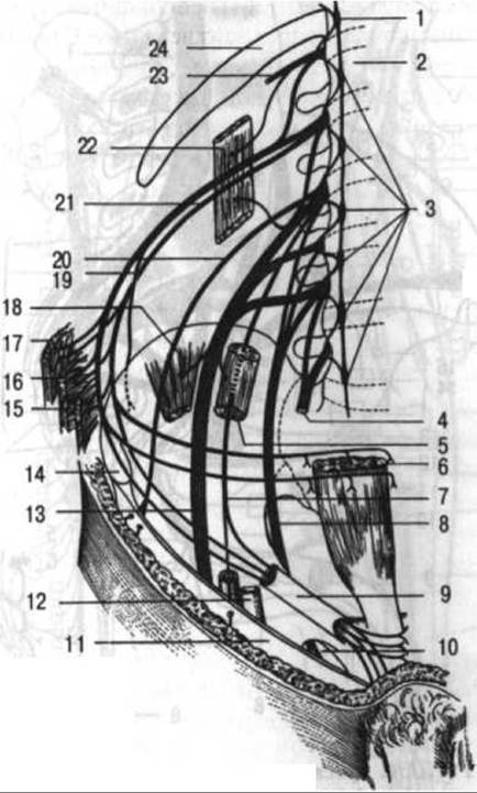
Fig. 129. Lumbar plexus:
1 - sympathetic trunk ganglion at the level of the 12th thoracic vertebra; 2 - 1st lumbar vertebra; 3 - sympathetic trunk; 4 - lumbosacral trunk; 5 - psoas major m.; 6 - rectus abdominis m.; 7 - genitofemoral n.; 8 - obturator n.; 9 - Inguinal Canal; 10 - obturator foramen; 11 - fascia; 12 - external iliac a.; 13 - femoral n.; 14 - anterior superior iliac spine; 15 - transversus abdominis m.; 16 - internal oblique m.; 17 - external oblique m.; 18 - iliacus m.; 19 - iliohypogastric n.; 20 - lateral cutaneous n. of thigh; 21 - ilioinguinal n.; 22 - quadratus lumborum m.; 23 - 12th intercostal n.; 24 - 12th rib (after Testut)
The branches of the sacral plexus include the following.
The superior and inferior gluteal nerves exit the pelvic cavity through the greater sciatic foramen, with the superior nerve emerging above the piriformis muscle and the inferior nerve below it. The superior gluteal nerve innervates the gluteus medius and minimus, while the inferior gluteal nerve supplies the gluteus maximus.
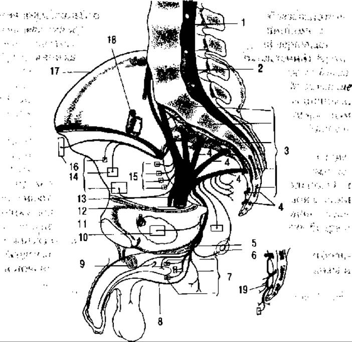
Fig. 130. Sacral and coccygeal plexuses:
1 — cauda equina; 2 — 5th lumbar vertebra; 3 — sacral vertebrae; 4 — anterior rami of sacral nn.; 5 — pudendal n.; 6 — inferior rectal nn.; 7 — perineal nn.; 8 — posterior scrotal nn.; 9 — corpora cavernosa; 10 - obturator internus m.; 11 — Pubic Symphysis; 12 — sciatic n.; 13 — posterior cutaneous n. of thigh; 14 — gluteal nn.; 15 — muscular branches; 16 — branches to fascia lata; 17 — iliac crest; 18 - external iliac vessels; 19 — Coccygeal plexus (after Testut)
The posterior cutaneous nerve of the thigh (see Fig. 131) exits the lesser pelvis through the infrapiriform foramen, runs down the posterior aspect of the thigh, and innervates its skin.
The sciatic nerve (see Fig. 131) is the largest nerve in The Human Body. It exits the lesser pelvis through the infrapiriform foramen and runs down the posterior surface of the thigh, deeply positioned in the groove between the biceps femoris (laterally) and the semimembranosus and semitendinosus muscles (medially). It gives off motor branches to these muscles. Near the superior angle of the popliteal fossa, the sciatic nerve divides into two major branches: the tibial nerve and the common fibular (peroneal) nerve.

Fig. 131. Diagram of the distribution of the nerves of the lower limb.
Left — posterior view: 1 — hip bone; 2 — piriformis m.; 3 — posterior cutaneous n. of thigh; 4 — sciatic n.; 5 — gluteal n.; 6 — branch to biceps femoris m.; 7 — common fibular n.; 8 — sural n.; 9 — medial plantar n.; 10 — lateral plantar n.; 11 — medial malleolus; 12 — Fibula; 13 — branches of tibial n.; 14 — popliteal a. and v.; 15 — branches to adductor mm. of thigh; 16 — branch to semimembranosus m.; 17 — branch to semitendinosus m. Right — anterior view: 1 — femoral n.; 2 — obturator n.; 3 — pubic bone; 4 — branch to pectineus m.; 5 — branch to gracilis m.; 6 — branches to adductor mm.; 7 — saphenous n.; 8 — branches to anterior mm. of leg; 9 — lateral dorsal cutaneous n. (of foot); 10 — branches to fibular mm.; 11 — common fibular n.; 12 — branches to quadriceps femoris m.; 13 — lateral cutaneous n. of thigh; 14 — branch to sartorius m.; 15 — branch to skin of thigh and genitalia (after Testut)
The tibial nerve (see Fig. 131) runs through the popliteal fossa and continues down the leg, innervating the triceps surae, tibialis posterior, flexor hallucis longus, and flexor digitorum longus muscles. Passing posterior to the medial malleolus, it enters the PLANTAR ASPECT OF the foot, where it divides into two branches — the lateral and medial plantar nerves, which supply the skin and muscles of the sole.
The common fibular nerve (Fig. 131) runs along the lateral border of the popliteal fossa, curves laterally and inferiorly around the neck of the fibula, and divides into two branches — the superficial and deep fibular nerves.
The superficial fibular nerve courses down the lateral surface of the leg, innervating the fibular muscles. Upon reaching the dorsum of the foot, it branches out within the skin, functioning solely as a cutaneous (sensory) nerve in this region.
The deep fibular nerve passes through the anterior muscle compartment of the leg, supplying the Anterior Group of muscles (tibialis anterior, long extensors of the toes). Like the superficial nerve, it continues onto the dorsum of the foot, where it innervates the dorsal foot muscles and, exclusively in the cleft between the 1st and 2nd toes, the skin.
The tibial and common fibular nerves anastomose with each other to form the sural nerve (Fig. 131), which supplies the skin. It descends along the posterior aspect of the leg, curves around the lateral malleolus, and continues onto the dorsum of The Foot as the lateral dorsal cutaneous nerve.
Thus, the sciatic nerve and its branches innervate the muscles of the posterior thigh, as well as all the Muscles of the Leg and foot; they also supply the skin of the lateral and posterior leg (with the medial side supplied by a branch of the femoral nerve — the saphenous nerve), the skin of the sole, and the skin of the dorsal and lateral surfaces of the foot (see Fig. 131).
The muscles of the anterior thigh are innervated by the femoral nerve, while the muscles of the medial thigh are supplied by the obturator nerve. Consequently, both the lumbar and sacral plexuses contribute to the innervation of the lower limb.
The branches of the sacral plexus also include the pudendal nerve. It passes through the greater sciatic foramen, innervating the skin and muscles of the Perineum as well as the external genitalia, and within the lesser pelvis, it gives off branches to the rectum, Urinary Bladder, Vagina, and Uterus.
The coccygeal plexus is formed by the 5th sacral and coccygeal nerves. Its branches supply the coccygeus muscle and the skin in the region of the coccyx.
Last update: 08/08/2026
Editorial and Educational Adaptation: This material has been compiled based on the primary/original source text. The project team performed an editorial review, corrected technical inaccuracies, structured sections, and adapted the content for an educational format.
What was processed:
- elimination of formatting defects (OCR errors, structural breaks, corrupted characters);
- editorial organization of content;
- standardization of terminology in accordance with academic sources;
- verification of factual statements against the original source text.
All mentions of the author, publication year, and origin of the primary text have been preserved in accordance with the source.