Human Anatomy (with the Fundamentals of Dynamic and Sports Morphology) - Ivanitsky M. F. 2008
The Doctrine of the Nervous System
Autonomic (Vegetative) Nervous System
Both names for this part of The Nervous system are conventional. Literally, the term "autonomous" means self-governing, while "vegetative" refers to plant life. Since the body Functions as a unified whole where all structures are interconnected, there are no truly self-governing Organs or systems within it.
It was previously believed that the action of the Autonomic (vegetative) nervous system extended exclusively to the so-called organs of vegetative life, which included all Internal Organs—specifically the digestive, respiratory, urogenital, and vascular systems, Endocrine glands, all other glands, and all smooth Muscle formations. However, the Autonomic Nervous System also innervates striated musculature, regulating its metabolic processes and tone. Thus, the autonomic nervous system participates in the innervation of all Organs of the body, remaining subordinate to the Central Nervous System, particularly the Cerebral Cortex.
Class="center">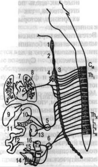
Fig. 132. Diagram showing The Emergence of sympathetic fibers and their areas of distribution
The autonomic nervous system comprises central and peripheral divisions. Its centers are located in the Spinal Cord and Brain (Figs. 132, 133). The Nerve Cells making up the autonomic system form ganglia situated both along THE Vertebral Column and within the organs they innervate.
The autonomic nervous system is divided into two major divisions: the sympathetic and the parasympathetic. They exhibit morphological, functional, and pharmacological differences.
Morphological differences lie in the fact that these two divisions are connected to different Regions of the central nervous system: the sympathetic division has its centers in the lateral horns of the thoracic and partially lumbar segments of the spinal cord, whereas the parasympathetic division is centered in the Midbrain, Medulla Oblongata, and sacral segments of the spinal cord. Another morphological distinction is the length of the axons of preganglionic and postganglionic Neurons. In the sympathetic division, preganglionic fibers originate from cells in the lateral horns of the spinal cord and terminate on cells forming the Sympathetic trunk—which runs along the vertebral column—as well as in ganglia located between the vertebral ganglia and the innervated organ. Postganglionic fibers travel to the target organs either as part of somatic nerves (such as limb nerves) or along Blood Vessels. Preganglionic fibers of parasympathetic nerves run within peripheral nerves directly to the innervated internal organs (Heart, Stomach, intestines, Urinary Bladder) and terminate there in ganglionic cells embedded in their walls, the so-called intramural ganglia. Unlike sympathetic fibers, parasympathetic postganglionic fibers are short, as the pathway from the nerve cells located within the organ itself to its Tissues is very brief.
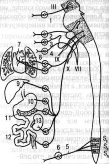
Fig. 133. Diagram showing the emergence of parasympathetic fibers and their areas of distribution:
1 — ciliary ganglion; 2 — pterygopalatine ganglion; 3 — otic ganglion; 4 — submandibular ganglion; 5 — pelvic splanchnic nerve; 6 — hypogastric plexus; 7 — heart; 8 — lung; 9 — Liver; 10 — stomach; 11 — Pancreas; 12 — Small Intestine; 13 — Kidney; III — Oculomotor nerve; IX — Glossopharyngeal nerve; X — Vagus nerve; VII — Facial Nerve; SA—SA — sacral segments (after V. N. Tonkov)
Functional differences consist in the fact that the sympathetic and parasympathetic Divisions of the autonomic nervous system sometimes exert opposing effects on the body. For instance, the sympathetic division innervates the pupillary dilator muscle, whereas the parasympathetic division innervates the pupillary constrictor (sphincter) muscle, and so forth.
Pharmacological differences involve the distinct Chemical composition of the neurotransmitter substances released upon excitation of sympathetic and parasympathetic nerves. For sympathetic neurons, this transmitter is norepinephrine, while for parasympathetic neurons, it is acetylcholine.
However, none of these three criteria of distinction is absolute, and the division of the autonomic system into sympathetic and parasympathetic is, in a sense, conventional.
Centripetal nerve fibers, which transmit impulses from the periphery to the center, equally "serve" both the somatic and the autonomic nervous system.
The Sympathetic division of the vegetative (autonomic) nervous system
The basis of the peripheral section of the sympathetic nervous system is formed by ganglia (see Fig. 132) that constitute the right and left sympathetic trunks, located on either side of the vertebral column. Adjacent superior and inferior ganglia are interconnected by interganglionic branches. Additionally, in the thoracic, lumbar, and sacral regions, there are connections between the ganglia of the right and left sides. Each ganglion is connected to the central nervous system (via white communicating branches consisting of preganglionic nerve fibers) and to the peripheral somatic system (via gray communicating branches consisting of postganglionic fibers). Finally, branches extend from each ganglion to the organs innervated by the sympathetic system. It should be noted that somatic sensory fibers are present within all sympathetic nerves and ganglia.
In the cervical region of the sympathetic division of the autonomic nervous system, there are only 3 ganglia, the largest of which is the superior cervical ganglion. It lies directly on the vertebral column at the level of the upper cervical vertebrae and anastomoses with the vagus nerve and Branches of the Cervical plexus. Branches extend upward from this ganglion (the internal carotid nerve) and downward (to the organs of the neck and The Heart). The internal carotid nerve runs toward the Internal Carotid Artery and, by branching, participates in The formation of the internal carotid plexus surrounding the artery. It is worth noting that one of the Characteristic Features of the distribution of the sympathetic division of the autonomic nervous system is the course of its branches along blood vessels. In this case, the continuation of the internal carotid plexus comprises all the plexuses accompanying the branches of the internal carotid artery. Thus, nerve fibers extending from the superior cervical ganglion accompany the Arteries supplying the brain, Salivary Glands, orbital structures, and other organs.
The middle cervical ganglion is significantly smaller than the superior one and may even be absent. At the same time, the middle cervical ganglion may split into smaller ones, meaning that the cervical portion of the sympathetic trunk can possess not 3, but a greater number of ganglia (up to 8).
The inferior cervical ganglion is small. Sometimes it fuses with the first thoracic ganglion of the sympathetic trunk to form the so-called cervicothoracic (stellate) ganglion. A well-defined anastomosis, termed the subclavian loop, exists between the middle and inferior cervical ganglia, encircling the Subclavian Artery. Branches extend from the stellate ganglion to the cords of the Brachial Plexus, along the course of which the sympathetic division of the autonomic nervous system is distributed to the upper limb (to blood vessels, Skin, and Muscles). The cervical ganglia give off branches to blood vessels and, through them, to the organs of the neck and thoracic cavity (specifically, to the aorta and the heart).
The thoracic region of the sympathetic division of the autonomic nervous system contains 10–12 ganglia. Its segmental Organization is more pronounced than in other regions. The thoracic ganglia are located on the heads of the Ribs and provide gray communicating branches to the intercostal nerves.
Two large nerves, termed splanchnic nerves, arise from the thoracic region. The greater splanchnic nerve originates from the 6th–9th thoracic ganglia of the sympathetic trunk. The lesser splanchnic nerve arises from the 10th–11th ganglia. Both of these nerves pass downward through the Diaphragm and enter the celiac (solar) plexus of the lumbar region. They contain not only sympathetic nerve fibers carrying centrifugal impulses, but also fibers conducting sensory impulses from internal organs to the spinal cord via its dorsal roots—that is, traveling in a centripetal direction.
The lumbar, sacral, and coccygeal regions of the sympathetic division of the autonomic nervous system feature ganglia that converge inferiorly, such that only a single unpaired coccygeal ganglion is formed in the region of the coccyx. Typically, there are 4 lumbar ganglia and 4 sacral ganglia. As already mentioned, the lumbar and sacral ganglia of the right and left sides anastomose with each other. They give rise to communicating branches directed toward the lumbar and sacral somatic nerves.
The celiac (solar) plexus (see Fig. 132) is situated at the level of the 1st lumbar vertebra and surrounds the celiac trunk (arising from the Abdominal Aorta). It consists of two large celiac ganglia, right and left, which lie on either side of the celiac trunk and anastomose with each other. Numerous branches extend from the celiac plexus, distributing themselves along the blood vessels.
Despite the fact that the celiac plexus is located quite deeply, blows to the epigastric region can severely traumatize it, which may reflexively induce a knockout state. In this process, the Nerve Impulse is transmitted via sensory fibers of the splanchnic nerves to the spinal cord and subsequently to the medulla oblongata, to the center of the vagus nerve. The excitation then travels centrifugally along the vagus nerve to The Heart and other organs. It should be added that the respiratory response to this stimulus is not always uniform: in some cases, breathing becomes more frequent, while in others it is sharply slowed down, even to the point of cessation.
Large ganglia of the lumbar region of the sympathetic division of the autonomic nervous system include the superior and inferior mesenteric ganglia, which correspond in their anatomical position to the points of Water/144.html">Origin of the superior and inferior mesenteric arteries from the aorta.
As previously mentioned, the sympathetic trunk is connected exclusively to the thoracic and lumbar segments of the spinal cord (see Fig. 132). Consequently, the ganglia of this trunk located in the cervical, sacral, and coccygeal regions have no direct connection to the spinal cord. This connection is established circuitously via preganglionic fibers that pass through the thoracic and lumbar ganglia and subsequently reach higher and lower sections of the sympathetic trunk via interganglionic branches.
The sympathetic plexus surrounding the abdominal aorta extends along its branches to the abdominal organs. Blood vessels of the limbs receive sympathetic innervation from fibers of adjacent somatic nerves. The median and sciatic nerves are particularly rich in sympathetic fibers. Along the gastrointestinal tract, the sympathetic plexus, together with branches of the Parasympathetic division of the autonomic nervous system, forms plexuses located beneath both the mucous membrane and the serous layer.
The parasympathetic division of the autonomic (vegetative) nervous system
The parasympathetic division of the autonomic nervous system is subdivided into cranial and pelvic divisions (see Fig. 133). In turn, the cranial division comprises the midbrain and medullary (Brainstem) components.
The midbrain division partially includes the oculomotor nerve (see p. 362). Its parasympathetic fibers enter the ciliary ganglion and proceed into the Eyeball. Here, they innervate the sphincter pupillae muscle, whereas the dilator pupillae muscle receives innervation from the sympathetic division of the autonomic nervous system, whose branches reach the ciliary ganglion from the plexus accompanying the ophthalmic artery.
The fibers of the medullary division of the parasympathetic autonomic nervous system travel within the facial (see p. 367), glossopharyngeal (see p. 316), and vagus (see p. 369) nerves. Parasympathetic Innervation of the HEAD organs is provided by postganglionic neurons of the ciliary, pterygopalatine, otic, and submandibular ganglia (see p. 366).
The vagus nerve is a mixed nerve. It contains a relatively larger proportion of parasympathetic fibers than the oculomotor and facial nerves, as the majority of these fibers are directed toward the internal organs. The parasympathetic fibers supply the gastrointestinal tract with impulses that enhance peristalsis and stimulate the secretion of digestive glands. To the heart, the vagus nerve delivers fibers that transmit impulses slowing the heart rate and constricting its blood vessels.
The pelvic division of the parasympathetic autonomic nervous system is relatively small. Its spinal centers are located in the lateral horns of only two to three sacral segments of the spinal cord. Nevertheless, this division participates in the formation of numerous autonomic nerve plexuses that innervate the abdominal and pelvic organs, including the descending, sigmoid, and rectum, the Uterus, the urinary bladder, and other pelvic viscera. The pelvic division supplies vasodilator fibers to the corpora cavernosa of the Penis. Within the walls of these organs, parasympathetic and sympathetic nerves form plexuses containing scattered intramural nerve cells.
Last update: 08/08/2026
Editorial and Educational Adaptation: This material has been compiled based on the primary/original source text. The project team performed an editorial review, corrected technical inaccuracies, structured sections, and adapted the content for an educational format.
What was processed:
- elimination of formatting defects (OCR errors, structural breaks, corrupted characters);
- editorial organization of content;
- standardization of terminology in accordance with academic sources;
- verification of factual statements against the original source text.
All mentions of the author, publication year, and origin of the primary text have been preserved in accordance with the source.