Human Anatomy - Kotsan I. Y. 2009
Peripheral nervous system
Spinal nerves
Lumbar plexus
The lumbar plexus (plexus lumbalis) is formed by a portion of the fibers from the anterior ramus of the twelfth thoracic nerve (ThXII), the anterior rami of the upper three lumbar nerves (LI-LIII), and the upper part of the anterior ramus of the fourth lumbar nerve (LVI) (Figs. 261, 262). The second part of the anterior ramus of the fourth lumbar spinal nerve descends into the pelvic cavity and, together with the anterior ramus of the fifth lumbar nerve (LV), forms the lumbosacral trunk.
Class="center">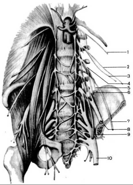
Fig. 261. Lumbar and sacral plexuses (on the left half of the specimen, the Muscles and part of the pelvic bones are removed); anterior view (according to M. R. Sapin):
1 — subcostal nerve; 2 — iliohypogastric nerve; 3 — ilioinguinal nerve; 4 — genitofemoral nerve; 5 — genital branch of the genitofemoral nerve; 6 — femoral branch of the genitofemoral nerve; 7 — lateral femoral cutaneous nerve; 8 — femoral nerve; 9 — obturator nerve; 10 — sciatic nerve
The lumbar plexus is located anterior to the transverse processes of the lumbar vertebrae and the quadratus lumborum Muscle, posterior to the psoas major muscle, and partially within the substance of the latter. The branches emerging from the lumbar plexus appear from beneath the lateral margin of the psoas major muscle or pierce it, running towards the anterior abdominal wall, the lower limb, and the external genitalia.
The Branches of the lumbar plexus include the following nerves: muscular branches, iliohypogastric nerve, ilioinguinal nerve, genitofemoral nerve, lateral femoral cutaneous nerve, femoral nerve, and obturator nerve.
The muscular branches (rr. musculares) are short, originating from the anterior rami of the lumbar nerves even before their interconnection, and innervate the quadratus lumborum, psoas Major and minor, and the lateral intertransversarii Muscles of the lumbar region.
The iliohypogastric nerve (n. iliohypogastricus) (ThXII-LI) emerges from beneath the lateral margin of the psoas major muscle and runs laterally across the anterior surface of the quadratus lumborum muscle. It then courses anteriorly and inferiorly, parallel to the subcostal nerve, within the substance of the lateral abdominal wall between the internal oblique and transversus abdominis muscles, innervating the lateral Abdominal muscles, and divides into two sensory terminal branches: the lateral and anterior cutaneous branches. The lateral cutaneous nerve (r. cutaneus lateralis) innervates the Skin of the upper lateral gluteal region. The anterior cutaneous nerve (r. cutaneus anterior) pierces the aponeurosis of the external oblique abdominal muscle above the superficial inguinal ring and innervates the skin of the abdomen above the pubic region.
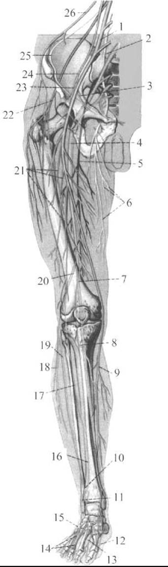
Fig. 262. Lumbosacral plexus and NERVES OF THE free lower limb (according to R. D. Sinelnikov)
1 — femoral nerve; 2 — obturator nerve; 3 — genital branch of the genitofemoral nerve; 4 — sciatic nerve; 5 — cutaneous branch of the femoral nerve; 6 — cutaneous branches of the obturator nerve; 7 — tibial nerve; 8 — medial sural cutaneous nerve; 9 — saphenous nerve; 10 — medial dorsal cutaneous nerve; 11 — intermediate dorsal cutaneous nerve; 12 — medial plantar nerve; 13 — lateral plantar nerve; 14 — dorsal digital nerves of the FOOT; 15 — lateral dorsal cutaneous nerve; 16 — sural nerve; 17 — lateral sural cutaneous nerve; 18 — superficial peroneal (fibular) nerve; 19 — deep peroneal (fibular) nerve; 20 — common peroneal (fibular) nerve; 21 — anterior cutaneous branches; 22 — lateral femoral cutaneous nerve; 23 — femoral branch of the genitofemoral nerve; 24 — genitofemoral nerve; 25 — ilioinguinal nerve; 26 — iliohypogastric nerve
The ilioinguinal nerve (n. ilioinguinalis) (ThXII-LI) emerges from beneath the lateral margin of the psoas major muscle, inferior to the iliohypogastric nerve, and runs almost parallel to it. Further on, the ilioinguinal nerve passes between the internal oblique and transversus abdominis muscles, innervates the lateral abdominal muscles, and enters the Inguinal Canal. Upon exiting the canal through the superficial inguinal ring, the nerve terminates in the skin of the pubic region, the Scrotum (anterior scrotal nerves), or the Labia Majora (anterior labial nerves). The anterior scrotal nerves (nn. scrotales anteriores) in males innervate the skin of the ROOT of the Penis, the anterior scrotum, the pubic region, and the inguinal region. The anterior labial nerves (nn. labiales anteriores) in females innervate the skin of the labia majora, the inguinal region, and the pubic region.
The genitofemoral nerve (n. genitofemoralis) (LII) passes through the substance of the psoas major muscle to its anterior surface and divides into two branches: the genital and femoral branches.
The genital branch (r. genitalis) pierces the posterior wall of the inguinal canal, where it lies posterior to the Spermatic Cord or the round ligament of the Uterus. In males, this branch innervates the cremaster muscle, the dartos tunic of the scrotum, the skin of the scrotum, and the skin of the upper medial aspect of the thigh. In females, the genital branch ramifies within the round ligament of the uterus and the skin of the labium majus.
The femoral branch (r. femoralis) enters the thigh through the vascular lacuna, lying lateral to the femoral artery, and innervates the skin of the upper medial aspect of the thigh immediately inferior to the inguinal ligament.
The lateral femoral cutaneous nerve (n. cutaneus femoris lateralis) (LI, LIII) emerges from beneath the lateral margin of the psoas major muscle, runs inferiorly along the anterior surface of the iliacus muscle, pierces the abdominal wall, enters the thigh, and descends along the lateral aspect of the thigh to the knee. This nerve innervates the skin of the inferoposterior gluteal region and the skin of the lateral aspect of the thigh down to the level of the knee joint.
The femoral nerve (n. femoralis) (LI-LIV) is the largest branch of the lumbar plexus; it emerges from beneath the lateral margin of the psoas major muscle and runs beneath the iliac fascia between the psoas major and iliacus muscles. The nerve then passes through the muscular lacuna into the thigh, where it divides into muscular branches, anterior cutaneous branches, and the saphenous nerve.
The muscular branches (rr. musculares) of the femoral nerve innervate the pectineus, sartorius, and quadriceps femoris muscles.
The anterior cutaneous branches (rr. cutanei anteriores) of the femoral nerve, numbering 3 to 5, pierce the fascia lata and innervate the skin of the anteromedial aspect of the thigh.
The saphenous nerve (n. saphenus) is a direct continuation of the femoral nerve. Initially, it lies alongside the femoral artery, then passes to the Medial surface of the leg, descending along it to continue onto the medial aspect of The Foot as far as the big toe. Functionally, this nerve is sensory, innervating the skin of the medial aspect of the knee joint, the Patella, the medial surface of the leg, and the medial border of the foot down to the big toe.
The obturator nerve (n. obturatorius) (LII-LIV) is the second largest branch of the lumbar plexus. Functionally, it is a mixed nerve. It emerges from beneath the medial margin of the psoas major muscle and descends along the lateral wall of the lesser pelvis. Passing through the obturator canal into the thigh, it gives off muscular branches (rr. musculares) to the adductor muscles of the thigh and divides into two terminal branches: anterior and posterior. The anterior branch (r. anterior) is a continuation of the main trunk of the obturator nerve; it passes between the adductor brevis and adductor longus muscles, innervating these muscles as well as the gracilis and pectineus muscles. Distally, the anterior branch continues as the cutaneous branch (r. cutaneus), which innervates the skin of the medial aspect of the thigh. The posterior branch (r. posterior) of the obturator nerve pierces the obturator externus muscle, lies between the adductor magnus and adductor brevis muscles, and innervates these muscles and the hip Joint Capsule.
The main nerves of the lumbar plexus are listed in Table 11.
Table 11 NERVES OF THE LUMBAR PLEXUS
|
Nerves |
Course of the nerve |
Branches |
Area of innervation |
|
Muscular branches (rr. musculares) |
Originate from the anterior rami of the plexus prior to their interconnection and run to the muscles |
Muscles: psoas major and minor, lateral intertransversarii of the lumbar region, quadratus lumborum |
|
|
Iliohypogastric nerve (n. iliohypogastricus) |
Emerges from beneath the lateral margin of the psoas major muscle, runs across the anterior surface of the quadratus lumborum muscle, pierces the transversus abdominis muscle, and extends toward the rectus abdominis |
Muscles: transversus abdominis, internal and external oblique abdominal, rectus abdominis; skin of the anterior abdominal wall above the pubic region and the upper lateral thigh |
|
|
Ilioinguinal nerve (n. ilioinguinalis) |
Runs parallel and inferior to the preceding nerve, then passes through the inguinal canal |
Anterior scrotal branches (in males). Anterior labial branches (in females) |
Skin of the inguinal region, pubic region, scrotum (in males), labium majus (in females), upper medial aspect of the thigh, external and internal oblique abdominal muscles, transversus abdominis muscle |
|
Genitofemoral nerve (n. genitofemoralis) |
Pierces the psoas major muscle, lies initially beneath its fascia, then courses anterior to it toward the internal inguinal ring, passes through the canal, and exits via the superficial inguinal ring together with the cremaster muscle, descending to the scrotum (genital branch) and inferior to the inguinal ligament (femoral branch) to the skin of the thigh in the region of the saphenous opening |
1. Femoral branch; 2. Genital branch |
Skin of the thigh inferior to the inguinal ligament Skin of the scrotum; Tunics of the Testis; cremaster muscle (in males); round ligament of the uterus, skin of the labium majus (in females) |
|
Lateral femoral cutaneous nerve (n. cutaneus femoris lateralis) |
Emerges from beneath the lateral margin of the psoas major muscle, runs toward the anterior superior iliac spine, where it enters the thigh |
Skin of the posterolateral aspect of the thigh (over the tensor fasciae latae), lateral aspect of the thigh down to the knee joint |
|
|
Obturator nerve (n. obturatorius) |
Descends inferiorly along the medial margin of the psoas major muscle, passing through the obturator canal into the thigh |
Hip joint capsule, skin of the medial aspect of the thigh in its lower half, obturator externus muscle, adductor muscles of the thigh, gracilis and pectineus muscles |
|
|
Femoral nerve (n. femoralis) |
The largest trunk of the lumbar plexus, passes between the psoas major and iliacus muscles and emerges onto the anterior surface of the thigh through the muscular lacuna |
1. Muscular branches; 2. Anterior cutaneous branches; 3. Saphenous nerve |
Muscles: quadriceps femoris, pectineus, sartorius. Skin of the anteromedial aspect of the thigh, skin in the region of the knee joint, anteromedial surface of the leg, dorsum and medial aspect of the foot down to the big toe |
Last update: 08/08/2026
Editorial and Educational Adaptation: This material has been compiled based on the primary/original source text. The project team performed an editorial review, corrected technical inaccuracies, structured sections, and adapted the content for an educational format.
What was processed:
- elimination of formatting defects (OCR errors, structural breaks, corrupted characters);
- editorial organization of content;
- standardization of terminology in accordance with academic sources;
- verification of factual statements against the original source text.
All mentions of the author, publication year, and origin of the primary text have been preserved in accordance with the source.