NEONATAL SURGERY - 1976
1. GENERAL PROVISIONS
5. Specifics of Surgical Intervention and Examination
Cytology/cytology/25.html">General characteristics and surgical technique in newborns
The outcome of a surgical intervention in a newborn infant depends, to a far greater extent than in other age groups, on the meticulous execution of surgical techniques and strict adherence to technical guidelines.
The General Principles of performing surgical interventions in children, including newborns, are outlined in the works of S. D. Ternovsky (1959), G. A. Bairov (1963), S. Ya. Doletsky and A. G. Pugachev (1968), Obermerdermaier (1959), Swenson (1970), and others. These publications provide a clear understanding of what needs to be done for a particular developmental anomaly, but a less distinct picture of how the surgical intervention should actually be performed.
Even in such manuals as "Pediatric Operative Surgery" edited by E. M. Margorin (1960), "Surgery of Developmental Anomalies in Children" edited by G. A. Bairov (1968), and "Pediatric Surgery (A Guide for Physicians)" by S. Ya. Doletsky and Yu. F. Isakov (1970), the specifics of surgical technique in newborns are not covered comprehensively enough. The only publication that briefly outlines the fundamental principles of pediatric surgical technique dates back to 1964.1
1 Doletsky S. Ya. Specifics of surgical technique in children. — Klinicheskaya Khirurgiya, 1964, no. 3.
The specifics of surgical technique are dictated by the anatomical, physiological, and topographical CHARACTERISTICS OF THE newborn Organism.
The newborn is brought into the operating room in an incubator. The optimal air Temperature in the operating theater is 23 — 27° C. The operating table must be equipped with a device for continuous warming of the infant during surgery, in the form of an automatically regulated electric heater or a special blanket (rubber mattress) circulating warm Water (Ferguson, 1962; Rickham, 1969). We prefer a simple rubber mattress filled with water warmed to 37° C prior to surgery. The mattress is wrapped in a sterile sheet. The infant is surrounded with a layer of cotton wool (leaving only the operative field exposed) and placed on the warm mattress. This method maintains the required body temperature of the infant for up to 2 hours—a duration sufficient for virtually all surgical Procedures.
The infant is positioned According to the upcoming surgery. Diatermoagulation electrodes and monitoring device sensors are attached.
The operative field is prepared by applying 1% iodine tincture and 70° alcohol twice. To reduce heat loss and prevent wound infection during surgery, the majority of foreign surgeons (Gross, 1970, et al.) recommend covering the operative field with a special sterile adhesive or plastic drape, through which the Skin incision is made. This technique prevents potential wound infection.
When dividing Tissues, manipulating Organs, and placing sutures in newborns, the surgeon must maintain a constant awareness of dealing with extremely delicate, fragile, and at times gelatinous tissues, touching which carries the risk of inadequate trauma. The difficulty of self-critically evaluating one's own manipulations in these cases stems from the apparent success of the completed surgery. When postoperative complications arise, their direct link to violations of surgical technique rules is far from always obvious.
The skin incision in newborn surgery is performed in several stages. Rapid, single-stage tissue division to the full depth is unjustified; Blood loss and traumatic stress resulting from deep, rough manipulations exacerbate the severity of the infant's condition. The division of subcutaneous adipose tissue and Muscle layers is performed using a fine diathermic needle. It is important to regulate the current so that "dry" tissue Separation occurs without charring the wound edges. Proper and systematic application of electrocoagulation significantly reduces blood loss, minimizes postoperative pain, and preserves tissue repair processes. Slow, phased, layer-by-layer tissue division allows the surgeon to visualize emerging Blood Vessels and perform "preventative" electrocoagulation before transection. This is best achieved by using ophthalmic forceps or a specialized bipolar forceps-shaped electrode. During surgery, the open portion of the wound is covered with warm, moist gauze Sponges to prevent subsequent infection. The Use of Kocher-type clamps is ruled out; they are heavy and traumatize the grasped tissues (Fig. 9, a, b). Sharp tissue dissection is preferable. Blunt dissection is carried out with careful, jerking movements that limit the possibility of straying from the correct anatomical plane. In some cases, tissue separation can be facilitated by preliminary hydraulic dissection with a 0.25% novocaine solution. To avoid novocaine intoxication, no more than 10 ml of the solution should be administered during surgery. Excess blood and fluid are removed from the wound using suction, avoiding the suction tip coming into direct contact with tissues.
Class="center">Fig. 9. Hemostasis by conventional and coagulation Methods.
Applying a hemostatic clamp to a bleeding vessel causes a significant zone of tissue crushing followed by necrosis. A capsule forms around the ligature, and a prolonged perifocal reaction develops (a). The use of electrocoagulation is accompanied by a smaller zone of carbonaceous coagulum, which is rapidly resorbed and replaced by Connective Tissue (b).


Among the factors influencing subsequent wound healing (trophism, Homeostasis, atraumatic intervention, use of adequate suture material, etc.), of particular importance is the surgeon's ability to select the optimal distance between sutures, the optimal depth of suture placement, and the correct degree of tension when tying knots (Fig. 10).
Fig. 10. Specifics of surgical technique in the newborn.
Unlike the skin of an adult (b), the skin of a newborn infant (a) features an abundant, delicate vascular network and a sparse elastic framework consisting of fine fibers. The newborn's Elastin is easily compressible and unable to withstand even relatively minor compression extending to the vascular network (c). The skin elastin of an adult (d) is more resilient.
The use of forceps in neonatal surgery (d) is accompanied by trauma to delicate tissues, primarily
vessels, leading to Hemorrhage (e). When placing sutures (g), if knots are tied until tissues are tightly approximated, ischemia (h) and tissue edema (i) occur, culminating in necrosis (k).
Proper suture placement (not only in the skin, but in any tissues and Organs of the newborn, and primarily the Esophagus, rectum, Ureters, and Urinary Bladder) involves abandoning forceps as a "grasping" instrument (l), tying knots with a certain strictly dosed slack (m, n) calculated to allow complete apposition of all wound layers after inevitable edema develops (o), but without vessel compression or tissue ischemia.

The skin edges of the wound are approximated with minimal tension whenever possible, as tension impairs Blood supply and reparative processes in the area. Fine synthetic threads or chromic catgut on an atraumatic needle (size № 0000 — 00000) are the suture material of choice. When operating on the digestive tract, blood vessels, Urinary Tract, and other Organ Systems, the use of atraumatic needles is mandatory. If there is any doubt about complete hemostasis, wound drainage is indicated by placing a thin rubber strip cut from a surgical glove into the subcutaneous tissue for 24 hours. For extensive wounds resulting from the excision of large tumors, sacrococcygeal teratomas, and similar pathologies, a system of tubes is placed at the Base of the wound, and continuous suction of wound secretions is maintained for the first 1 — 2 postoperative days (Fig. 11). This simple technique effectively prevents fluid accumulation in the wound and promotes faster healing.
Fig. 11. Postoperative wound drainage.
Active suction from extensive wounds prone to the accumulation of blood, Lymph, and tissue fluid is performed using double-lumen perforated tubes connected to a system generating a negative pressure of — 20 — 40 mm water Column.
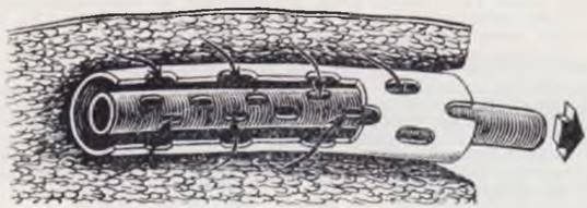
In recent years, various types of plastics utilized as temporary and permanent prostheses have gained increasing acceptance in neonatal surgery (Heiss, 1970; Lister, 1971).
Protecting a newborn's postoperative wound with a gauze dressing is inconvenient. The dressing easily detaches, soils quickly, and gets soaked with urine. Instead, we apply a paste of the following composition to the postoperative wound: Brill. green 2.2 g; Spir. Vini rectif. 96° 10 g; Formalini 10 g; Ol. ricini 5 g; Collodii 100 g.
The paste dries rapidly, forming a dense, elastic film that adheres securely to the underlying skin, remains airtight, and allows continuous monitoring of the wound condition.
Surgical Approaches
The desire to minimize tissue trauma and prevent potential postoperative complications drives surgeons to constantly refine surgical approaches in neonatal operations. The fundamentally correct strategy in this pursuit is to make skin incisions along Langer's lines and use alternating directions when dividing deeper tissues. Adhering to these principles drastically reduces The rate of complications associated with wound dehiscence and yields excellent cosmetic results.
Surgical approaches to thoracic organs. Lateral thoracotomy is employed for Operations on the thoracic organs in newborns. This approach is less traumatizing than the posterolateral one, provides excellent access to the lung ROOT, esophagus, and Mediastinum, does not hinder the anesthesiologist, and preserves pulmonary ventilation. For optimal wound healing, the skin is incised vertically along the midaxillary line. Mismatching the directions of the skin incision and the intercostal muscle split serves as reliable prophylaxis against wound dehiscence, which is of particular importance in surgeries for acute Purulent diseases of the Lungs and Pleura.
Currently, we utilize two approaches: transpleural thoracotomy with a vertical skin incision (for pulmonary diseases and mediastinal tumors) and extrapleural thoracotomy with skin displacement (for esophageal atresia and isolated tracheoesophageal fistulas).
Transpleural approach. The infant is placed on the healthy side with a roll positioned beneath the lower Ribs. The arm on the operative side is secured in maximum abduction using adhesive tape or a soft gauze cuff.
The skin incision is made along the midaxillary line, measuring 6 — 7 cm from the lower edge of the axilla (Fig. 12, a). Hemostasis is achieved using a diatermocoagulator and fine forceps. The skin and subcutaneous tissue are bluntly dissected 2 — 3 cm toward the Sternum and spine at the level of the corresponding intercostal space (most commonly the fourth to sixth, depending on The Nature of the surgery). The latissimus dorsi muscle (m. latissimus dorsi) is retracted toward the spine using a blunt hook. After ligation or electrocoagulation of the lateral thoracic artery and vein (a. et v. thoracales laterales) at the same level, the serratus anterior muscle (m. serratus anterior) is bluntly separated along the fiber direction using two forceps to expose the intercostal space. The intercostal Muscles are carefully incised in a limited area with a scalpel or electrocautery, and the pleura is opened with a scalpel. Further division of the intercostal space is performed along the upper border of the lower rib under the guidance of a finger introduced into the pleural cavity. If an intercostal vessel is injured, bleeding is controlled by electrocoagulation. The ribs are spread using a neonatal rib spreader, after which the wound edges are surrounded with sponges soaked in an antibiotic solution. In patients with Pleural Empyema and adhesions, the ribs are spread only after the lung surface has been sufficiently mobilized; otherwise, expanding the wound edges may cause rupture of the pulmonary tissue.
Wound closure is performed in reverse order. The ribs are approximated with 1 — 2 circumferential sutures using chromic catgut or No. 000 nylon. A single row of interrupted, closely spaced No. 0000 nylon or catgut sutures is placed along the edge of the m. serratus anterior. The Second layer of sutures closes the subcutaneous tissue, and the third closes the skin.
The described approach allows for fairly unhindered manipulation of all thoracic organs. The tissue elasticity of newborns makes it possible to avoid rib transection and resection, even when excising large mediastinal tumors or dealing with extensive pleural adhesions.
Extrapleural approach. The skin incision is made 2 cm below the chosen intercostal space on the lateral chest wall, extending 1.5 — 2 cm beyond the anterior and posterior axillary lines (Fig. 12, b). The upper flap is dissected upward to the overlying intercostal space, where further division of the chest wall tissues is carried out in the same manner as in the transpleural approach. The greatest challenge is dividing the intercostal muscles without breaching the pleura. After exposing the pleural leaf with a moist dissecting sponge, the parietal pleura is gently and evenly detached from the periosteum of the upper rib up to the dome of the pleura using sweeping, delicate motions. The mediastinum is exposed down to the lung hilum, extending from the spine to the sternum. In the second stage, the pleura is detached from the underlying ribs. The arch of the azygos vein is not ligated. The BOUNDARIES OF THE pleural sac dissection prove sufficient for free rib spreading and manipulation of the mediastinal organs. Upon completion of the Procedure, the re-expanded lung presses the pleura against the chest wall, eliminating The Need for its additional fixation.
Fig. 12. Surgical approaches in newborns.
Vertical skin incision for the lateral approach (a). For extrapleural thoracotomy, the skin incision is made 2 cm below the selected intercostal space (b). Diagram of upper (c) and lower (d) midline laparotomy, and intersecting approaches for other abdominal surgeries (e).

Closure of the thoracotomy wound is performed in the same manner as for the transpleural approach.
Unlike the transpleural method, extrapleural lateral thoracotomy causes less trauma to the lung tissue, prevents drying of the pleural leaves, and helps avoid infection of the pleural cavity in the event of complications involving the esophageal anastomosis.
Surgical approaches to abdominal organs. Due to the limited reparative capacity of newborn tissues—especially in premature infants—the rate of wound dehiscence and subsequent Evisceration following abdominal surgeries is higher than in older children, all other things being equal. Consequently, the choice of surgical approaches to the abdominal organs is of critical importance. In recent years, most pediatric surgeons abroad (Rickham, 1969; Gross, 1970) have abandoned midline incisions in favor of transverse approaches for neonatal abdominal operations, as the latter significantly reduce the incidence of postoperative wound dehiscence and evisceration. Classical transverse approaches involve the transection of all layers of the abdominal wall in a transverse direction, including the rectus abdominis muscles. Even when adhering to meticulous surgical technique, dividing the muscle layers is traumatic and frequently accompanied by significant blood loss. Less traumatic approaches that reliably prevent postoperative complications—developed in our clinic—involve a transverse incision of the skin and subcutaneous tissue followed by muscle splitting along the fiber direction or midline incision of the aponeurosis. We consider such approaches to be the method of choice for abdominal surgeries in neonates.
When correction of most Developmental anomalies of the duodenum and Small Intestine is required, or in cases of large bowel perforation, the Abdominal cavity is entered via a cruciate (cross-shaped) approach (Fig. 12, c).
The infant is positioned supine with a roll under the lumbar region. The skin and subcutaneous tissue are incised transversely 2 — 2.5 cm above the umbilicus. The ends of the incision extend approximately 1 cm beyond the lateral border of the rectus muscles on both sides. Following meticulous electrocoagulation of bleeding vessels, the skin and subcutaneous layer are undermined downward to the umbilicus and upward to the xiphoid process. Bleeding vessels are coagulated while being grasped with fine forceps. The wound edges are retracted with blunt hooks, and the aponeurosis is incised along the midline from the xiphoid process to the umbilicus. This approach provides unhindered access to manipulate all abdominal organs, with the exception of the terminal sigmoid colon. Upon completion of the intra-abdominal phase of the operation, the aponeurosis and Peritoneum are closed in a single layer with sturdy nylon sutures. A second layer of sutures is placed in the skin and subcutaneous tissue. If abdominal drainage is necessary, the drainage tubes are brought out through a separate stab wound. If there is any uncertainty regarding complete hemostasis, a thin rubber drain can be inserted into one corner of the wound for the first 15 — 20 hours postoperatively. With meticulous hemostasis, fluid collections do not form.
The recommended approach, featuring intersecting suture rows, prevents wound edge separation and, notably, evisceration. Over the past 4 years, we have operated on newborns exclusively using the transverse cruciate approach and have observed no instances of evisceration, even among the most critically ill septic patients.
THE PRINCIPLE OF cross-access with transverse division of the skin and subcutaneous tissue is also successfully applied in other operations (Fig. 12, d)—repair of diaphragmatic hernias, pyloric stenosis, gastrostomy, appendectomy, surgeries for inguinal hernias, cryptorchidism, etc. An exception is made for interventions regarding Hirschsprung's disease, where a paramedian or transverse approach with division of the right rectus abdominis muscle is used.
The advantages of the recommended approaches also lie in the fact that they provide excellent cosmetic results. Incisions along skin creases heal with a fine scar that can be difficult to detect after a few years. Approaches involving cross-division of tissues require a certain degree of surgical experience. Non-compliance with the rules for performing the approach limits manipulation capabilities and complicates the surgical procedure. Approaches to retroperitoneal organs are the same as in older children.
In Conclusion, it should be noted that compliance with the rules of surgical technique in newborns significantly reduces the incidence of postoperative complications and improves the Outcomes of surgical Treatment.
Let us dwell on the principles of performing the specialized examinations and surgical interventions most commonly used in neonatal surgery.
Methods for examining the Urogenital System
The need to examine the urinary tract in newborns arises when Congenital Malformations of the urinary tract are suspected, in cases of secondary involvement, and in combinations of congenital malformations (A. Ya. Pytel, 1966; S. Ya. Doletsky, 1970; Bischoff, 1964).
Urological examination is indicated for clinical manifestations of urinary tract infection, Hematuria, azotemia, renal enlargement, lumbar masses, and persistent bladder distension. Assessment of urinary tract anatomy and function is necessary for lower urinary tract malformations (bladder exstrophy, epispadias, etc.).
Secondary Changes in the urinary tract provide grounds for suspecting Displacement of the urinary tract or its potential obstruction by space-occupying lesions (hydrocolpos, sacrococcygeal teratomas, retroperitoneal tumors, etc.) and also serve as an indication for specialized examinations.
Examinations of newborns include the entire complex of methods used in urological practice. Their Structure/175.html">Implementation is specific due to the anatomical and PHYSIOLOGICAL CHARACTERISTICS OF the newborn's body. These features must be taken into account when determining indications and contraindications for the examination, and when selecting the method.
The examination begins with inspection, Percussion, and Palpation. Palpation of the renal regions is performed with the infant in the supine position with hips and knees flexed. Valuable diagnostic data are obtained in cases of Renal Tumors, Hydronephrosis, Polycystic Kidney Disease, etc. A distended bladder most often indicates infravesical obstruction or a neurogenic bladder.
Radiological Research Methods. Uroradiological examination of newborns begins with a plain radiograph of the Urinary System. Modern equipment is used that allows radiography with a short exposure time—up to 0.03 s—so that image quality is not degraded by the infant's respiratory movements. A significant cause of difficulty during abdominal radiography in newborns and infants is meteorism, making prior bowel preparation mandatory. On the evening before the examination, a hypertonic enema using a 5 % sodium chloride solution is prescribed. The enema is repeated in the morning 2 hours before the examination. Care is taken to prevent air from entering the intestine along with the solution.
A plain radiograph of the urinary system makes it possible to determine the position, contours, and size of the Kidneys, as well as the presence of additional tissues. A decrease in kidney size is a sign of hypoplasia, while irregular contours point to secondary nephrosclerosis (scarring). An increase in kidney size is in most cases due to the presence of cysts or renal tumors. Displacement occurs in cases of dystopia. Assessment of the Skeletal System is mandatory: some patients are found to have spina bifida occulta or the absence of the sacrococcygeal articulation, which are the underlying causes of urinary system alterations.
Excretory urography provides an idea of the function and morphological changes of the kidneys. The examination is indicated when a urinary tract disease is suspected. A contraindication is renal failure accompanied by a high level of residual nitrogen and low urine specific gravity. Triiodinated agents are used—60–80% hypaque solution, urotrast, urographin, triiotrast, verographin, etc. They are administered to newborns at a dosage of 3–3.5 ml per 1 kg of the child's weight.
The contrast agent is injected into the Veins OF THE HEAD, neck, dorsum of the hand, or the antecubital fossa. It is advisable to administer the contrast solution by drop infusion combined with 5% glucose from two bottles, one containing the contrast agent and the other the glucose solution. After venipuncture, the glucose solution with a few drops of the contrast agent added is injected first to determine the infant's reaction. Then, the contrast agent is administered slowly (60 drops per minute), regulating the amount and rate of infusion. Radiographs are taken at appropriate intervals. During film development, the drop infusion of the glucose solution is continued, and if repeated urography is necessary, the contrast agent is injected again without resorting to a new venipuncture. The first radiograph is taken at 3 minutes, repeated ones at 8 and 15 minutes, and thereafter according to the schedule. If venipuncture is impossible, the contrast agent can be administered intramuscularly, intraosseously, or subcutaneously. A simple, though not always reliable, method is intestinal administration according to S. B. Potashnikova. Up to 40–50 ml of a 60% radiopaque solution is administered intrarectally to the child. The highest quality urograms are obtained via intravenous administration.
Infusion urography, which has become widespread in recent years, is based on the fractional administration of a contrast solution, making it possible to obtain all phases of urodynamics and avoid retrograde pyelography. The contrast agent in a 10% glucose solution is administered intravenously by drip over 10 minutes at 5–8 ml per 1 kg of the child's weight. Indications, contraindications, and radiotechnical conditions are the same as for excretory urography.
Excretory urography in newborns provides a clear picture of changes not only in the kidneys, but also in the ureters, bladder, and Urethra. Other Methods of radiological Diagnosis of urinary system disorders in newborns are used only when excretory urography data are insufficient to make a diagnosis.
Retrograde pyelography is practically possible only in females. Simultaneous bilateral catheterization is hazardous. Administer 0.5–1 ml of a 20% contrast solution. If clear diagnostic data are absent on the excretory urogram, retrograde administration of the contrast solution into the urethra and bladder is indicated.
Urethrography is indicated for urethral developmental anomalies, Valves, strictures, fistulas, etc. A contraindication is an inflammatory process in the urethra. Ascending and descending urethrography are distinguished. 20% contrast solutions are used.
Ascending urethrography is performed with the child in the supine position. One leg must be flexed at the knee and hip joints. To clearly fill the posterior urethra, descending urethrography is performed during urination after prior Introduction of 40–60 ml of a 20% contrast solution into the bladder. If spontaneous urination is impossible, the physician induces urination by applying pressure to the suprapubic region.
Cystography. Indications for cystography include bladder developmental anomalies, megaureter, ureterocele, and bladder tumors. A contraindication is an acute inflammatory process in the urethra and urinary bladder.
The technique of retrograde cystography consists of introducing 40–60 ml of a 20% contrast solution or oxygen into the bladder via a catheter. After introducing the contrast agent, the catheter is removed and radiographs are taken before and after urination, which allows the detection of passive and active reflux.
Normally, the urinary bladder is oval in shape with clear, smooth contours. In atony, it assumes a pagoda (tower-like) shape. Passive and active reflux are detected, and the bladder neck is elongated and dilated. Diverticula produce an additional shadow, and tumors are identified as filling defects.
Cystoscopy in newborns is extremely difficult due to the small diameter of the urethra. The technique does not differ from that in older children. The examination is performed under anesthesia.
Radioisotope renography. The method is based on the intravenous administration of small amounts of a tracer—the isotope 131I labeled with hippuran. The uptake of the radioactive substance by the kidneys and its excretion in the urine are recorded by special electronic counters that measure counts per minute. The resulting curve is called an isotope renogram. Indications for the examination are broad: Pyelonephritis, hydronephrosis, uremia, polycystic kidney disease, etc. There are practically no contraindications to the examination (O’Neill et al., 1972; Winter et al., 1968). Radioisotope renography is not a primary diagnostic method, but in combination with others, it provides valuable information for assessing renal functional capacity.
Renal biopsy. Renal biopsy is among modern diagnostic methods that enable the in vivo detection of subtle morphological and histochemical changes in the organ. Renal biopsy is indicated for Glomerulonephritis, pyelonephritis, systemic renal disorders, hematuria, etc. Renal biopsy in newborns is not yet widely used. The procedure is performed via an open approach with limited exposure of the organ.
In addition to the aforementioned techniques, other methods may be used in newborns, such as retroperitoneum, tomography, renal angiography, etc.
Methods for Investigating The Vascular System
The successful development of neonatal surgery is largely indebted to the establishment of a comprehensive diagnostic approach involving vascular studies. Below
are the most common techniques used to examine the vessels of various organs.
Arteriography. Indications for the procedure include Vascular Diseases, malformations, and vascular tumors. In neonates, the study is performed using an open technique, which involves injecting a contrast agent directly into an artery surgically isolated from surrounding tissues. For extremity arteriography, the proximal brachial or femoral artery is utilized; in cases of aneurysms, the contrast medium is injected directly into the aneurysmal sac. Contrast solutions of 50 — 70% concentration are employed at an injection rate of 5 — 7 ml/s. The first radiograph is taken after half of the contrast volume has been administered. Arteriography allows for a precise diagnosis of vascular wall alterations and the state of peripheral blood flow.
Venography. Contrast venography is used to assess the condition of veins in a range of pathologies. Indications include vascular Malformations of the extremities, tumors, edema, and varicose veins. A distinction is made between intravenous (direct) and intraosseous (indirect) venography. Intravenous venography predominantly visualizes superficial veins. Intraosseous venography is indicated when evaluating the deep Venous system is necessary. Low-concentration contrast agents—up to 35%—are used at an injection rate of 2 — 4 ml/s. Phlebography makes it possible to determine the condition of the vascular vessel wall, the degree of vascular obliteration, and the extent of venous system involvement.
Splenoportography. Examination of the portal venous system in newborns is performed exclusively under general anesthesia.
The patient is placed in the supine position with the left arm abducted upward. Strict adherence to asepsis is mandatory. The skin at the puncture site is prepared with alcohol; the use of iodine tincture is discouraged, as it promotes enhanced X-ray absorption during subsequent splenoportography. The non-enlarged or mildly enlarged Spleen is punctured under general anesthesia during a period of apnea in the 9th–10th intercostal space along the middle or posterior axillary line. The puncture requires a needle with a stylet at least 10 cm in length and an inner lumen diameter of at least 0.8 mm. In cases of significant Splenomegaly, the puncture is performed through the anterior abdominal wall 1 to 3 cm below the costal margin. The first step involves piercing the abdominal wall until a characteristic "give" or loss of resistance is felt, at which point the needle enters the free peritoneal cavity. The needle is then carefully advanced anteriorly and inserted about 1 cm into the splenic parenchyma. When properly positioned, upon removal of the stylet, dark blood drips from the needle lumen. The needle is connected to a Waldmann manometer to measure intrasplenic pressure. Afterwards, the needle hub is attached to a syringe pre-filled with a contrast solution warmed to 37°C (15 ml of a 65 — 70% solution). Without altering the needle position, the surgeon rapidly injects the entire dose of the contrast medium into the splenic tissue within 4 — 5 seconds. For single-shot splenoportography in newborns, the X-ray exposure is made 6 — 7 seconds after THE START OF the injection. If a serial angiograph is available to produce serial splenoportograms, the first radiograph is taken after half of the contrast agent has been administered, followed by 4 — 5 exposures at 1-second intervals.
In a newborn with normal portal Circulation, the splenoportogram reveals a contrast depot within the spleen. The 1st and 2nd-order branches forming the splenic vein, the splenic vein trunk, the portal vein, and its intrahepatic ramifications are clearly visualized (Fig. 13). No reflux of contrast medium into the collateral Branches of the portal vein is observed. In cases of impaired portal circulation and The Development of portal Hypertension syndrome, the splenoportogram demonstrates changes depending on the specific form of portal hypertension, which are identical to those seen in adults and older children.
Fig. 13. Normal splenoportogram of a newborn. Contrast depot in the spleen. The splenic vein, portal vein, and intrahepatic portal branches are opacified.

When performing a splenoportography, the surgeon is not entirely immune to various complications and failures. Among these in newborns is a through-and-through puncture of the spleen, resulting in the accidental injection of the contrast medium into the free peritoneal cavity. Extravasation of contrast into the peritoneal cavity is not dangerous and typically resolves rapidly. A much more formidable complication is splenic rupture followed by hemorrhage. Performing the splenic puncture under general anesthesia during apnea helps prevent such a complication.
Due to The small size of the spleen, performing splenoportography in neonates presents considerable challenges even for experienced investigators. Therefore, in recent years, when measuring portal pressure and opacifying the portal venous system have been necessary, umbilical vein portomanometry and portohepatography have gained widespread acceptance.
Umbilical Vein Portomanometry and Portohepatography. Accessing the portal vein via the umbilical vein is employed not only for portomanometry and portohepatography, but also for the prolonged infusion of various medications directly into the Liver.
The human umbilical vein lies between the transversalis Fascia of the abdomen and the peritoneum, coursing upward from the umbilicus to enter the liver in the umbilical fissure region, where it empties into the left branch (98%) or directly into the main trunk (2%) of the portal vein (A. D. Nikolsky, 1965). By the time of birth, the length of the umbilical vein reaches 70 mm, and the inner diameter at its junction with the portal vein is 6.5 mm.
The newborn's umbilical vein exhibits The structure of a standard venous vessel. The walls and lumen are clearly defined. However, by the 10th postnatal day, Atrophy of the muscle fibers and proliferation of connective tissue are already noticeable. By the end of the 3rd week of life, signs of venous wall atrophy, particularly near the umbilicus, are pronounced. Throughout the entire neonatal period, the umbilical vein can be isolated from surrounding tissues, bougie-dilated, and utilized as an access route to the portal venous system.
In neonates and infants, the PROJECTION OF THE vein onto the anterior abdominal wall corresponds to the midline of the abdomen (Fig. 14). The extraperitoneal segment of the umbilical vein is sufficiently well-defined in the majority of patients.
Fig. 14. Projection of the umbilical vein onto the anterior abdominal wall in a newborn (a) and an adult (b). The midline is indicated by equal signs.

The procedure is carried out under general anesthesia with the infant in the supine position. A 3-cm skin and subcutaneous tissue incision is made along the midline 3 cm above the umbilicus. The aponeurosis is incised, and within the preperitoneal adipose tissue, the umbilical vein cord is located and secured with a vessel loop (stay suture). Extraperitoneal Isolation of the vein is performed. The venous wall is incised with a scalpel until the lumen is exposed. Recanalization of the lumen is accomplished using a ureteral catheter. Successful bougienage is indicated by the appearance of blood in the venous lumen after the dilator is withdrawn; in cases of high portal pressure, this blood emerges in frequent drops. A plastic catheter with a cannula is then inserted into the lumen of the vein, and portal pressure is measured. Subsequently, during a period of apnea, the contrast medium is injected into the vein (5 ml of a 65 — 70% solution). The radiograph is exposed 4 — 5 seconds after the start of the injection.
On the umbilical vein portosulcatogram (portohepatogram) under normal portal circulation, the junction of the umbilical vein with the portal vein is clearly visualized, and a sharp image of the intrahepatic ramifications of the portal vein is obtained. As a rule, the vascular architecture of all liver segments is opacified, regardless of whether the umbilical vein drains into the main trunk or the left branch of the portal vein (Fig. 15, a, b). No reflux of contrast into the portal branches is observed. Hepatic vessel opacification on portohepatograms obtained via umbilical vein injection is sharper than on splenoportograms. In most cases, portal branches up to the 6th–7th order are visualized, with the vessels extending almost to the liver edge without abrupt termination. This is facilitated by the lower degree of contrast dilution by blood compared to splenoportography. The clearer visualization of small intrahepatic portal branches—superior to that seen in splenoportograms—increases the diagnostic resolution of portohepatography in detecting small focal lesions (particularly hepatic tumors), making portohepatography the method of choice in such cases.
Fig. 15. Normal umbilical vein portohepatograms in newborns. a — main-type branching of the portal vein; b — dispersed-type branching of the portal vein.
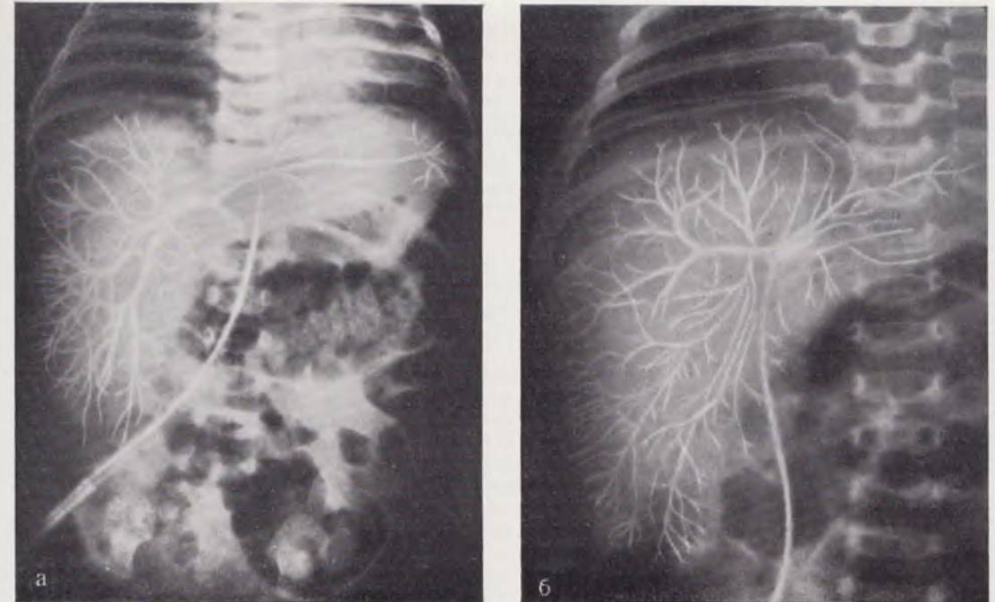
In cases of impaired portal circulation, the portohepatogram demonstrates retrograde opacification of portal vein branches and collateral blood flow pathways (Fig. 16).
Fig. 16. Portohepatogram of a 6-month-old infant. The intrahepatic vascular pattern is markedly impoverished. All Vessels of the portal system and an extensive collateral network are opacified. Diagnosis: biliary atresia; Biliary Cirrhosis of the liver.

Transumbilical aortography is a method used for contrast visualization of the aorta and its branches. Contraindications include the infant's critical general condition and hypersensitivity to iodine-containing agents.
The procedure is performed under anesthesia. Through a skin incision in the inguinal region, after dissecting the aponeurosis of the external oblique abdominal muscle within the preperitoneal tissue, the umbilical artery is identified, isolated with a ligature, and incised. A catheter is introduced into the arterial lumen and advanced under fluoroscopic guidance to the target position. During a period of apnea, the contrast medium is injected and radiographs are taken. Once the catheter is withdrawn, the artery is ligated.
Transumbilical aortography eliminates the need to catheterize any major magistral vessel, thereby significantly reducing the risk of vascular complications.
Renal angiography. This contrast-enhanced imaging method of the renal vasculature expands the capabilities of topographical Diagnostics for lesions of the renal parenchyma, renal vessels, and various retroperitoneal disorders (Fig. 17). The procedure is indicated in cases of persistent hematuria, hypertension, suspected renal vascular pathology, Congenital Renal Anomalies, hydronephrosis, polycystic kidney disease, etc. Contraindications include infant hypersensitivity to iodine, exudative diathesis, and blood dyscrasias.
Fig. 17. Aortogram of a 28-day-old infant. The catheter has been advanced through the left umbilical artery into the aorta. The right renal artery is not opacified. Diagnosis: Aplasia of the right kidney.

The procedure is performed under general anesthesia. Radiopaque catheters of the Ödman or Cournand type are used for catheterization. The contrast medium is injected under a pressure of 5 atm (2 ml of a 70% solution per 1 kg of the infant's body weight). The renal vessels are opacified by catheterizing the aorta via either the umbilical or femoral Arteries. In newborns, the former approach is preferred.
In the transfemoral approach, the femoral artery is exposed layer-by-layer through a skin incision in the upper third of the thigh. Tourniquets are placed around both ends of the vessel. A U-shaped nylon suture is placed on the artery, within the bounds of which a longitudinal arteriotomy or puncture is performed. A probe is inserted into the aorta up to the level of the renal artery origins and connected to a pneumatic injector. Serial angiograms are obtained during a period of apnea. After removing the catheter from the artery, the U-shaped suture is tied off. Access to the renal vessels via the umbilical arteries is designated as transumbilical renal angiography (Y. P. Terekhov, Y. A. Tikhonov, V. G. Hakobyan, 1966). With this technique, access to the aorta is analogous to that described above for transumbilical aortography.
Endoscopic diagnostic methods
Timely diagnosis of thoracic and abdominal disorders is currently achieved by correlating medical history, the patient's clinical status, and the results of laboratory and functional examinations. Direct visual inspection of a pathologically altered organ using one of the endoscopic techniques plays a well-established role in establishing the diagnosis. Laryngotracheobronchoscopy, esophagoscopy, laparoscopy, thoracoscopy, and cystoscopy are the most frequently employed procedures.
Tracheobronchoscopy. Inspection of the Trachea and Bronchi in newborns is performed for both diagnostic and therapeutic purposes. In recent years, with the advancement of neonatology and critical care, along with an expansion of indications for early surgical intervention, the need for bronchoscopy in neonates has substantially increased.
Indications. Bronchoscopy in newborns is indicated for airway obstruction of the trachea and bronchi resulting from various etiologies (aspiration of Amniotic Fluid, mucus, blood, meconium, gastric contents, or saliva in cases of tracheoesophageal fistula; pus from inflamed areas of the lung, etc.). Bronchoscopy enables the diagnosis of congenital malformations of the trachea and bronchi (tracheo- and bronchobronchial fistulas, pulmonary agenesis, tracheal and bronchial stenosis). Crucial diagnostic data are obtained in cases of tracheoesophageal fistulas. In diagnostically challenging cases, tracheobronchoscopy allows for confirming or ruling out the diagnosis of a tracheoesophageal fistula. Furthermore, bronchoscopic equipment is utilized to perform bronchography.
The procedure is performed under general anesthesia using a Friedel ventilating bronchoscope or a domestic DB-type bronchoscope (tubes 4 and 5). The bronchoscope is introduced into the trachea under laryngoscopic control following lung hyperventilation during a period of apnea. As the scope passes through the vocal cords, the bronchoscope tube is rotated 90° along its axis so that the beveled edge corresponds to the longitudinal axis of the glottis. Once the tube is in the trachea, the laryngoscope is removed. Subsequently, the tube is advanced only under direct Vision through a clearly visible, unobstructed lumen. This is particularly crucial when performing bronchoscopy in neonates, as blind, forceful advancement of the tube in an infant can lead to severe trauma, including rupture of the tracheal wall. The tube is advanced to the tracheal bifurcation. Secretions are cleared from the trachea and main bronchi using a suction catheter, after which inspection can commence. The main bronchi are examined by positioning the tip of the tube just above the bifurcation while gently turning the infant's head to the corresponding side. Advancing the tube further into distal segments is not recommended, as the diameter of the main bronchi is significantly narrower than the tube; attempting to push the bronchoscope into these branches can result in mucosal injury or bronchial wall perforation. Following inspection of the main bronchi, the trachea is examined by slowly and gradually withdrawing the tube. The anterior, right lateral, posterior, and left lateral walls are systematically evaluated while the distal tip of the bronchoscope traces a spiral path. Prior to removing the bronchoscope, residual secretions are aspirated from the trachea and Oral Cavity.
Complications during bronchoscopy can be divided into those caused by procedural manipulations and those associated with anesthesia. When using combined anesthesia with muscle relaxants, complications related to bronchoscopic technique occur only in cases of rough, traumatic tube manipulation or when attempting to force the tube into a bronchus of inappropriate caliber.
Esophagoscopy. The design of modern esophagoscopes is optimized for the direct visualization of the entire esophageal wall. In our country, Brünings-type esophagoscopes (Brunnings, 1910) with proximal illumination are the most widely used. Special small-caliber tubes are employed for esophagoscopy in newborns, though a pediatric ventilating bronchoscope tube can also be utilized.
Indications. Visual examination of the esophagus in newborns is indicated for achalasia, congenital short esophagus, and suspected tracheoesophageal fistulas.
The patient is positioned supine with a small roll placed beneath the shoulders. Under endotracheal anesthesia, the endotracheal tube is positioned in the corner of the Mouth opposite to the intended insertion site of the esophagoscope (typically the left corner). The esophagoscope is introduced under continuous visual control. When using a laryngoscope to displace the base of the Tongue with its blade, the esophageal inlet is clearly visualized, making the introduction of the esophagoscope straightforward. If a laryngoscope is not used, the esophageal inlet is located using anatomical landmarks. The first landmark is the uvula. After passing the uvula, the esophagoscope is brought into a near-vertical position and advanced along the midline until the second anatomical landmark—the epiglottis—comes into view. Next, the tip of the tube is tilted slightly backward and forward to reach the third anatomical landmark, the inferior pharyngeal constrictor or the "upper esophageal sphincter" (cricopharyngeus). During apnea, this sphincter typically gapes open and readily allows the passage of the esophagoscope. Upon traversing the upper esophageal sphincter, the esophagoscope enters the cervical esophagus, the lumen of which has a slit-like shape. Guided by the center of this slit, the scope is slowly advanced down the esophagus while carefully inspecting the esophageal wall. Tracheoesophageal fistulas are most frequently located on the anterior-left wall of the esophagus at the 10 to 11 o'clock position. During Respiration, the fistula opening can occasionally be seen ballooning outward from a puff of air. The lumen of the thoracic esophagus is funnel-shaped, and the esophagoscope tube is advanced toward the center of this funnel. Detailed mucosal inspection is carried out by lateral displacements of the tube. Normally, the esophageal mucosa is pale pink with longitudinal folds. In cases of achalasia complicated by food stasis, inflammatory mucosal changes are frequent, whereas mucosal erosion is rarely observed in the newborn period. In select cases, esophagoscopy permits the identification of esophageal duplication, diverticula, and other anomalies. Following mucosal inspection, the esophagoscope is withdrawn.
Complications during esophagoscopy are largely dependent on the quality of anesthesia and the operator's experience. Operators should first master esophagoscopic technique in adults or older children. Inadequate anesthesia may lead to spasm of the upper esophageal sphincter and subsequent mucosal trauma as the scope is forced through the spastic segment. Injury to the posterior pharyngeal wall and cervical esophagus can occur if the infant's head is hyperextended too abruptly.
The use of fiberoptic endoscopes significantly broadens the diagnostic capabilities for various pathologies.
Laparoscopy. Abdominal endoscopy1 allows for the direct visualization of Internal Organs, objective photographic documentation of pathological changes, and the performance of various adjunctive diagnostic procedures under visual control. This method was pioneered by the Russian scientist D. O. Ott (1901). In neonates, laparoscopy was first applied by A. B. Okulov (1964) and Beato (1965).
1 Various terms for this method are found in the literature: ventroscopy, organoscopy, abdominoscopy, peritoneoscopy, and laparoscopy.
Indications. Laparoscopy is employed when simpler diagnostic methods prove insufficient to establish a diagnosis or clarify the morphological features of pathological processes within the abdominal cavity.
In newborns, laparoscopy is most frequently indicated for Disorders of the portal venous system (jaundice and ascites of unclear Etiology, congenital biliary malformations, hepatomegaly and splenomegaly of various origins), intra-abdominal tumors, specific pelvic organ pathologies in female infants, and disorders of sex development (Hermaphroditism) to determine genetic sex. Less commonly, laparoscopy is utilized to investigate retroperitoneal diseases and intestinal malformations. It can also be valuable in diagnosing closed abdominal birth trauma. The application of this method in acute inflammatory intra-abdominal conditions remains controversial to this day.
Contraindications to laparoscopy are classified into general and local. General contraindications include cardiovascular and respiratory failure, decreased prothrombin time, prolonged bleeding time, coagulopathies, and thrombocytopenia. Local contraindications comprise acute diffuse Peritonitis, extensive intra-abdominal adhesions, and diaphragmatic hernias. These contraindications must also be kept in mind when performing adjunctive diagnostic procedures under direct vision, such as targeted liver biopsy, splenoportography, or cholangiography.
The method consists of several stages. The First stage involves establishing an artificial pneumoperitoneum by puncturing the anterior abdominal wall with a special needle at a standard point along the midline, 1 cm below the umbilicus, using a safety ligature (Fig. 18, a, b). Nitrous oxide, carbon dioxide, or helium is introduced into the abdominal cavity. The optimal amount of gas for creating a pneumoperitoneum is determined by the level of intra-abdominal pressure, which in newborns should not exceed 20 — 25 mmHg. The Optical System of the laparoscope is introduced into the abdominal cavity by puncturing the abdominal wall with a trocar at a standard point located 2 — 3 cm to the left of the midline and inferior to the umbilicus, utilizing a safety ligature. Next, the abdominal cavity is inspected and photographed (Fig. 19, a, b, c). If necessary, the inspection is supplemented by intra-abdominal instrumental palpation of the organs, targeted liver biopsy, splenoportography, and cholangiography, the indications for which are established during the abdominal endoscopy (Fig. 20, a, b, c).

Fig. 18. Technique for establishing pneumoperitoneum and introducing the laparoscope. a — points for creating pneumoperitoneum; b — introduction of the laparoscope optical system under the control of a safety ligature.

Fig. 19. Position of the laparoscope during examination of The Liver and Gallbladder (a), Stomach (b), and spleen (c).
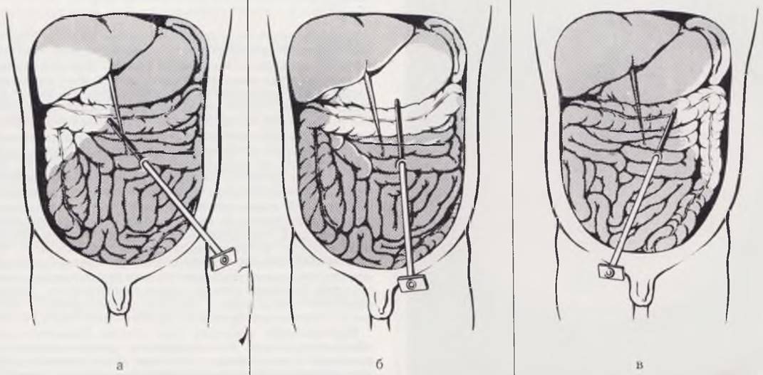
Fig. 20. Manipulations under laparoscopic guidance. a — instrumental palpation and puncture of the spleen; b — needle biopsy of the liver; c — photolaparoscopy, with the puncture needle inserted into the right lobe of the liver. Diagnosis: chronic hepatitis.

The procedure is performed under general halothane anesthesia using muscle relaxants during the most critical moments of the manipulation (punctures of the anterior abdominal wall, photography, biopsy, splenoportography, cholangiography, etc.).
Complications. The initial puncture of the abdominal wall to establish an artificial pneumoperitoneum is always more hazardous than subsequent punctures. The introduction of the optics is carried out only when the anterior abdominal wall is separated from the intra-abdominally located organs by a distinct gas cushion. Additional punctures for liver biopsy, splenoportography, etc., are performed under direct visual control, which eliminates the possibility of injuring intra-abdominal organs.
Complications may occur at any stage of the laparoscopic examination. Hemorrhage may arise from the abdominal wall puncture sites or if the tips of the needle (for creating pneumoperitoneum) or trocar (for introducing the optics) penetrate underlying internal organs. Gas embolism is extremely rare; it occurs when the tip of the pneumoperitoneum needle enters the bloodstream or when gas is forced into parenchymal organs.
Abdominal endoscopy is safe in itself. At this stage of the examination, complications are associated with the use of additional diagnostic manipulations.
The possibility of these complications does not diminish the value of laparoscopic examination. Adherence to the methodology and techniques of this complex diagnostic intervention, along with careful monitoring of the newborn in the post-manipulation period, can reduce the number of complications to a minimum.
Thoracoscopy. This method for examining the pleural cavity and the organs contained within it was proposed in 1910 by Jacobeaus. In newborns, thoracoscopy has been used since 1969 (V. G. Geldt).
Indications. The examination is performed for pleural complications of pulmonary diseases, Spontaneous pneumothorax, and pyopneumothorax. In rare cases, indications for thoracoscopy arise in cases of Cysts and tumors of the lung and mediastinum. Thoracoscopy is used for both diagnostic and therapeutic purposes. In spontaneous pneumothorax, it helps identify the cause of lung collapse. In patients with pyopneumothorax, it determines the degree of destruction of the lung tissue, the size of the bronchial fistula, the presence and Nature of the effusion, and the degree of lung reexpansion. Therapeutic measures can be performed during thoracoscopy, such as lysis of adhesions, removal of fibrin, irrigation of the pleural cavity, etc.
Thoracoscopy in newborns is performed in the operating room under endotracheal halothane anesthesia with nitrous oxide and oxygen. The infant is positioned on the healthy side. The procedure consists of four stages: establishing an artificial pneumothorax, introducing the thoracoscope and inspecting the pleural cavity, photographing during thoracoscopy, and intracavity instrumental palpation. A domestic thoracoscope with lateral optics or an East German-produced endoscope with lateral optics and a 135° angle of view is used in pediatric patients.
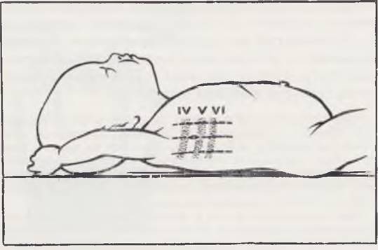
The site for pleural puncture is chosen far away from the pathologically altered lung and pleura. Air is introduced into the pleural cavity under manometric pressure control. The most convenient point for introducing the thoracoscope is located along the midaxillary line in the 6th or 5th intercostal space (Fig. 21). The trocar is inserted into the pleural cavity with caution to avoid injuring the lung. After thoracentesis is performed, the stylet is removed and the thoracoscope is introduced through the trocar cannula. The pleural cavity is inspected sequentially, starting from the upper sections. Examination of the pleural cavity in pyopneumothorax is difficult due to the limited space and the presence of fibrinous deposits. Prior to the examination, the pleural cavity is evacuated of pus. Revision begins at the site of the greatest accumulation of fibrin, which indirectly indicates the localization of pulmonary destruction.
Fig. 21. Patient position during thoracoscopy and the site of thoracoscope insertion.
To obtain additional data on the consistency and mobility of the organ under investigation, a special probe-manipulator is used. Through it, additional instillation of gas or solutions and aspiration of fluid are possible (Fig. 21, b). The probe-manipulator can be used to push the lung aside or break down loose adhesions. Upon completion of the thoracoscopic examination, gas is removed from the pleural cavity. A suture is placed on the chest wall wound.
Complications are possible at any stage. Anesthesia-related complications include respiratory depression, laryngospasm, prolonged apnea, and rupture of a bulla during Artificial ventilation. When establishing an artificial pneumothorax, the needle may accidentally enter the lung tissue, a blood vessel, or the abdominal cavity; these complications are detected using a manometer. During the procedure itself, bleeding may occur from damaged adhesions, intercostal vessels, or vessels of the lung root. After removal of the thoracoscope and wound closure, subcutaneous emphysema may develop if air is incompletely evacuated from the pleural cavity.
Complications can be avoided by strictly following the rules of thoracoscopic examination and handling the thoracoscope and manipulator with care.
Technique of the Most Common Surgical Interventions
In various diseases and congenital malformations in newborns, surgeons frequently face the need to perform surgical interventions such as pleural cavity drainage and irrigation, tracheotomy, gastrostomy, etc. The technique of these operations in newborn infants has specific features that we will discuss in greater detail.
Pleural cavity drainage. The pleural cavity in newborns is drained when air, pus, or blood accumulates within it. The need for this manipulation most commonly arises in necrotizing Pneumonia complicated by pyothorax or pyopneumothorax, in tension spontaneous pneumothorax, and following surgeries on the thoracic organs and Diaphragm.
Insertion of a drainage tube is performed under aseptic conditions via thoracentesis or thoracotomy. Thoracentesis requires a trocar, a tube of appropriate diameter, a scalpel, and a needle with a needle holder and a nylon (or silk) thread to secure the drain to the skin. During thoracotomy, the drain is introduced using a curved clamp with sharply pointed jaws. Drainage of the pleural cavity in newborns is carried out using silicone or polyvinyl chloride tubes with a lumen diameter of 2 — 3 mm. A wide lumen and 4 — 5 openings on the segment of the drainage tube inserted into the pleural cavity ensure reliable system performance and the aspiration of thick pus, fibrin clots, or blood.
Drainage of the pleural cavity in newborns is usually performed under local anesthesia. The infant is placed in a lateral position on a roll, which helps widen the intercostal spaces and provides counter-pressure. The drain is inserted in the 6th — 7th intercostal space along the mid-axillary or posterior axillary line. One must account for the high standing of the diaphragm in cases of abdominal distension.
For thoracentesis, a 0.5 cm skin incision is made with a scalpel. The trocar stylet with the cannula fitted over it is inserted with a drilling motion along the up
per edge of the underlying rib (Fig. 22, a), having previously displaced the skin relative to the rib to avoid injuring the intercostal artery. After perforation of the chest wall, the stylet is withdrawn, and the cannula is directed toward the pleural dome (Fig. 22, b). The drainage tube is advanced into the pleural cavity to the marked level and, after removal of the cannula, is fixed to the skin with a nylon suture placed at the edges of the incision. If necessary, the thoracentesis site is sealed with adhesive tape.
During thoracotomy, a 1 cm skin incision is made. The end of the tube is grasped with a clamp and introduced into the pleural cavity with a twisting motion, directing it upward. The tube is set to the required depth according to the mark and secured to the skin.
When draining an open pleural cavity during surgery, the tube is grasped by the jaws of a clamp passed through the chest wall and brought out from the inside to the outside under visual control.
The drainage tube is connected to an aspiration system (Fig. 22, c). Chest radiography is advisable after surgery to monitor THE POSITION OF the drain. The optimal position is considered to be along the posterolateral or posterointernal surface of the pleural cavity up to the pleural dome. In cases of encysted pyopneumothorax, the position of the tube depends on the Location OF THE cavity.
Fig. 22. Pleural cavity drainage. a — puncture of the chest wall; b — direction of insertion of the drainage tube; c — placement of the drain and diagram of its connection to the aspiration system.

Unsatisfactory function of the pleural drain is most commonly a result of the drainage tube collapsing or kinking. In the event of a kink, it is advisable to pull the tube slightly. If the end of the tube has fallen into the sinus, incomplete lung expansion and The formation of loculations in the upper PARTS OF THE pleural cavity may occur. In such cases, the tube should be withdrawn and repositioned correctly. A frequent complication is bleeding from a damaged intercostal artery or lung tissue. With prolonged drainage of the pleural cavity in newborns, tissue necrosis develops around the drainage tube, and the system loses its hermetic seal. To prevent severe complications—such as chest wall Phlegmon and rib Osteomyelitis—the duration of drainage is shortened whenever possible, and repeat thoracentesis is performed in a healthy area if necessary. In valvular pneumothorax or pyopneumothorax, subcutaneous emphysema may develop if the drainage tube has a narrow lumen. To eliminate this, the drainage tube is connected to an active aspiration system or replaced with a wider tube.
Pleural lavage. The indication for continuous irrigation (lavage) of the pleural cavity in newborn infants is purulent Pleurisy—pleural empyema. Antiseptics (0.05 — 0.1% rivanol solution), nitrofurans (aqueous furacilin solution 1:5000, 0.1% furagin K solution), or broad-spectrum Antibiotics in isotonic sodium chloride solution are typically used.
Irrigation of the pleural cavity is carried out through a thin polyethylene or polyvinyl chloride catheter—a micro-irrigator (0.4 — 0.7 mm in diameter)—which is inserted in the 2nd intercostal space along the midclavicular line through the lumen of a Dufour needle or via the Seldinger technique and secured with adhesive tape to the skin previously treated with collodion. A drip set with the irrigating fluid is connected to the micro-irrigator. The amount varies from 1.5 to 2.5 liters per day depending on the rate of administration. The irrigating fluid is aspirated through a drainage system connected to a water-jet pump. The primary condition is strict compliance between the volume of solutions introduced and removed.
A contraindication to applying a lavage system is the presence of large bronchopleural fistulas. In such cases, the irrigating fluid enters the patient's bronchi, causing persistent coughing and vomiting.
Tracheostomy. Indications for tracheostomy in newborns arise in cases of mechanical upper airway obstruction, croup, foreign bodies, weakened cough reflex, neuromuscular diseases, Central Nervous system disorders, etc.
In infants with respiratory failure, tracheostomy may be combined with artificial ventilation.
Tracheostomy in a newborn is performed under general anesthesia with an endotracheal tube inserted into the trachea. This ensures a calm surgical procedure and significantly reduces the likelihood of complications. Performing the operation without anesthesia is permissible only in emergency situations when asphyxia occurs outside a hospital Setting.
The infant's position is supine, with a roll under the shoulders and the head hyperextended (Fig. 23, a). The child's head is placed on a ring cushion and held in the midline position. The skin incision is made horizontally along one of the neck creases (Fig. 23, b). The subcutaneous tissue and Cervical fascia are also incised horizontally. Thorough hemostasis is performed, and the wound edges are retracted. In the midline, the sternohyoid muscles are carefully separated in a longitudinal direction using forceps. In high tracheotomy, The Thyroid Gland is pushed downward and remains at the lower angle of the wound; in low tracheotomy, the thyroid gland is pushed upward. By blunt and sharp dissection, the tissue over the trachea is carefully incised. To allow free manipulation, the trachea should be freed by one ring above and below the intended incision site. Steping back 1 — 2 mm from the midline to the right and left, stay sutures made of nylon threads are applied to the trachea (Fig. 23, c), which help stabilize the trachea during the operation and facilitate the insertion and removal of the cannula in the postoperative period. The anterior wall of the trachea is secured with stay sutures, and two rings are incised longitudinally (Fig. 23, d). If an endotracheal tube is present in the trachea, the tip of the scalpel rests against it, thereby eliminating the risk of cutting the posterior wall. Using the stay sutures, the wound edges are retracted, the endotracheal tube is withdrawn to the upper edge of the tracheal wound, and the tracheostomy cannula is then carefully inserted into the lumen (Fig. 23, e). If there is no endotracheal tube, sputum and blood-tinged mucus are forcefully expelled from the trachea at the moment the rings are incised; wound edge infection is possible. Further manipulations are complicated due to the continuous expulsion of sputum. To facilitate the introduction of the cannula into the trachea, prior insertion of a wound dilator is necessary (Fig. 23, e). After the insertion of the tracheostomy cannula, its correct positioning is verified. The wound is closed in layers. The stay sutures are brought out near the cannula and secured over gauze pledgets without cutting the ends of the threads. Several layers of gauze are placed under the flange of the tracheostomy tube. The tube is secured around the neck using gauze ties.
A cannula with a 3 mm diameter and a removable inner tube is used as a tracheostomy tube in newborns. During artificial ventilation, an inflatable cuff is placed on the end of the cannula to eliminate the space between the cannula and the trachea.
Fig. 23. Stages of tracheostomy. a — patient's position; b — incision lines; c — trachea fixed with stay sutures, endotracheal tube in the tracheal lumen; d — incision of two rings, endotracheal tube prevents damage to the posterior wall; e — endotracheal tube withdrawn, cannula inserted into the trachea.

Postoperative radiographic control of the correct positioning of the tube in the trachea is required. Radiographs are taken in anteroposterior and lateral projections.
Postoperative care for the tracheostomized infant is described above.
A newborn with a tracheostomy must be cared for by specially trained and experienced nurses.
Tracheostomy complications are divided into operative and postoperative (Holinger et al., 1956). Operative complications occur more frequently in newborns than in older children. These include hemorrhages, pneumothorax, pneumomediastinum and subcutaneous emphysema, injury to the posterior wall of the trachea and esophagus, and respiratory arrest. Late complications appear at various intervals in the form of wound suppuration, Pulmonary Atelectasis, cannula obstruction, tracheal ulceration and bleeding from eroded vessels, cannula dislodgement, tracheal stenosis, delayed stoma healing, and tracheomalacia. Prolonged retention of the tube complicates the decannulation process.
Gastrostomy. In pediatric surgical clinics of our country, gastrostomy in newborns is used for very narrow indications, primarily to ensure Nutrition in esophageal atresia, etc. Most surgeons view gastrostomy as a forced procedure. There is a widespread opinion regarding frequent complications associated with gastrostomy placement in newborns. Not infrequently, they are one of the main causes of infant mortality.
Foreign pediatric surgeons (Rickham, 1969; Gross, 1970, et al.), in contrast, adopt a very broad indication for gastrostomy and employ it in the majority of abdominal surgeries. They emphasize that the widespread use of gastrostomy has significantly improved treatment outcomes for severe congenital anomalies such as esophageal atresia, intestinal malformations, diaphragmatic hernias, and large omphaloceles, among others.
Thus, Meeker (1958) believes that gastrostomy is indicated in newborns: a) to protect proximal and distal anastomoses, b) for abdominal decompression, and c) when enteral nutrition must be established in patients with esophageal malformations.
Strict adherence to the technique of gastrostomy creation is of paramount importance. The method of choice is the one that is technically simplest.
The patient is placed in the supine position. The skin and subcutaneous tissue are incised horizontally at the level of the costal margin to the left of the midline, with an incision length of 3 cm. The Muscles of the anterior abdominal wall are separated layer by layer, and The Stomach is brought into the wound. Two purse-string sutures are placed on the anterior wall of the stomach at the boundary of its upper and middle thirds (the knots of the purse-strings should face opposite directions). In the center of the inner purse-string, the full thickness of the gastric wall is incised with a scalpel. A specialized catheter or a thin Pezzer catheter is introduced into the wound, and the purse-string sutures are tied over it. They should not be pulled too tight. An additional puncture of the abdominal wall is made 2 cm below and to the left of the skin incision, through which the catheter is brought out. The stomach is secured to the peritoneum from the abdominal cavity side with 2 — 3 sutures (Fig. 24). The catheter is anchored to the edge of the skin incision. The abdominal wall is closed tightly layer by layer. In the postoperative period, the child remains in the supine position,
the gastrostomy catheter is secured to the incubator without being clamped, and is covered with a gauze pad on top.
Fig. 24. Gastrostomy in a newborn. An uncomplicated course is ensured by two loosely tied purse-string sutures, careful fixation of the stomach to the abdominal wall, reliable anchoring of the gastrostomy tube to the skin, and an open management approach in the postoperative period.

The parietal placement of the catheter in this method of gastrostomy helps prevent trauma to the gastric mucosa. Bringing the catheter out through a separate puncture ensures optimal wound healing and prevents the leakage of gastric contents. A permanently open catheter drains air from the stomach, which facilitates abdominal decompression. The ability to administer feeding via the gastrostomy protects the esophageal anastomosis. The protection of distal anastomoses is achieved primarily through air drainage. It has been shown that one of the main causes of abdominal distension and intestinal paresis following abdominal surgery is the child swallowing air during sucking motions. Preventing gastric distension helps reduce intra-abdominal pressure after surgeries for large diaphragmatic hernias and omphaloceles. Feeding is performed according to general guidelines 2 — 4 hours after the creation of the gastrostomy.
Once the need for the gastrostomy has passed, the catheter is removed. The wound at the catheter site typically closes spontaneously.
We have performed gastrostomies using this technique in 48 children, with no complications observed in any of the cases. We limit the indications for gastrostomy, utilizing it primarily in cases of esophageal atresia requiring prolonged preoperative preparation, and following intestinal anastomoses when the surgeon is uncertain of the anastomosis's reliability.
Esophagostomy. Indications for esophagostomy in a newborn typically arise in cases of esophageal atresia, when a two-stage surgical intervention is planned due to the severity of the patient's condition or specific anatomical factors. The patient is placed supine with a bolster under the shoulders; the head is hyperextended and turned to the right.
The incision is made along the anterior border of the sternocleidomastoid muscle, parallel to the skin crease, at the midpoint between the upper edge of the thyroid Cartilage and the jugular notch. Following the incision of the skin and subcutaneous tissue, the first fascia and the platysma are divided parallel to the anterior border of the muscle. Hemostasis is achieved, and the sternocleidomastoid muscle is mobilized from the surrounding tissues and retracted anteriorly. The omohyoid, sternohyoid, and sternothyroid muscles are also mobilized and retracted medially. The fifth cervical fascia at the base of the wound is opened after palpating the vertebrae and tracheal rings. The esophagus lies in the space between the spine and the trachea. The periesophageal tissue is bluntly separated to expose the wall of the esophagus, which exhibits longitudinal striation. The length of the esophageal segment accessible to surgical intervention in a newborn does not exceed 2 cm. Mobilization of the esophagus is facilitated by the preliminary insertion of an elastic probe into its lumen. After isolating the wound with sponges, the esophageal wall is transected transversely and sutured to the edges of the skin wound through all layers using a single row of interrupted nylon sutures. In high-atrisia cases, it is often possible to isolate the blind end of the esophagus and bring it to the surface, whereas with a sufficiently low blind segment, it is advisable to mobilize the esophagus as extensively as possible to prevent the formation of a long blind pouch.
Our experience demonstrates that a significant simplification and acceleration of the surgery can be achieved through a simple maneuver — the preliminary insertion of a metal urethral catheter into the esophagus, which greatly facilitates and speeds up locating the esophagus and separating it from the surrounding tissues.
Abdominal paracentesis (puncture) in newborns is used for diagnostic purposes when purulent-inflammatory conditions or intra-abdominal hemorrhage are suspected. In recent years, we have frequently resorted to this procedure in diagnostically complex cases. The use of a trocar in newborns and infants carries the risk of injuring the intestinal wall due to continuous abdominal distension. Therefore, we have abandoned the classical technique. The child is placed in the supine position. The abdominal cavity is punctured at a point on the midpoint of the line connecting the umbilicus to the anterior superior iliac spine. If there are signs of localized peritonitis, the puncture is performed on the contralateral side. Under local anesthesia with a 0.25% novocaine solution (5 ml), a point skin incision is made using the tip of an ophthalmic scalpel. Next, the layers of the abdominal wall are carefully spread using the jaws of a mosquito clamp, the peritoneum is ruptured, and the resulting wound is dilated. Perforated polyvinyl chloride tubes, fitted one inside the other, are introduced into the abdominal cavity between the instrument's jaws. The Contents of the abdominal cavity either drain spontaneously or are gently aspirated with a syringe. We have observed no complications when using this described technique.
Last update: 10/08/2026
Editorial and Educational Adaptation: This material has been compiled based on the primary/original source text. The project team performed an editorial review, corrected technical inaccuracies, structured sections, and adapted the content for an educational format.
What was processed:
- elimination of formatting defects (OCR errors, structural breaks, corrupted characters);
- editorial organization of content;
- standardization of terminology in accordance with academic sources;
- verification of factual statements against the original source text.
All mentions of the author, publication year, and origin of the primary text have been preserved in accordance with the source.