Tuberculosis - I.T. Piatnochka 2005
Methods of examination of tuberculosis patients
Methods of radiological diagnostics
X-ray Examination is one of the primary Diagnostic Methods for Tuberculosis and non-specific respiratory diseases. The following X-ray diagnostic modalities are employed: fluoroscopy, radiography, fluorography, tomography, computed tomography, targeted radiography, bronchography, fistulography, angiopulmonography and bronchial arteriography, pleurography, kymography, and polygraphy.
Fluoroscopy is performed in various patient positions and across different respiratory phases, allowing for the assessment of diaphragmatic function and cardiac pulsation, the Selection of an optimal puncture site, and preparation for targeted radiography, bronchography, fistulography, angiopulmonography, and other Procedures.
Nowadays, pulmonary fluoroscopy is generally performed infrequently, more often utilizing equipment equipped with image intensifiers. An initial survey fluoroscopy is conducted to determine the shape of the chest, the transparency and width of the lung fields, the localization and dimensions of the mediastinal and cardiac shadows, as well as the mobility of the diaphragmatic domes and anterior rib segments. Following the survey fluoroscopy, the screen field is narrowed using a Diaphragm to enable a more detailed examination of the lung tissue Structure and pathological changes.
Radiography makes it possible to detect and capture morphological lung structures on X-ray film that remain invisible during fluoroscopy, document and archive X-ray images, and compare them dynamically. Crucially, the patient's radiation exposure dose is significantly lower than that associated with fluoroscopy.
The drawback of radiography is its static nature, which precludes the assessment of organ function. However, The Use of serial radiography and cine-radiography largely compensates for this limitation.
Normally, the left lung is narrower and longer than the right, while the mediastinal Organs are located between the medial ends of the clavicles, against the Background of the sternal and spinal shadows.
The right lung comprises 3 lobes (upper, middle, lower), whereas the left comprises 2 (upper and lower). The lobes (upper and lower) are separated by interlobar fissures. The oblique interlobar fissure follows a similar course in both the right and left Lungs: extending obliquely downward and anteriorly from the level of the fourth thoracic vertebra to its intersection with the seventh rib. On the right side, the horizontal fissure, running from the level of the attachment of the fourth rib to the Sternum to its intersection with the oblique interlobar fissure, separates the upper lobe from the middle lobe. Each lung contains 10 segments; however, the left lung occasionally presents with only 9 segments (Fig. 7).
The tuberculosis process most frequently localizes in segments 1, 2, and 6, and less commonly in the anterior and basal segments. To precisely determine the localization of the process, a lateral projection radiograph must also be performed.
Lung roots (hila). The shadows of the hila are located in the medial sections of the lungs, adjacent to the cardiac shadow, spanning a vertical distance of two intercostal spaces from the III rib downward, and gradually blending into the pulmonary vascular pattern.
The anatomical substrate of the hila consists of major arterial and venous vessels, Bronchi, groups of Lymph Nodes, Connective Tissue containing Lymphatic vessels, and neural trunks. Radiologically, three components are distinguished within the ROOT: the HEAD, body, and tail. The head of the root is formed by the shadows of the arches of the main Branches of the pulmonary artery and is located at the level of the III rib or the III intercostal space. The body of the root is formed by the shadows of the descending part of the pulmonary artery trunk and other vessels. The lower tail section is formed by the shadows of the inferior Veins and transitions into the pulmonary pattern of the lower lung zones.
Class="center">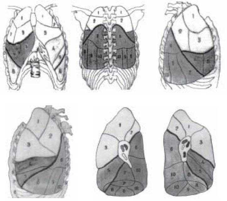
Fig. 7. Lobar and segmental STRUCTURE OF THE lungs.
Segments of the right lung: 1 - apical; 2 - posterior; 3 - anterior; 4 - lateral; 5 - medial; 6 - superior (lower lobe); 7 - medial basal; 8 - anterior basal; 9 - lateral basal; 10 - posterior basal. Segments of the left lung: 1 - apical; 2 - posterior; 3 - anterior; 4 - superior lingular; 5 - inferior lingular; 6 - superior (lower lobe); 8 - anterior basal; 9 - lateral basal; 10 - posterior basal.
The pulmonary pattern is formed by the branching of pulmonary artery and vein vessels, which is why it is also referred to as the vascular pattern. In various pathological processes within the lungs, the vascular pattern may become accentuated or blurred. Occasionally, a major vessel positioned transversely can project a round shadow mimicking a focal lesion. To clarify The Nature of this shadow, the patient should be examined in multiple projections.
The primary radiological shadows observed in Pulmonary Tuberculosis include focal (up to 1 cm in diameter), infiltrative (greater than 1 cm), annular, and linear shadows. According to their dimensions, focal shadows are classified as small (up to 2 mm in diameter), medium (3–5 mm), and large (6–10 mm), while in terms of density, they are classified as low, medium, and high intensity.
A shadow exceeding 1 cm in diameter is termed an infiltrate or Tuberculoma.
Before proceeding with the analysis of a radiograph, its technical quality must be evaluated. This is carried out in a specific sequence: completeness of the examined object coverage (the radiograph must capture the entire chest cage, from the apices to the costophrenic angles); patient positioning during the Procedure (with correct positioning, the distance between the medial ends of the clavicles and the spinous processes of the third thoracic vertebrae is symmetrical, and the scapular shadows are projected outward beyond the lung fields); clarity and contrast (defined as the sharp delineation of every detail across various tonal shades); and finally, radiographic "penetration" or "hardness" (at optimal penetration, the upper three to four thoracic vertebrae are clearly visible).
Survey radiography is performed in straight (anteroposterior and posteroanterior), lateral, and oblique projections. A lateral projection radiograph is utilized to specify the localization and Nature of the pathological process.
Targeted radiography is performed on a restricted area of the lungs with the patient positioned to obtain the most optimal image of pathological changes obscured by the bony structures of the Thorax.
Tomography is a section-by-section examination of a specific organ, specifically the lungs. It allows for a detailed study of The structure of a pathological formation at a precise optimal depth, which is selected based on the results of lateral radiography or fluoroscopy.
Computed tomography is based on the mathematical analysis of X-ray absorption intensity by Tissues of varying densities, converting this data into an image that displays cross-sections of The Human Body at different levels (Fig. 8). Computed tomography enables the precise localization and extent of pulmonary and mediastinal pathological processes to be determined, while also revealing minor abnormalities within the Pleura and intrathoracic lymph nodes.
Fluorography is the primary method for mass population screenings, offering high throughput and substantial diagnostic information when performed in multiple projections. However, the radiation dose to the examinee is somewhat higher than in standard radiography, which is why survey radiography is preferred for children.

Fig. 8. Normal computed tomography scans of the chest (after Gabuniya R.I. et al., 1983).
a - at the level of the sternoclavicular joint: 1 - right brachiocephalic vein; 2 - Trachea; 3 - common carotid artery; 4 - left brachiocephalic vein; 5 - Esophagus; b - at the level of the tracheal bifurcation: 1 - descending artery; 2 - main bronchi; 3 - trunk of the right pulmonary artery; 4 - SUPERIOR VENA CAVA; 5 - pulmonary trunk; 6 - pulmonary artery; 7 - esophagus; c - 2 cm below the tracheal bifurcation: 1 - descending aorta; 2 - left main bronchus; 3 - right main bronchus; 4 - right pulmonary artery; 5 - superior vena cava; 6 - ascending aorta; 7 - pulmonary trunk; 8 - pulmonary artery; d - above the diaphragmatic dome: 1 - descending aorta; 2 - right atrium; 3 - right ventricle; 4 - left ventricle.
Bronchography is used to examine the bronchial tree, detect Bronchiectasis (including its extent and Morphology), and identify cavities. The procedure is performed on an empty Stomach, usually under local anesthesia, after which a contrast agent (such as propyliodone or sulfoyodlipol) is administered through a catheter under fluoroscopic guidance, followed by radiography in two projections.
Fistulography. This method is used to evaluate patients with various thoracic fistulas (thoracic and thoracobronchial). A contrast agent (iodolipol, or oily and aqueous solutions of propyliodone) is injected into the fistula, and radiographs are taken in the required projections.
Pleurography is primarily performed in patients with Empyema of the pleura to precisely determine its boundaries. First, the empyema content is aspirated, after which a radiopaque contrast agent (propyliodone, urografin, or verografin) is injected into the cavity, and radiographs are taken in multiple projections.
Angiopulmonography and bronchial arteriography are radiopaque imaging methods used to study the pulmonary Vessels of the lesser (angiopulmonography) and greater (bronchial arteriography) circuits of the Circulatory system. Angiopulmonography involves the catheterization of the right Heart chambers and the pulmonary artery under fluoroscopic control, followed by the injection of a contrast agent and a series of radiographs. The purpose of this study is to diagnose thrombosis and Pulmonary Embolism, as well as to assess the degree of pneumofibrosis.
Bronchial arteriography involves catheterization, contrast enhancement, and radiography of the bronchial Arteries and their branches. The main indications for this procedure are recurrent Pulmonary Hemorrhage and Hemoptysis of an unknown origin.
Kymography and polygraphy are used to assess the mobility of the diaphragm and The Heart.
Ultrasound examination (Ultrasonography). In respiratory diseases, ultrasound is mainly used to examine the pleura, right ventricular function, and pulmonary artery pressure. Ultrasonography is not used for lung parenchyma imaging because air filling within the lungs distorts lesion contours. The ultrasound method is employed to clarify pleural conditions, specifically for differentiating between fluid and solid masses.
Ultrasound examination, particularly two-dimensional echocardiography, is used to monitor the right ventricular index and the thickness of the anterior wall of the right ventricle, as well as to determine the time interval between the closure of the tricuspid valve and the opening of the pulmonary trunk valve. The obtained data demonstrate the degree of correlation between hemodynamics and pulmonary function parameters.
Magnetic Resonance imaging (MRI). This method is used for patient evaluation because it provides sufficient contrast between the mediastinal adipose tissue, solid masses, and vascular structures, allowing lesions to be identified without intravenous contrast administration. Limitations of the method include the inability to detect calcification and insufficient information regarding the lung parenchyma.
CONTROL QUESTIONS
1. Methods of X-ray examination of the lungs and indications for their use.
2. Criteria for evaluating the technical quality of radiographs.
3. Advantages of radiography over fluoroscopy.
4. What determines the normal pulmonary vascular pattern on a radiograph?
5. Anatomical Structure and radiographic appearance of the lung root in a healthy individual.
6. Lobar and segmental structure of the lungs and their localization on anteroposterior (straight) and lateral radiographs.
7. What four types of pathological shadows do you know in pulmonary tuberculosis?
8. Most frequent localization of the tuberculous process in the lungs.
TESTS
1. The primary method for detecting pulmonary tuberculosis during mass population screenings.
A. Fluoroscopy
B. Computed tomography
C. Bronchography
D. Photofluorography
E. Targeted radiography
2. To confirm the presence of bronchiectasis, it is necessary to perform:
A. targeted radiography
B. plain radiography
C. fistulography
D. tomography
E. bronchography
3. What is meant by a focal shadow?
A. An opacity up to 0.2 cm in diameter
B. An opacity 0.2–0.4 cm in diameter
C. An opacity 0.5–1.0 cm in diameter
D. An opacity up to 1.0 cm in diameter
E. An opacity from 1.0 to 2.0 cm in diameter
4. X-ray examination of patient P., 29 years old, reveals a low-intensity opacity up to 1 cm in diameter with indistinct contours under the right clavicle. Determine the type of pathological shadow:
A. focal
B. infiltrative
C. focal-infiltrative
D. ring-shaped
E. linear
5. The most frequent segmental localization of Secondary forms of pulmonary tuberculosis:
A. I, II, III
B. II, III, IV
C. III, V, VI
D. I, II, VI
E. II, III, X
6. To confirm the presence of fluid in the pleural cavity, one performs:
A. two-view fluorography
B. tomography
C. bronchography
D. laterography
E. targeted radiography
7. When were X-rays discovered?
A. In 1882
B. In 1895
C. In 1944
D. In 1951
E. In 1965
8. How many criteria are used to evaluate the technical quality of a plain radiograph?
A. 1
B. 2
C. 3
D. 4
E. 5
9. How many parts (radiologically) does the root of the lung consist of:
A. 1
B. 2
C. 3
D. 4
E. 5
10. How many segments can the left lung have:
A. 8-11
B. 8-12
C. 9-10
D. 9-11
E. 9-12
11. To confirm a mild form of tuberculous bronchial adenitis, it is necessary to perform:
A. Targeted radiography
B. Bronchography
C. Tomography
D. Lateral projection radiography A Fluorography on inhalation and exhalation
12. The percentage of pulmonary tuberculosis patients in Ukraine detected through mass fluorographic screening examinations.
A. 5 %
B. 15 %
C. 25 %
D. 35 %
E. 50 %
PROBLEMS
1. A routine chest X-ray of a 10-year-old patient reveals a medium-density paracardiac shadow on the right with a central area of translucency.
Determine: a) the nature of the shadow; b) the localization of the process; c) which morphological changes correspond to this X-ray picture.
Answer: a) infiltrate; b) a lateral projection radiograph is required; c) infiltrate with breakdown.
2. In a 25-year-old female patient Z., an X-ray examination revealed a low-density pathological shadow up to 1 cm in diameter with blurred contours above the right clavicle. The patient's general condition is good.
a) What type of pathological shadow is this?
b) What is The activity of the tuberculous process?
3. An X-ray of a 6-year-old boy shows an widened tumor-like root of the left lung.
a) Which radiological syndrome do such pathological changes belong to?
b) What additional radiological examinations need to be performed?
4. A ring-shaped shadow is detected below the left clavicle on the chest X-ray.
a) Determine the segmental localization of the pathological process.
b) What X-ray methods can be used to clarify the localization of this shadow?
5. What pathological formations in the lungs can correspond to a ring-shaped shadow:
1... 2... 3...
6. A fluoroscopic examination reveals a non-homogeneous shadowing with blurred margins and a central radiolucency below the right clavicle.
a) Which radiological syndrome does this pathological shadow correspond to?
b) Segmental localization.
c) Name 2–3 diseases with a similar radiological presentation.
Last update: 10/08/2026
Editorial and Educational Adaptation: This material has been compiled based on the primary/original source text. The project team performed an editorial review, corrected technical inaccuracies, structured sections, and adapted the content for an educational format.
What was processed:
- elimination of formatting defects (OCR errors, structural breaks, corrupted characters);
- editorial organization of content;
- standardization of terminology in accordance with academic sources;
- verification of factual statements against the original source text.
All mentions of the author, publication year, and origin of the primary text have been preserved in accordance with the source.