Tuberculosis - I.T. Pyatnochka 2005
Methods for examining patients with tuberculosis
Laboratory studies
The source of human infection is individuals and animals suffering from tuberculosis who shed Mycobacterium tuberculosis. Materials used to detect M. tuberculosis include sputum, bronchial washings, feces, urine, pus from fistulas, pleural exudate, CEREBROSPINAL FLUID, as well as punctates and biopsy specimens from various Organs and Tissues.
Examination of sputum for M. tuberculosis is of major epidemiological and clinical significance. If sputum is absent or scanty, expectorants, irritant aerosol inhalations, or bronchial lavage are prescribed.
Methods for Detecting Mycobacteria. 1. Bacterioscopic method is one of the primary approaches for detecting M. tuberculosis, encompassing conventional bacterioscopy, flotation, and fluorescent Cell/15.html">Microscopy. Conventional bacterioscopy is universally accessible, simple, and rapid to perform. In smears stained by the Ziehl-Neelsen method, M. tuberculosis is detected when at least 50,000 microbial bodies are present per 1 mL of pathological material. According to the "Instruction on Bacteriological Diagnosis of Tuberculosis Infection" by the Ministry of Health of Ukraine (Order No. 45 dated February 6, 2002), M. tuberculosis can be identified at a concentration of 5,000–10,000 bacterial Cells per 1 mL of pathological material. Under the Microscope, M. tuberculosis appears as red rods against a blue Background.
The flotation method (enrichment or concentration of M. tuberculosis in a small volume facilitated by droplets of gasoline, benzene, xylene, or toluene On the surface of the flask ring) is employed when the pathological material contains a low concentration of M. tuberculosis or when conventional bacterioscopy yields negative results. Utilizing the flotation method detects M. tuberculosis 10–15% more frequently than direct bacterioscopy.
Fluorescent microscopy is based on the ability of M. tuberculosis, stained with fluorochromes, to fluoresce under ultraviolet light and to be examined at low magnification. This increases the sensitivity of the method by 15–30% compared to direct bacterioscopy and by 10% compared to the flotation method. Overall, fluorescent microscopy allows for the detection of M. tuberculosis when approximately 1,000 cells are present per 1 mL of material.
2. The bacteriological method involves inoculating sputum or other material, after preliminary special Processing, onto nutrient media (solid, Blood-based, or semi-synthetic) (Fig. 9). The solid Lowenstein-Jensen egg medium is most commonly used. To detect an M. tuberculosis culture, 20–100 microbial bodies per 1 mL of sputum are sufficient. The first colonies appear on the 14th–30th day of incubation. A negative
result is recorded only after 2.5–3 months from inoculation. This detection method makes it possible to determine their viability, virulence, group identity (differentiation from acid-fast Saprophytes and atypical M. tuberculosis), species affiliation, and susceptibility to anti-mycobacterial drugs. Additionally, bacteriological findings allow for a quantitative assessment of bacterial shedding: scanty (up to 20 colonies on the nutrient medium), moderate (20 to 100 colonies), and massive (over 100 colonies).
Thus, the culture result should reflect not only qualitative characteristics (positive or negative) but also a quantitative assessment (colony count). To this end, it is recommended to follow Table 1 (National Tuberculosis Program, 2000).
Class="center">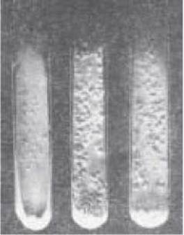
Fig. 9. Culture of Mycobacterium tuberculosis on solid egg medium.
Table 1 WHO Scheme for evaluating the results of cultural examination for M. tuberculosis
|
Colony count |
Result evaluation |
Characteristics |
|
No growth |
Negative |
|
|
1-19 colonies |
Positive (indicate number of colonies) |
Isolated colonies (scanty bacterial shedding); bacterioscopy of pathological material reveals almost no mycobacteria |
|
20-100 colonies |
1 + |
Moderate bacterial shedding; bacterioscopy of pathological material reveals isolated mycobacteria in each microscopic field or single ones in the preparation, but no fewer than five |
|
100-200 colonies More than 500 colonies (confluent growth) |
2 + 3 + 4 + |
Massive bacterial shedding; bacterioscopy shows 10 or more mycobacteria in each microscopic field |
|
Overgrowth by opportunistic microflora |
Overgrowth — repeat inoculation |
3. Biological method involves inoculating guinea pigs, which possess high sensitivity to M. tuberculosis, with sputum or other pathological material. The biological assay is the most sensitive method for detecting M. tuberculosis, as tuberculosis develops in laboratory animals following the administration of material containing fewer than 5 microbial bodies per 1 mL. However, it should be noted that M. tuberculosis strains resistant to chemotherapeutic drugs, particularly isoniazid, may be avirulent to guinea pigs. Therefore, detecting M. tuberculosis in pathological material requires a combination of microbiological examination methods. Overall, prior to initiating Treatment, a comprehensive bacteriological evaluation must be performed: direct sputum bacterioscopy three times, or in the absence of sputum, a threefold examination of material following provocative inhalations or bronchial lavage; in case of negative results, a threefold examination using the flotation method on CSC; and three sputum cultures on nutrient media with mandatory determination of M. tuberculosis susceptibility to anti-mycobacterial drugs. Subsequently, a twofold analogous examination is conducted monthly until bacterial shedding ceases, followed by monthly single examinations of the material via microscopy and culture until the end of Chemotherapy (if M. tuberculosis is present, drug susceptibility testing of M. tuberculosis is performed).
Currently, immunological and molecular-Genetic Methods are employed for the etiological identification of tuberculosis. The most promising among these are enzyme-linked immunosorbent assay (ELISA) and radioimmunoassay methods for detecting mycobacterial Antigens, which rely on The Use of Monoclonal Antibodies. The molecular-genetic Polymerase Chain Reaction (PCR) method involves detecting mycobacterial DNA in biological material (sputum, tissues; lavage, pleural, or cerebrospinal fluid, etc.). The assay is based on the Amplification of a specific DNA fragment of M. tuberculosis. PCR is a highly sensitive and rapid method for the Laboratory Diagnosis of tuberculosis. The identification of M. tuberculosis in biological material containing 1–10 cells per sample can be accomplished within 3–6 hours. Performing PCR requires specialized test systems and laboratories.
The administration of anti-mycobacterial drugs leads to The Development of drug resistance in M. tuberculosis. Primary and secondary drug resistance are distinguished.
Primary drug resistance refers to the resistance of M. tuberculosis in newly diagnosed patients who have not yet received treatment, arising As a result of infection with resistant strains of M. tuberculosis.
Secondary drug resistance develops during the course of prolonged, irrational anti-mycobacterial therapy.
According to WHO data, the frequency of primary drug resistance to any given drug averages 10.4%, while secondary resistance is 36%. In Ukraine, primary drug resistance exceeds the WHO average by a factor of two, and secondary resistance by 1.5 times.
Determining the susceptibility of M. tuberculosis to anti-tuberculosis drugs is of paramount importance for treatment tactics, adjusting anti-mycobacterial therapy, and disease prognosis. The susceptibility of M. tuberculosis to anti-tuberculosis drugs is defined by the minimum concentration of the drug that inhibits the growth of M. tuberculosis on a nutrient medium. M. tuberculosis is considered susceptible to a given drug if fewer than 20 colonies grow in the test tube, provided there is abundant growth in the control. A culture is considered resistant if more than 20 colonies grow. M. tuberculosis is deemed resistant if growth occurs at the following drug concentrations per 1 mL of nutrient medium: isoniazid 1 mcg, rifampicin 20 mcg, streptomycin 5 mcg, ethambutol 2 mcg, PAS 1 mcg, thioacetazone 2 mcg, cycloserine 50 mcg, and all other drugs 30 mcg.
Blood examination. The primary causes of alterations in the peripheral blood of tuberculosis patients are intoxication and Hypoxia. In early forms of tuberculosis, the hemogram is normal or shows minor deviations. In recent years, hypochromic anemia has been increasingly observed; however, in chronic, protracted courses of a widespread specific process accompanied by increasing pulmonary insufficiency and hypoxia, a compensatory increase in the erythrocyte count and Hemoglobin level is possible. Overall, more severe Clinical forms of tuberculosis are characterized by mild leukocytosis (9.0–15.0×10⁹/L), an increased percentage of band neutrophils, lymphopenia, monocytopenia, eosinopenia, accelerated ESR, as well as a decrease in the albumin-globulin ratio (normal is 1.5) with a relative increase in a2 and y-globulins (normal: a1 – 4, a2 – 8, ß – 12, y – 16).
Urine examination. In patients with pronounced tuberculous intoxication, proteinuria, erythrocytes, and leukocytes may be observed in the urine. As intoxication subsides, pathological Changes in the urine disappear. Nevertheless, in the presence of Amyloidosis of internal organs, particularly the Kidneys, hyposthenuria, persistent proteinuria, an elevated number of erythrocytes and leukocytes in the urine, and the appearance of casts are characteristic features.
1. The CAUSATIVE AGENT OF tuberculosis, its properties and species.
2. Forms of Variability in Mycobacterium tuberculosis and their clinical significance.
3. Reversion of Mycobacterium tuberculosis and its clinical significance.
4. Atypical mycobacteria, their properties, and clinical significance.
5. Methods for detecting Mycobacterium tuberculosis and their diagnostic value.
6. Most characteristic blood and urine abnormalities in Pulmonary Tuberculosis.
TESTS
1. Which BIOCHEMICAL COMPONENTS OF MTB are responsible for their resistance to acids, alkalis, and alcohols?
A. Proteins
C. Lipids
E. Mineral salts
2. The primary carriers of antigenic properties in MTB are:
A. proteins
B. carbohydrates
C. lipids
D. polysaccharides
E. mineral salts
3. The L-form of mycobacteria refers to:
A. a vaccine strain of MTB
B. non-visual forms of MTB
C. atypical MTB
D. MTB that have partially lost their Cell wall
E. filterable forms of MTB
4. The cause of primary drug resistance in MTB is:
A. untimely detection of tuberculosis
B. delayed detection of tuberculosis
C. irregular intake of antimycobacterial drugs
D. treatment with inadequate doses of chemotherapy agents
E. infection with resistant strains of MTB
5. The frequency of primary MTB resistance in tuberculosis patients:
A. 0.5-1 %
B. 2-5 %
C. 10-14%
D. 15-20 %
E. 25-30 %
6. The frequency of secondary MTB resistance to antimycobacterial drugs in tuberculosis patients:
A. 1-5 %
B. 5-10 %
C. 10-20 %
D. 20-40 %
E. 50-60 %
7. What is primary MTB resistance?
A. MTB resistance in newly diagnosed patients who have not yet been treated with antimycobacterial drugs
B. MTB resistance in patients with primary forms of tuberculosis
C. MTB resistance in patients with chronic forms of tuberculosis
D. MTB resistance in patients with tuberculosis relapses
E. MTB resistance in patients with minor forms of pulmonary tuberculosis
8. Which MTB species is the most pathogenic to humans?
A. M. africanum
B. M. avium
C. M. bovis
D. M. tuberculosis
E. M. kansasii
9. Mycobacteriosis refers to diseases caused by:
A. L-forms of mycobacteria
B. M. tuberculosis
C. acid-fast saprophytes
D. atypical mycobacteria
E. antimycobacterial drug-resistant strains of MBT
10. On the 3rd day of sputum inoculation onto a solid medium for MBT detection, colonies appeared. This indicates:
A. growth of rapidly multiplying mycobacteria
B. growth of highly virulent mycobacteria
C. growth of atypical mycobacteria
D. growth of non-specific microflora
E. growth of L-forms of mycobacteria
11. Which of the following sputum characteristics is most typical for patients with pulmonary tuberculosis?
A. muco-purulent, odorless, 10-50 mL per day
B. purulent with a pungent, unpleasant odor, rusty in color, up to 500 mL
C. purulent, odorless, up to 300 mL
D. muco-watery, 50-100 mL
E. purulent-bloody with an unpleasant odor, 100-150 mL per day
12. Timeframe for the appearance of Mycobacterium tuberculosis growth on solid nutrient media:
A. on days 2-3
B. on days 7-14
C. in 3-4 weeks
D. in 3-5 months
E. in 6 months
13. What percentage of tuberculosis cases in humans is caused by M. bovis?
A. 1-2 %
B. 3-5%
C. 10-20 %
D. 25-30 %
E. 35-50 %
14. According to Runyon's Classification, the third group of atypical mycobacteria includes:
A. M. bovis
B. M. africanum
C. M.aque
D. M. avium
E. M. tuberculosis
PROBLEMS
1. Patient S., aged 6, was admitted to the anti-tuberculosis dispensary with a provisional diagnosis of Primary tuberculosis complex of the lower lobe of the right lung.
Outline a plan for: a) radiological; b) immunological; c) laboratory; d) Bacteriological examination OF the patient to confirm the clinical diagnosis.
Answer: a) tomogram at the level of the tracheal bifurcation; b) Mantoux test with 2 TU; c) complete blood count and urinalysis; d) sputum smear microscopy and culture at least 3 times.
2. A newly diagnosed pulmonary tuberculosis patient exhibits resistance of Mycobacterium tuberculosis to isoniazid (1 mcg/ml) and streptomycin (10 mcg/ml).
a) what is this type of resistance called;
b) What is the management strategy.
3. A patient is diagnosed with focal Tuberculosis of the apical segment of the upper lobe of the right lung in the infiltration phase. There is no sputum production.
Outline a plan for: a) sputum collection; b) bacteriological examination.
4. A patient has been admitted to the hospital with a 10-year history of tuberculosis, irregular treatment history, and mycobacteria resistant to isoniazid (1 mcg/ml) and rifampicin (20 mcg/ml).
a) what is this type of resistance called;
b) What are the causes of its development;
c) tactics of further management.
5. Sputum culture was inoculated onto Lowenstein-Jensen medium.
a) when colonies of Mycobacterium tuberculosis may appear;
b) how long it takes to wait for the final laboratory report.
Last update: 10/08/2026
Editorial and Educational Adaptation: This material has been compiled based on the primary/original source text. The project team performed an editorial review, corrected technical inaccuracies, structured sections, and adapted the content for an educational format.
What was processed:
- elimination of formatting defects (OCR errors, structural breaks, corrupted characters);
- editorial organization of content;
- standardization of terminology in accordance with academic sources;
- verification of factual statements against the original source text.
All mentions of the author, publication year, and origin of the primary text have been preserved in accordance with the source.