MEDICAL BIOLOGY, HUMAN ANATOMY, PHYSIOLOGY AND PATHOLOGY - Ya.I.Fedoniuk 2010
BIOLOGY
CHAPTER 2. BIOCOENOTIC LEVEL OF ORGANIZATION OF LIFE AND HUMAN PLACE IN IT
2.4. MEDICAL HELMINTHOLOGY
Laboratory Diagnostics of Helminthiases
Mature helminths residing within the host's body constantly lay eggs or larvae. Depending on the helminth localization, these are either released into the external environment or accumulate in the Blood, Lymph, and other Tissues. The detection of specific helminths, their eggs, or larvae constitutes the core of Laboratory Diagnostics for helminthiases. Eggs and larvae are most commonly excreted into the external environment with feces (stool). Therefore, fecal examination, or coprological analysis (from Greek kopros - feces), is the most frequently performed Procedure. The primary Methods applied for this purpose include helminthoscopy, helminthoovoscopy, and helmintholarvoscopy.
Helminthoscopy. Helminthoscopy is a method of laboratory diagnostics of helminthiases based on the detection of intact helminths or their fragments. This is a macroscopic method performed without a Microscope, using a magnifying Glass only when necessary. Macroscopic examination of feces is utilized either to detect helminths expelled after deworming or to identify proglottids or tapeworm fragments that periodically detach from the helminth's body and are excreted into the external environment with feces.
The technique of helminthoscopic examination is as follows. Small portions of stool are mixed with Water and examined under good illumination in glass trays with black paper placed underneath; helminths and any suspicious white formations are removed using tweezers or a pipette for further study. The sequential sedimentation method is more commonly used in helminthoscopy. For this, the entire stool sample is mixed with water in glass cylinders, allowed to settle for a few minutes, the liquid is decanted, and the sediment is refilled with water and stirred. This procedure is repeated several times. Once the liquid becomes clear, it is decanted, and the sediment is examined in small portions in a glass tray or Petri dish against a dark Background.
Helminthoovoscopy is a method of laboratory diagnostics of helminthiases based on the detection of helminth eggs in feces (Figs. 2.41, 2.42).
Class="center">
Fig. 2.41. Flukes
1 - Nanophyetus; 2 - cat Liver fluke; 3, 4 - lancet liver fluke (immature and mature); 5 - Echinostoma; 6 - lung fluke; 7 - Metagonimus; 8 - Fasciola; 9 - Schistosoma japonicum; 10 - Schistosoma haematobium; 11 - Schistosoma mansoni; 12 - taeniids; 13 -
dwarf tapeworm (low egg count); 14 - broad fish tapeworm.
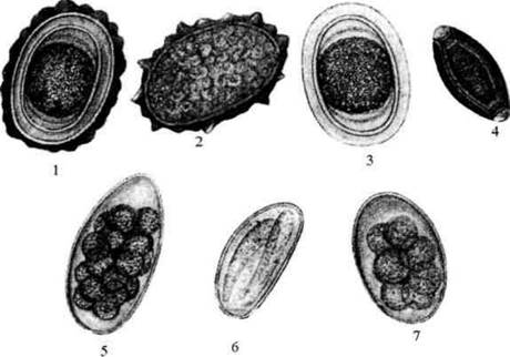
Fig. 2.42. Nematode eggs:
Ascaris lumbricoides: 1 - fertilized, with a protein coat; 2 - unfertilized, with a protein coat; 3 - fertilized, without a protein coat; 4 - whipworm; 5, 6 - pinworm: freshly passed and with a larva; 7 - hookworms
Native smear method. A pea-sized particle of feces is placed on a glass slide (6 x 9 cm) in a 50% aqueous solution of glycerin, mixed with a wooden applicator stick, insoluble coarse particles are removed, and the preparation is examined under a microscope without a coverslip. Eggs of all helminth species can be detected in a native smear. More accurate methods are those based on concentrating the eggs contained in a sample into a small volume (so-called enrichment methods). These include Fulleborn's method, Kalantarian's method, etc.
Fulleborn's method. This method is based on the flotation of helminth eggs in a saturated sodium chloride solution (specific gravity 1.2). About 2.5-5 g of feces is placed in a tall porcelain or glass beaker (50-100 mL), and saturated sodium chloride solution is gradually added while thoroughly mixing the feces. The solution is added almost to the brim. Wooden applicator sticks, fresh each time, are most convenient for mixing. Large particles that float to the surface are removed, and the solution is left to stand for 45 minutes. During this time, due to the difference in specific gravity, the lighter eggs float and gather in the surface film, while large insoluble fecal particles settle to the bottom. Using a wire loop bent at a right angle, the film is skimmed off, transferred onto a microscope slide, covered with a coverslip, and examined under a microscope. At least four preparations are made. The loop is sterilized over a spirit lamp. While effective, this method fails to float eggs of trematodes, taeniids, and the broad fish tapeworm (as they are heavier), as well as unfertilized Ascaris eggs (whose shell is permeable to salts). Therefore, In addition to the surface film, the sediment must also be examined, which complicates the procedure.
Kalantarian's method is a slightly modified version of Fulleborn's method. A saturated sodium nitrate solution (specific gravity 1.38) is used instead of saturated sodium chloride solution. This method allows the flotation of broad fish tapeworm eggs and unfertilized Ascaris eggs as well. The disadvantage of this method is the higher cost of sodium nitrate compared to sodium chloride.
There are also helminthovroscopic Methods based on THE PRINCIPLE OF egg sedimentation in a solution whose specific gravity is lower than that of the eggs.
Pinworms lay their eggs in the perianal region; therefore, a specific diagnostic technique—the perianal swab method—is used for the Laboratory Diagnosis of enterobiasis. A cotton swab tightly wound on a wooden stick and moistened with a 50% aqueous glycerin solution is gently scraped across the perianal folds. The stick with the swab is placed in a dry test tube and sent to the laboratory. There, the eggs are washed off the swab using 1-2 drops of 50% aqueous glycerin solution, and 4 preparations are made on two glass slides. Alternatively, a wooden spatula (such as a trimmed matchstick) moistened with a 50% aqueous glycerin solution or transparent adhesive tape can be used. In addition to pinworm eggs, this method also detects beef tapeworm eggs.
Helmintholarvoscopy is a method for detecting helminth larvae. In certain helminthiases (such as strongyloidiasis), larvae rather than eggs are excreted with feces, and the Baermann technique is used to detect them. This method relies on the thermotactic migration of helminth larvae toward warmth. A glass funnel fitted with a rubber tube and a pinchcock at its narrow end is fixed in a stand. 5-10 g of feces is placed on a sieve lined with double-layered gauze. The sieve is immersed in the funnel, which is filled with warm water (40-45 °C) so that the lower part of the sieve is submerged. The larvae actively migrate from the feces into the warm water and concentrate in the rubber tube. After 4 hours, the water from the tube is drained into a centrifuge tube and centrifuged. The supernatant is discarded, and the sediment is examined under a microscope.
Last update: 08/08/2026
Editorial and Educational Adaptation: This material has been compiled based on the primary/original source text. The project team performed an editorial review, corrected technical inaccuracies, structured sections, and adapted the content for an educational format.
What was processed:
- elimination of formatting defects (OCR errors, structural breaks, corrupted characters);
- editorial organization of content;
- standardization of terminology in accordance with academic sources;
- verification of factual statements against the original source text.
All mentions of the author, publication year, and origin of the primary text have been preserved in accordance with the source.