IMMUNOLOGY TEXTBOOK - Mercury Podillia 2013
HIV INFECTION: IMMUNOPATHOGENESIS, IMMUNODIAGNOSIS, IMMUNOCORRECTION
Laboratory Diagnostics of HIV Infection and AIDS
Laboratory Diagnostics are based on the detection of viral markers and specific Antibodies in biological fluids.
The following Methods are used to establish a Diagnosis:
1. Polymerase Chain Reaction (PCR) is a highly sensitive method for detecting viral RNA.
2. Hybridization assay (HA) involves searching for specific Nucleic Acids and quantifying them based on binding to a DNA probe.
3. Immunofluorescence assay (IFA) detects Antigens in Blood Leukocytes. The method is specific, yet has low sensitivity.
4. Enzyme-linked immunosorbent assay (ELISA) is a highly sensitive method for detecting HIV antibodies in the serum of a patient or carrier.
5. Immunoblotting is a confirmatory test. This method detects antibodies to one or more HIV envelope or core Proteins. A result is considered positive if antibodies are detected against any two of the three major HIV antigens — p24, gp41, and gp120 (or gp160).
6. Flow cytometry enables the determination of lymphocyte subpopulations and the detection of phenotypic markers that characterize Changes in the functional state of Cells.
Determination of HIV-1 and HIV-2 antibodies in blood. Antibodies to HIV-1 and HIV-2 are normally absent in blood serum. Antibody testing serves as the primary method for the Laboratory Diagnosis of HIV infection. This method is based on ELISA (sensitivity >99.5%, Specificity >99.8%). HIV antibodies appear in 90–95% of infected individuals within 3 months post-infection, in 5–9% within 6 months, and in 0.5–1% at later stages. During the AIDS stage, antibody titers may decrease until they disappear entirely. Upon receiving a positive result for HIV antibodies, the test should be repeated 1 or 2 times—preferably using a diagnostic kit from a different lot—to avoid false-positive results. A result is deemed positive if both of two or two of three assays clearly detect antibodies.
Western blot for HIV viral protein antibodies in blood serum. Antibodies to HIV viral proteins are normally absent in blood serum. The ELISA method for HIV antibody detection acts as a screening test. When a positive result is obtained, the Western blot method is used to confirm its specificity. This involves counter-current Immunoelectrophoresis in a gel, reacting the patient's serum antibodies with various viral proteins separated by molecular weight via Electrophoresis and transferred onto nitrocellulose. Antibodies to viral proteins gp41, gp120, gp160, p24, p18, p17, and others are identified. The detection of antibodies to any of the Glycoproteins — gp41, gp120, or gp160 — is considered a positive result. If antibodies to other viral proteins are detected, the result is considered indeterminate, and the individual should be re-tested twice, at 3 and 6 months. The absence of antibodies to specific HIV proteins indicates that the ELISA yielded a false-positive result. Nevertheless, when interpreting immunoblotting results in clinical practice, one must follow the manufacturer's instructions provided with the test kit.
Protein p24 in blood serum. The p24 antigen is normally absent in blood serum. The p24 antigen is a protein of the HIV nucleocapsid shell. The stage of primary manifestations following HIV infection is the result of the onset of active viral Replication. The p24 antigen appears in the blood 2 weeks post-infection and can be detected by ELISA between 2 and 8 weeks. After 2 months from the onset of infection, the p24 antigen disappears from the blood. Later in the clinical course of HIV infection, a secondary rise in blood p24 levels occurs, coinciding with The Development of AIDS. Existing ELISA test kits for p24 antigen detection are used for the early screening of blood Donors and infants, predicting the progression of AIDS, and monitoring therapy in these patients. The ELISA method boasts high analytical sensitivity, allowing the detection of HIV-1 p24 antigen in blood serum at concentrations of 5–10 pg/mL and less than 0.5 ng/mL for HIV-2, along with high specificity. However, it should be noted that blood p24 levels are subject to individual variation, meaning that only 20–30% of patients can be identified by this assay during the early post-infection period.
IgM and IgG Class antibodies to the p24 antigen appear in the blood starting from the 2nd week, peak within 2–4 weeks, and persist at this level for varying durations: IgM class antibodies last for several months, disappearing within a year of infection, whereas IgG antibodies can persist for years.
The algorithm for diagnosing HIV infection is presented in Fig. 9.
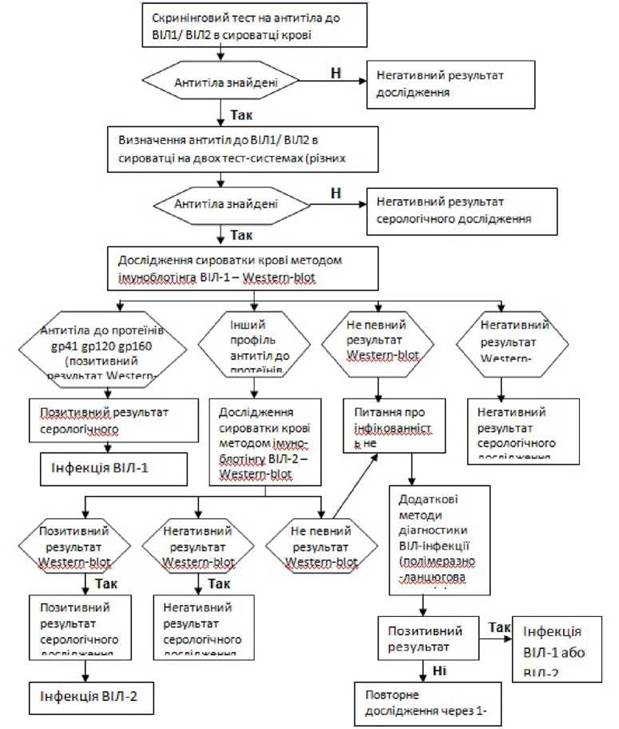
Fig. 9. Algorithm for diagnosing HIV infection.
Last update: 13/08/2026
Editorial and Educational Adaptation: This material has been compiled based on the primary/original source text. The project team performed an editorial review, corrected technical inaccuracies, structured sections, and adapted the content for an educational format.
What was processed:
- elimination of formatting defects (OCR errors, structural breaks, corrupted characters);
- editorial organization of content;
- standardization of terminology in accordance with academic sources;
- verification of factual statements against the original source text.
All mentions of the author, publication year, and origin of the primary text have been preserved in accordance with the source.