NEONATAL SURGERY - 1976
1. GENERAL PROVISIONS
4. Preoperative Preparation. Anesthesia. Postoperative Management
Preparing a newborn for surgery, ensuring adequate pain relief, and providing proper postoperative management are of paramount importance for the Outcomes of surgical intervention.
Preoperative Preparation
The primary objectives of preoperative preparation include maintaining an optimal microenvironment, preventing manifestations of hemorrhagic syndrome, correcting Homeostasis parameters, and preventing vomiting and gastric aspiration. Implementing these measures is particularly crucial in infants presenting with toxicosis, exicosis, Peritonitis, hyperthermia, etc. It is essential to maintain continuity in the therapeutic measures initiated at the maternity hospital and continued during The transport of the newborn to the surgical department.
Issues regarding the establishment of a microenvironment, the Prevention and Treatment of aspiration Pneumonia, and fluid calculations for parenteral administration are discussed in the section “Peculiarities of Postoperative Management.”
Upon admission of the infant to the department, their condition is evaluated. The widespread Introduction of rapid laboratory and micro-diagnostic Methods into clinical practice allows for obtaining fairly complete information regarding the newborn's homeostasis status within a short preoperative evaluation period. Based on the experience of the neonatal surgery department, the most essential Laboratory tests are determined to be:
1) Hemoglobin, hematocrit (using micromethods), leukocyte count, and platelet count. Determining the erythrocyte count is not mandatory, as hematocrit serves as a more precise indicator;
2) plasma electrolytes: sodium, potassium, calcium;
3) total protein and protein fractions;
4) clotting and bleeding times;
5) Blood glucose, urea, and residual nitrogen;
6) acid-base balance and blood gases: pH, BE (or alkaline reserve), pCO2, and pO2 of capillary blood;
7) urinalysis (sugar, protein, urea, sediment Cell/15.html">Microscopy).
Chest radiography is performed in all cases.
Blood Coagulation testing is indicated regardless of the infant's condition. In 80% of newborns, blood clotting time is prolonged. This parameter is significantly reduced, in particular, During the first 4 days of life due to vitamin K and prothrombin deficiency. The blood clotting time on the first day of life averages 5 min 20 s, on the 4th day — 5 min 15 s, and on the 8th day — 4 min 8 s (A. A. Orekhova, 1953; N. V. Potanin, 1965, et al.). Intramuscular administration of Vikasol (1 – 2 mg/kg per day) the day before or immediately prior to surgery helps enhance blood clotting and prevents tissue Hemorrhage during the Procedure and in the early postoperative period.
Anesthesia
Anesthesia in newborns represents the most complex and critical area of pediatric anesthesiology. General anesthesia in the youngest infants should be administered by an experienced anesthesiologist. In the absence of an anesthesiologist, local anesthesia combined with enhanced premedication (phenothiazines, neuroleptics, analgesics) is employed as an exception.
Any painful or complex procedure, as well as diagnostic investigations in newborns, must be performed under general anesthesia, and when indicated, with sufficient Muscle relaxation and adequate pulmonary ventilation. The multi-component anesthesia techniques developed at our clinic have enabled the virtual elimination of anesthesia-related neonatal mortality.
Almost all modern multi-component anesthesia regimens can be utilized. Anesthesia is administered taking into account the anatomical and PHYSIOLOGICAL CHARACTERISTICS OF the newborn's body. A newborn's Tongue is relatively large, whereas the epiglottis is small, rigid, and triangular in shape. These features complicate tracheal intubation. A curved blade (Macintosh) fails to bring the epiglottis into the field of view; therefore, a specialized straight blade, with the distal third curved at an angle of 25–30°, is available for newborns.
The lability of the respiratory center and the weakness of the Respiratory Muscles necessitate broadening the indications for controlled mechanical ventilation. Surgical interventions lasting longer than 30 minutes are advised to be performed under anesthesia with controlled mechanical ventilation.
A condition specific to newborns is the potential development of Hypoxia during controlled ventilation. Such a complication is rare and is associated with a patent foramen ovale and a functioning ductus arteriosus. As a result of controlled mechanical ventilation, pressure in the Pulmonary Circulation increases, leading to a shunt of deoxygenated blood through the foramen ovale or the ductus arteriosus. Discontinuing controlled mechanical ventilation and transferring the patient to spontaneous or assisted breathing resolves the hypoxia.
Premedication. Administering standard premedication consisting of analgesics (promedol) and anticholinergics (atropine) under modern multi-component anesthesia conditions in newborns is unjustified. The administration of analgesics such as promedol in patients with severe hypoxia, or atropine in cases of dehydration, tachycardia, and hyperthermia, is contraindicated. Antihistamines are used when septic processes develop. Clinical experience has demonstrated the advisability of expanding the indications for modern multi-component anesthesia in newborns without premedication. Intravenous administration of atropine is indicated only in cases of pronounced hypoxia, bradycardia, and Cardiac Arrhythmias occurring during anesthesia.
Anesthetic Equipment. Anesthesia Technique. Standard anesthesia machines equipped with special attachments and systems are used for anesthesia in newborns. Modern adult equipment sets include circle absorber systems. An Ayre's T-piece system for spontaneous or controlled breathing can be adapted to any machine. Domestic industry mass-produces the “Narkon DP” carbon dioxide absorption attachment for anesthesia machines to perform general anesthesia in newborns and infants.
The advantages of the pendulum system include minimal resistance to breathing, as well as the conservation of heat and warmth when working with an absorber. Its disadvantages are the relatively large size of the absorber and the inconvenience of securing the system near the patient's HEAD. The circulating attachment retains the benefits of the pendulum system (heat and moisture preservation, minimal breathing resistance) and is convenient to operate.
Ayre's system is assembled from Components of the anesthesia machine set; it is lightweight and easily secured. The T-piece offers virtually no resistance to breathing at flows up to 12 L/min. When performing controlled mechanical ventilation, a manometer or a safety valve is integrated into the system—
a valve that is easily fashioned from a Bobrov apparatus filled with Water. The safety valve vents excess pressure of the anesthetic gas mixture. A disadvantage of Ayre's system is the loss of heat and moisture during high flow rates of dry and cooled gases. Lewis and Spoerel (1961) recommend administering an anesthetic gas mixture flow equal to twice the patient's minute ventilation when using Ayre's system while maintaining spontaneous breathing. Under these conditions, air entrainment from the open arm of the T-piece does not exceed 25%. V. M. Yuryevich and A. S. Perelmutr (1973), when using Ayre's system, calculate the anesthetic gas mixture flows using the following formulas:
a) for artificial pulmonary ventilation:
Q = v (1 + a),
б) for spontaneous breathing:
Q = 1,5 v (1 + a),
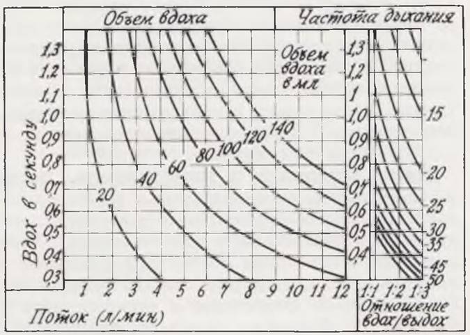
where Q is the total gas flow rate According to the flowmeter (gas flow) in liters per minute; v is the minute ventilation of the Lungs in liters per minute, and a is The ratio of expiration time to inspiration time. For newborns during spontaneous breathing, this value is 1, whereas during controlled breathing, it is advisable to consider it equal to 2.
When changing the inspiratory-to-expiratory ratio, Keuskamp (1963) recommends using a special table to calculate the total gas flow from the flowmeter (Fig. 8).
Class="center">Fig. 8. Table for calculating total gas flow based on flowmeter readings (after Keuskamp).
Rees's system allows for controlled ventilation using a bag. This system preserves the advantages of Ayre's system, but lung inflation is performed manually, more smoothly, and without the risk of tearing the lung tissue with a high flow of the anesthetic gas mixture. To prevent hypercapnia, a gas flow of at least twice the child's minute volume is established.
Tracheal intubation is performed after abolishing spontaneous breathing with muscle relaxants. The Pediatric Surgery Clinic of the Central Institute for Advanced Medical Training (CIALMT) has developed a technique for suppressing spontaneous breathing prior to tracheal intubation not by using muscle relaxants, but by deepening anesthesia with halothane and providing assisted followed by controlled ventilation. This approach allows laryngoscopy and tracheal intubation to be performed against the Background of apnea and good muscle relaxation without administering muscle relaxants. When performing general anesthesia with anesthetics other than halothane, the administration of depolarizing muscle relaxants is indicated prior to laryngoscopy.
Tracheal intubation under deep halothane anesthesia while preserving spontaneous breathing is performed only in cases where the insertion of the endotracheal tube into the Trachea is complicated by anatomical deformities in the face and neck region, inflammatory edema, or tumor masses in the Larynx and trachea causing stridulous breathing. In these situations, abolishing spontaneous breathing with muscle relaxants and unsuccessful attempts at tracheal intubation can lead to severe hypoxia, while deformations and compression of the Airways make it difficult to provide controlled ventilation via an anesthesia machine mask.
Laryngoscopy is performed with the patient in a horizontal position. A pillow under the shoulders is optional. After advancing the laryngoscope to the Base of the epiglottis, the assistant gently pushes the larynx backward from the outside. Efforts are made not to capture the epiglottis with the laryngoscope blade.
Smooth, reinforced, and variable-caliber tubes of the Cole type are used for intubation. According to V. M. Yuryevich and A. S. Perelmutr (1973), Cole tubes can reduce expiratory resistance by more than a factor of 2. Recently, plastic thermoplastic tubes have been used, which adapt their configuration to the shape of the larynx and trachea under the Influence of the child's body heat. Plastic tubes do not irritate the mucous membrane of the larynx and trachea, and they reduce the risk of developing laryngitis, tracheitis, and post-intubation edema. For prolonged oro- or nasotracheal intubation, which is undertaken in newborns for therapeutic purposes (in cases of respiratory depression or airway obstruction),
as well as for connection to a mechanical ventilator lasting longer than 4 hours, only plastic tubes should be used. Table 8 presents the main tube sizes for newborns.
Table 8. Tube sizes for newborns and infants in the first 3 months of life
Age of children |
Tube sizes |
|||
outer diameter (mm) |
Charrière scale no. |
length for oral intubation (cm) |
length for nasal intubation (cm) |
|
Premature infants |
2.5 — 3.5 |
8 — 12 |
8 — 9 |
10 — 12 |
Newborns |
3.5 — 4.0 |
12 — 13 |
9 — 10 |
12 — 14 |
1 month |
4.0 — 4.5 |
13 — 15 |
10 — 11 |
13 — 15 |
2 » |
4.5 — 5.0 |
15 — 17 |
10.5 — 11.5 |
14 — 16 |
3 » |
5.0 — 5.5 |
17 — 18 |
11.0 — 12.0 |
14 — 17 |
Packing of the Oral Cavity is not mandatory. The tubes are secured with two strips of adhesive plaster.
Absolute indications for tracheal intubation include anesthesia for surgeries in the thoracic cavity, face, and neck regions, as well as abdominal and limb operations lasting more than 30 minutes. The smaller the child, the greater the indication for tracheal intubation and controlled or assisted ventilation during anesthesia.
After each anesthesia, endotracheal tubes are cleaned in soapy water, thoroughly rinsed with tap water, sterilized in diocide for 30 minutes, and then rinsed with running water for 10–15 minutes. Thirty minutes before anesthesia, the tubes are placed in a furacilin or rivanol solution (1:1000) or in a 0.6% levomycetin solution prepared in 0.6% ethyl alcohol. Immediately before intubation, the tubes are rinsed with a sterile isotonic sodium chloride solution. Nowadays, single-use endotracheal tubes sterilized by gamma rays and packaged in sterile bags are commercially available. In such cases, additional sterilization is not required.
Choice of anesthetic and muscle relaxant. Almost all anesthetics, muscle relaxants, and anesthesia protocols used in adults can be applied to newborns. Our experience, encompassing over 1,500 anesthetics in newborns, allows us to consider halothane anesthesia with nitrous oxide and oxygen as the method of choice. Halothane causes minimal irritation to the respiratory tract and lung tissue, does not provoke hypersecretion of the salivary and bronchial glands, and reduces the risk of postoperative pneumonia. Halothane can be used for almost all surgical interventions. It is hazardous only in cases of pronounced hypotrophy (pyloric stenosis) and ongoing hemorrhage, as it induces peripheral vasodilation, which—in the presence of a significant circulating blood volume deficit—leads to a drop in blood pressure and a reduction in venous return to The Heart. In such instances, halothane may be used provided that adequate concurrent transfusions of blood, plasma, or other colloidal solutions are administered. Neuroleptics make it possible to perform anesthesia in the sickest patients with severe homeostasis disorders. The average dose of Thalamonal (fentanyl + droperidol) for premedication is 0.06 mL/kg of the child's body weight intramuscularly 40 minutes before surgery. For induction into neuroleptanalgesia, Thalamonal is used at a dose of 0.2 mL/kg. Anesthesia is maintained by repeated administrations of fentanyl (0.1–0.06 mL/kg every 30 minutes). Droperidol is used at a dose of 0.1–0.07 mL/kg every 1 to 1 1/2 hours. After the completion of anesthesia, respiratory analeptics such as nalorphine (0.1–0.2 mL) or bemegride (50–75 mg) are administered intravenously.
The Use of neuroleptanalgesia in newborns requires further study and strict individualization of drug dosages, as clinical experience has shown that upon completion of neuroleptanalgesia, nearly 1/3 of newborns exhibited pronounced respiratory depression, frequently necessitating prolonged mechanical ventilation.
Infants in the first month of life are relatively resistant to succinylcholine-type drugs; therefore, larger doses calculated per kilogram of the child's body weight are required for myoplegia (Table 9). The best route of administration for muscle relaxants is intravenous. In emergency situations or for single administrations, such as for a brief examination, injections of succinylcholine into the tongue or sublingual region can be recommended. Intramuscular administration of muscle relaxants with or without hyaluronidase is also feasible1.
1 A detailed Description of the methodology for intramuscular administration of depolarizing muscle relaxants with lidase was published by us in the journal Surgery (Khirurgiya, 1967, no. 7).
Table 9. Dosage of muscle relaxants in newborns
Drug |
Route of administration |
||
intravenous (mg/kg) |
intramuscular (mg/kg) |
intramuscular with lidase (mg/kg) |
|
Succinylcholine initial dose |
3 (1% solution) |
5 (5% solution) |
3 (5% solution) |
repeat doses |
1,5 (1% solution) |
3 (5% solution) |
2 (5% solution) |
Tubocurarine initial dose |
0,4 |
0,6 |
0,5 |
Repeat doses |
For each repeated dose, decrease the amount by 1/4 |
||
The main disadvantages of succinylcholine include its short duration of action, The Need for frequent repeat administrations, the risk of a "dual block", prolonged apnea when the infant cools down during anesthesia, and acid-base balance disorders (acidosis).
According to Payne (1955) and V. A. Kovanev et al. (1970), newborns exhibit increased sensitivity to tubocurarine, reacting to its administration similarly to patients with myasthenia gravis. At the same time, the availability of an antidote (proserine) for this group of muscle relaxants, the possibility of infrequent dosing (once every 30–40 minutes), and a favorable ganglion-blocking effect do not preclude the use of tubocurarine. Decurarization using proserine and atropine (intravenously) is mandatory, but only after the initial signs of spontaneous breathing return. Administering tubocurarine to newborns with severe hemodynamic disorders, acrocyanosis, or metabolic acidosis is hazardous due to the risk of prolonged postoperative apnea.
The protocol for decurarization following tubocurarine administration: intravenous administration of atropine (0,02 mg/kg), followed in 5 minutes by intravenous proserine (0,05 mg/kg). If the effect is insufficient, proserine is administered repeatedly.
Termination of anesthesia. After discontinuing anesthetic inhalation, patients are ventilated with pure oxygen (100%) for at least 10 minutes, and the nasorotopharynx is cleared. Once muscle tone, swallowing, and cough Reflexes return, the trachea is cleared via the endotracheal tube, and lung sounds are auscultated during spontaneous breathing. Oxygen inhalation is repeated (5–10 minutes), a suction catheter is passed through the endotracheal tube, and the tube is removed from the trachea under laryngoscopic visualization while simultaneously aspirating mucus from the trachea and oropharynx. Immediately after anesthesia, the infant is placed in an incubator pre-set to the required Temperature, humidity, and oxygen concentration parameters.
The most frequent anesthetic complication in newborns is prolonged apnea, which typically arises from intraoperative cooling, inadequate replacement of blood loss or fluids, decompensated metabolic acidosis, analgesic overdosage, the residual effects of neuroleptics (fentanyl), or anesthetic overdosage. Respiratory depression may also stem from hypoglycemia, which is common in infants during their first days of life. The recovery of striated muscle tone and the appearance of Moro-1 and Moro-2 reflexes point to central respiratory depression rather than anesthetic overdosage or residual muscle relaxant effects. If spontaneous breathing fails to recover adequately after warming the infant, administering sodium bicarbonate, and performing decurarization, extubation is deferred, and the newborn is placed in an incubator with the endotracheal tube left in place. In cases of profound central respiratory depression, mechanical ventilation using the VITA-1 apparatus is continued until adequate spontaneous breathing is fully restored.
Table 10 outlines the primary anesthesia management protocols for newborns.
Table 10. Anesthesia protocols for newborns
Premedication |
Induction |
Intubation |
Maintenance |
Emergence |
None |
Halothane + nitrous oxide + oxygen |
Halothane + oxygen, without relaxants |
Halothane + nitrous oxide + oxygen, without relaxants |
Oxygen |
Atropine + promedol |
Hexenal intravenously + nitrous oxide + oxygen |
Same with muscle Oxygen + succinylcholine |
relaxants Nitrous oxide + oxygen + tubocurarine |
Oxygen + decurarization |
Neuroleptananalgesia + nitrous oxide + oxygen |
Oxygen + succinylcholine |
Neuroleptananalgesia + nitrous oxide + oxygen + relaxants |
Oxygen + respiratory analeptics: nalorphine, bemegride |
|
Cyclopropane + nitrous oxide + oxygen |
Oxygen + succinylcholine |
Cyclopropane + nitrous oxide + relaxants |
Oxygen + decurarization after tubarine |
|
Ether + nitrous oxide + oxygen |
Oxygen + succinylcholine |
Ether + nitrous oxide + oxygen + relaxants |
Oxygen |
1 Atropine is contraindicated in hyperthermia, tachycardia, and dehydration.
Particular aspects of postoperative management
During the postoperative period, individualized nursing care and the prevention of potential complications are of paramount importance. Primary focus is directed toward establishing an optimal microclimate, preventing and managing respiratory failure and intestinal paresis, providing adequate analgesia, and maintaining fluid therapy.
Microclimate. Cooling is particularly dangerous for the infant. Unaccounted fluid losses during surgery resulting from exposure of the abdominal or thoracic cavities (and subsequent cooling) amount to approximately 50 mL/h in a newborn. In undernourished infants, thermal insulation is inadequate, which further increases heat loss. Hypothermia carries a high risk of pneumonia and sclerema. An optimal microclimate can be maintained within an incubator (Table 11).
Table 11. Incubator microclimate parameters according to infant weight
Infant weight (g) |
Incubator temperature (°С) |
Humidity (%) |
Up to 1400 |
35 — 36 |
90 — 100 |
» 1700 |
32 — 35 |
60 |
» 2000 |
30 — 32 |
60 |
Over 2000 |
28 — 29 |
60 |
Prolonged exposure of a non-hypoxic newborn to an atmosphere with an elevated oxygen concentration (exceeding 40%) may lead to Optic nerve damage up to complete blindness (retrolental fibroplasia); however, in the presence of hypoxia, the oxygen concentration may be increased until hypoxia is fully resolved.
If an incubator is not available, the infant is placed in a heated crib or a standard crib surrounded by warm-water bottles (34 — 36 °C) alongside oxygen inhalation.
Prevention and treatment of respiratory failure. One of the most frequent postoperative complications is obstructive respiratory failure. To prevent this, periodic aspiration of thick mucus from the oropharynx using a sterile catheter is indicated. Following thoracic surgeries, laryngoscopy or bronchoscopy and toilet of the tracheobronchial tree under direct visualization are performed daily. These Procedures are carried out under halothane anesthesia. Chest physiotherapy, specifically gentle Percussion with the fingertips, is effective, as is frequent repositioning of the infant in bed. For pronounced pneumonia, broad-spectrum Antibiotics are administered intravenously.
Progressive respiratory failure and uncorrectable metabolic acidosis serve as indications for mechanical ventilation (Lees, 1971). However, the outcomes of prolonged mechanical ventilation have remained unsatisfactory to date. For instance, according to Battersby (1971), out of 24 newborns who received postoperative mechanical ventilation, only two survived. The poor results of prolonged Artificial ventilation are attributed to the Anatomical and physiological characteristics of newborns as well as significant design challenges in ventilators built for small infants.
Treatment of intestinal paresis. Impaired passage of food masses leading to intestinal paresis and vomiting is a frequent postoperative complication, particularly following gastrointestinal surgeries. Aspiration of vomitus results in aspiration pneumonia; therefore, vomiting in a newborn is an absolute indication for passing a gastric tube and continuously evacuating Stomach contents. In the absence of plastic tubes, a thin rubber tube can be inserted intermittently to aspirate stagnant contents every 1,5 — 2 hours. Following aspiration, The Stomach is lavaged with room-temperature isotonic sodium chloride solution (40 — 60 mL), and 5 — 10 mL of a 5% glucose solution or breast milk is introduced. Intestinal paresis warrants cautious digital rectal examination, placement of a rectal tube for 10 minutes every hour, hypertonic microenemas (5 — 10 mL of a 5% sodium chloride solution), and rectal administration of 5 — 10 mL of glycerin. Periodically placing the infant in a prone position is also effective.
Administration of analgesics. Analgesic therapy is mandatory in the postoperative period. Preference is given to agents that do not cause respiratory depression in the infant. A favorable effect is achieved by intramuscular administration of analgin up to 15 — 20 mg/kg every 4 — 6 hours. This drug provides complete analgesia and prevents The Development of hyperthermia. Following major, traumatic operations, promedol (1 — 1,5 mg) may be administered every 6 hours in combination with antihistamines such as suprastin, diphenhydramine (dimedrol), or pipolfen.
Management of hyperthermia. Temperatures exceeding 38 °C manifest clinically as tachypnea, cyanosis, and restlessness in the infant. If the temperature rises above 39 °C, generalized seizures may develop. For moderate fever up to 38 — 38,5 °C, pyramidon is indicated at 10 mg/kg orally or intramuscularly, or analgin at up to 15 — 20 mg/kg; these agents may be combined. In cases of persistent or worsening hyperthermia, pipolfen (diprasine) or propazine (promazine) is administered intramuscularly at a dose of 1 — 3 mg/kg. Fifteen to twenty minutes after administering these medications, the Skin is rubbed with 50 — 75-degree alcohol to induce cutaneous hyperemia and enhance heat dissipation. The same effect can be achieved using circular mustard plasters. If these measures prove ineffective, physical cooling is indicated: an ice pack applied to the head and gastric lavage with cold water via a tube. In dehydrated infants, hyperthermia will not respond to treatment until the circulating blood volume is restored.
Prevention and treatment of sclerema. Sclerema typically develops as a result of pre- or intraoperative cooling. Dehydration, prolonged hypoxia, and impaired peripheral microcirculation are also frequent causes. The prognosis is invariably serious. Early administration of glucocorticoid Hormones—hydrocortisone (4 — 5 mg/kg per day) or prednisolone (1 mg/kg per day)—is vital for treatment. An essential condition for successful recovery is prolonged nursing of the infant within an incubator.
Postoperative management of patients after thoracic surgery. The most crucial aspects of managing a thoracic patient include proper analgesia for 4 to 6 days in the postoperative period and the prevention of aspiration. Immediately following surgery, a gastric tube is inserted and maintained until intestinal transit is restored. Thick secretions are suctioned from the oropharynx every 2 to 3 hours. The humidity in the incubator is increased up to 100% to prevent desiccation of the tracheobronchial mucosa. The oxygen concentration is maintained at 40%, though in the event of hypoxia, the oxygen percentage may be increased to levels that eliminate cyanosis. Pleural cavity suction is set to a negative pressure of 5 — 10 cm H2O. Daily chest radiography is performed to monitor lung expansion.
Postoperative management of patients after intestinal surgery. The gastric tube is left in place for at least 48 h. Stagnant contents are periodically aspirated. If peristalsis is not restored within 24 h after surgery, neostigmine (proserine) 0.05 mg/kg is administered intramuscularly three times at 30-minute intervals. Alternatively, continuous intravenous infusion of neostigmine at a dose of 0.1 mg/kg over 24 h can be performed. The absence of spontaneous bowel movements 48 h postoperatively is an indication for a cautious digital rectal examination, irrigation of the rectum with Ringer's solution containing glycerin, or a glycerin microenema. Simultaneously, chest vibratory massage is performed to prevent Atelectasis, along with gentle massage and passive movements of all extremities at least twice a day.
Infusion Therapy
Infusion therapy in newborns presents a complex challenge. This is due to the immaturity of their water-electrolyte balance and excretory systems. Infusion therapy is administered to cover baseline physiological requirements and provide parenteral Nutrition, restore circulating blood volume deficits, achieve rehydration in cases of dehydration, and correct acid-base balance.
Meeting Baseline Physiological Requirements. Parenteral Nutrition. Water and Electrolyte METABOLISM in an infant is more intensive than in an adult. Over the course of 24 hours, a newborn undergoes a water turnover equal to 14% of body weight (or 33% of extracellular fluid). The total body water content is significantly higher in newborns than in adults. The influx of endogenous water resulting from the Breakdown and Oxidation of nutrients amounts to 14 ml/kg. In preterm infants, the volume of extracellular fluid is 50%, in term newborns — 40%, and in adults — only 20%. The body surface area relative to body weight in newborns is 2.5 times greater than in adults.
When a newborn is cared for outside an incubator, insensible water loss ranges from 40 to 100 ml per 24 h. Elevated body temperature causes an increase in insensible water loss up to 150 — 170 ml. When housed in an incubator, insensible water loss during the first 1 — 2 days of life does not exceed 15 ml/kg. Renal function in newborns is immature. Normal urine output values are presented in Table 12.
Table 12. Daily Urine Output in Children Under 1 Year of Age
Child's Age |
Urine Volume (ml) |
1 — 2 days |
30 — 60 |
3 — 10 days |
100 — 300 |
10 days — 2 mo |
250 — 450 |
2 mo — 1 yr |
400 — 500 |
During the first 5 — 7 days of life, the infant maintains a low fluid requirement, particularly premature infants weighing less than 2500 g. Only later do baseline requirements increase to 60 — 80 ml/kg. To calculate the baseline parameters of infusion therapy without accounting for pathological losses, Table 13 can be used.
Table 13. Daily Newborn Requirements for Essential Nutrients (according to Schulte, Hotes, 1964)
Age (DAYS) |
Water (ml / kg per 24h) |
Sodium (mEq / kg) |
Potassium (mEq / kg) |
Amino Acids (g / kg) |
Plasma (ml / kg) |
Fats (g/kg) |
1 — 3 4 — 8 |
40 — 60 80 — 100 |
1 — 1.5 1.5 — 2 |
— 0.5 — 1 |
— 1 — 3 |
2 (starting from the 3rd postoperative day), 1 (for premature infants) |
|
Older than 8 |
100 — 200 |
2 |
0.8 — 1.5 |
2 — 4 |
15 ml daily |
2 — 4 |
Electrolytes are prescribed only after urinary output is restored. During surgery and immediately afterward, an electrolyte-free isotonic glucose solution is used. During the first 2 days, sodium and potassium ions are not administered to newborns unless there are losses through the gastrointestinal tract. Starting from the 14th day of life, the daily requirement for potassium is 2 — 4 mEq/kg of body weight. Intravenous administration of potassium in the form of potassium chloride solution is advisable in glucose with Insulin (1 unit of insulin per 4 g of dry glucose). Potassium requirements are calculated based on a 7.5% potassium chloride solution, 1 ml of which contains 1 mEq of potassium. However, during intravenous administration, the concentration of potassium chloride should not exceed 1%. To achieve this, no more than 13 ml of 7.5% potassium chloride is added to every 100 ml of glucose solution. Accounting for fluid losses allows for the timely replenishment of electrolyte deficits. Table 14 presents data on the electrolyte content of the main gastrointestinal fluids (secretions) in newborns and infants.
Table 14. Electrolyte Content in Digestive Tract Secretions (in milliequivalents per liter)
Electrolytes |
Saliva |
Gastric Juice |
Pancreatic Secretion |
Small Intestinal Juice |
|
Potassium Sodium Chloride |
30 10 20 — 160 |
4 — 16 45 20 — 160 |
5.2 145 100 |
4.6 141 77 |
5.1 105 99 |
Newborns and premature infants are characterized by low blood sugar levels, which may be associated with increased hydrocortisone production (Kenny, 1963). At the same time, some authors attribute hypoglycemia to infant starvation (Euler, 1964). In the first 3 days of life, blood sugar concentration is below 30 mg%; on subsequent days, it is no more than 40 mg%. Clinically, neonatal hypoglycemia manifests with various symptoms, including cyanosis, lethargy, apnea, seizures, and coma. To treat hypoglycemia, a 15% glucose solution at 75 — 85 ml/kg per day is administered intravenously (Cornblath, 1963). After 48 h, the dose can be increased to 100 ml/kg of a 10 — 15% glucose solution in 0.25 N saline. The lack of effect within 12 h from THE START OF glucose administration is an indication for prescribing hydrocortisone at a dose of 5 mg/kg per day.
Disease and surgical intervention accelerate catabolic processes; therefore, the early administration of agents that cover the energy demands of the growing Organism is essential. The daily requirement to cover baseline Energy Expenditure is 80 — 100 kcal/kg. During prolonged parenteral nutrition, the administration of glucose solutions alone does not provide a sufficient number of calories. In such cases, fat emulsions are added (intralipid 10 — 20%, lipofundin 10%), while protein requirements are met by administering crystalline amino acids (aminosol, alvesin) and plasma. Lipofundin is prescribed at a rate of 1 — 4 ml of solution per 1 kg of body weight per day. The preparation is administered in 3 — 4 divided doses simultaneously with amino acid solutions. Insulin is added to the fat emulsions at a rate of 3 — 4 units per milliliter of the preparation. Currently, prolonged parenteral nutrition for up to 400 days is feasible (Wilmore et al., 1970).
Restoration of Circulating Blood Volume Deficits. The circulating blood volume (CBV) in a normal newborn is 85 ml/kg, and in a 1-month-old infant — 75 ml/kg (Weibenbacher, 1970). The hematocrit decreases from 65 in the first month of life to 31 by the 3rd month. The hemoglobin level is 110 — 112 (17 g%). CBV consists of globular volume (i.e., formed elements — erythrocytes, leukocytes) and plasma volume, or Blood Plasma proper. When evaluating the components of CBV, great importance is attached to total protein levels and the albumin-to-globulin ratio (albumin-globulin coefficient). The normal protein content in a newborn is 5.5 — 6 g%, and the albumin-globulin ratio (A/G) is 1.5 — 2.5. According to Hartig (1969), inadequate albumin content leads to impaired fluid diffusion from the vascular bed into Cells and vice versa, dramatically increasing the risk of Shock development.
Replenishment of blood loss during surgery is best performed under the control of colorimetric measurements or gravimetry. Rickham (1969) believes that blood loss amounting to at least 5 — 7% of the expected CBV should be replaced in a newborn during surgery. The estimated blood loss of 10% of CBV is replaced from the moment surgery begins (skin incision). Blood stored for no more than 3 — 5 days is transfused. In massive transfusions (more than 50 ml), 1 ml of 10% calcium gluconate and 1 ml of 4% sodium bicarbonate solution are administered intravenously for every 25 — 30 ml of stored blood.
Restoration of CBV is characterized by the normalization of hourly urine output (5 — 15 ml/h in newborns). During rehydration therapy, the dynamics of central venous pressure (CVP) are of great importance. For newborns, normal central venous pressure values are 30 — 50 mm H2O.
Rehydration therapy in cases of dehydration. All pathological losses are measured and replaced with an equivalent amount of glucose-electrolyte solutions ("drop by drop"). When assessing the severity of water-electrolyte metabolism disorders, consideration is given to the patient's appearance, hematocrit, plasma electrolytes, total protein, residual nitrogen, creatinine, daily and hourly urine output, and its specific gravity. Clinically, dehydration is characterized by weight loss, decreased skin elasticity and turgor, diminished moisture of the mucous membranes and tongue, the appearance of "thirst fever," weakened pulse volume, and altered consciousness. Table 15 outlines the Clinical symptoms of various degrees of patient dehydration. When administering infusion therapy for rehydration, depending on the degree of dehydration, the baseline requirements listed in Table 13 can be utilized. The more severe the dehydration, the greater The amount of fluid and electrolytes administered to the patient to correct the imbalances. Depending on the degree of dehydration, baseline requirements are increased by 40 — 80 — 120%. Mild (grade I) dehydration can be corrected by administering increased amounts of fluids orally, provided the patient's gastrointestinal function is preserved. Intravenous infusions are required only in moderate (grade II) and severe (grade III) degrees. Rehydration therapy begins with the intravenous administration of plasma or colloidal plasma substitutes: polyglucin, gelatinol, hemodez. Their volume is determined by the formula: volume of insensible water loss + minimum urine output + 5 — 10% of body weight (depending on the degree of exicosis).
Table 15. Clinical Symptoms of Various Degrees of Dehydration (according to Hartig, 1969)
Degree of Dehydration and Operability |
Clinical Symptoms |
Fluid Volume for Rehydration |
Mild (weight loss up to 5%). Conditionally operable. Operable after 4 — 6 hours of preparation |
Irritability. Oral mucosa and tongue are dry. Face is flushed, skin is warm, turgor is slightly reduced, thirst is not always present |
50 ml/kg |
Moderate (weight loss from 5 to 10%). Emergency inoperable. Conditionally operable after 4 — 6 hours of preparation |
Anxiety. Frightened, agitated facial expression. Oral mucosa and tongue are very dry, sunken eyes, decreased intraocular pressure. Skin is pale with a marked reduction in turgor. Thirst, fever, tachycardia, altered mental state, distinct oliguria. Piercing cry, sunken fontanelle |
75 ml /kg |
Severe (weight loss greater than 10%). Emergency inoperable. Conditionally operable after 24 — 48 hours of preparation |
Appearance of a critically ill patient. Unresponsiveness to surroundings, areflexia, hypotonia. Oral mucosa and tongue are very dry. Deeply sunken, shiny eyes; soft eyeballs; skin is pale, cold with cyanosis and acrocyanosis. Extreme loss of turgor. Rapid, weak pulse. Severe oliguria or anuria. Paralytic ileus. Increased muscle tone, seizures, high temperature, sunken fontanelle, piercing cry |
100 — 200 ml/kg |
Mild (grade I) dehydration occurs during vomiting and the aspiration of small quantities of gastric and intestinal juices during gastrointestinal drainage. Moderate (grade II) degree is observed in marked impairment of intestinal transit with the aspiration of large amounts of gastrointestinal secretions. Severe (grade III) dehydration is associated with total or near-total fluid losses from the upper gastrointestinal tract.
During rehydration therapy, blood is transfused adequately to the blood loss (surgical or preoperative). In anemia that has developed insidiously in a premorbid state, repeated administrations of stored blood at a dose of 10 — 15 ml/kg daily or every other day are indicated until hemoglobin and hematocrit values normalize. Average plasma doses are 15 — 20 ml/kg per day, taking into account the total required fluid volume. Of the total calculated daily fluid intake, blood, plasma, and other colloidal solutions constitute about 1/3, while the remaining 2/3 consist of 10 — 15% glucose solution and electrolyte solutions.
To ensure an even distribution of the infused fluid volume throughout the day, it is recommended to calculate the drip rate per minute using the following formula:
Fluid volume (L)
over 24 h X 14 = number of drops per minute.
Correction of acid-base balance. In newborns, metabolic acidosis requires emergency treatment. This is achieved using sodium bicarbonate solution or the organic buffer trisamine (THAM, tris-buffer). One milliliter of 8.4% sodium bicarbonate solution contains 1 mEq of bicarbonate. The requirement for an alkaline solution is calculated after assessing acid-base parameters: pH, BE, EB, pCO2. The value is then determined using the formula:
Volume in milliliters of 8.4% solution
NaCO3 = BE X body weight (kg) X 0.5.
When administering a 4% sodium bicarbonate solution, the 0.5 coefficient is omitted from this formula. Acidosis correction is more effective when bases are infused dropwise in a glucose solution (5 – 10%). The approximate infusion rate is 1 drop per 1 kg per minute. Initially, the solution is infused at double speed. Acid-base monitoring is scheduled every 2 h initially, and subsequently every 6 – 8 h. As soon as normal pH values are reached, soda infusion is discontinued. Hypocalcemia and post-acidotic seizures may develop following acidosis correction. Therefore, at the end of the alkaline solution infusion, it is recommended to administer 1 –
2 mg/kg of a 10% calcium gluconate solution.
Methods of infusion therapy. Medications and fluids in newborns are generally administered intravenously. Subcutaneous administration is hazardous due to the risk of purulent-inflammatory complications. For infusions lasting no longer than 24 hours, standard puncture of the Veins OF THE head or extremities is used. A butterfly needle and a fine transitional catheter facilitate needle fixation in the vein. For prolonged intravenous infusions, venipuncture, venous cutdown, and catheterization via puncture are employed.
Catheterization of peripheral veins or central veins — such as the jugular, subclavian, or femoral — is performed using the Seldinger technique. Peripheral vein puncture reduces the risk of infection, and the absence of a skin incision allows for repeated use of the vein. However, peripheral vein catheterization does not fully resolve The Challenge of prolonged infusions. Inflammatory complications frequently occur after 24 – 36 hours of catheter placement in a peripheral vein, necessitating its removal. Central venous catheterization is free from this drawback. In recent years, we have widely utilized subclavian vein puncture catheterization.
The technique of subclavian vein puncture was first described by Aubaniac in 1952, though its widespread application began later (Smith, 1965). Subclavian vein puncture is feasible regardless of the patient's condition and age. Our clinical practice includes experience with subclavian catheterization in premature infants weighing from 1200 g. The subclavian vein does not collapse even in the most severe hypovolemia. Exceptions include anatomical variations and Developmental anomalies of major venous trunks. Under normal conditions, the constant anatomical and topographical position of the vein, fixed behind the inner edge of the clavicle, along with its significant diameter (2 – 3 mm in newborns), allows for high-volume infusions. Rapid blood flow promotes the dilution of hypertonic and irritating solutions and prevents thrombosis. The anatomical Location OF THE vein minimizes the risk of external contamination.
Supraclavicular and infraclavicular approaches are used for subclavian vein puncture and long-term catheterization. In newborns, the puncture site is located along the midclavicular line, 0.3 – 0.5 cm below the clavicle. The supraclavicular point corresponds to the same anatomical landmarks, only positioned above the clavicle. Puncture is performed using a short-beveled needle, 4 – 5 cm in length and with an outer diameter of 1 – 1.2 mm. Typically, a Bier needle is used for subclavian puncture in infants. The needle attached to a syringe half-filled with isotonic sodium chloride solution is advanced forward, upward, and inward while continuously applying gentle negative pressure to the plunger until a trickle of blood appears. The angle between the needle and the clavicle is 30 – 40°, and the inclination angle relative to the chest wall plane is 20 – 30°. Difficulties sometimes arise when advancing the nylon guidewire into the vein. This is presumably because, due to the small diameter and perpendicular direction of the needle, the wire abuts the posterior wall of the vein (a similar situation can easily be modeled using polyethylene tubing of appropriate diameter). To facilitate the smooth entry of the stylet into the vascular lumen, the angle of the needle relative to the clavicle is slowly reduced to 10 – 15°, aligning the needle nearly parallel to the clavicle. After this maneuver, the guidewire advances smoothly into the vein. Forcible insertion of the stylet is strictly unacceptable; its movement must be free and resistance-free, as a resilient guidewire can perforate the venous wall. The correct position of the catheter is verified daily by aspirating the syringe attached to the catheter until a blood return appears. To prevent thrombosis, the catheter is flushed 3 – 4 times daily with a heparin solution (20 – 30 IU per 1 ml) or a prednisolone solution (10 – 15 mg).
The techniques for puncture catheterization of the femoral and jugular veins are identical. The femoral vein puncture site is located 1 cm below Poupart's ligament, medial to the pulsating femoral artery. The jugular vein is punctured in the middle third of the neck under visual guidance.
Errors in puncture and puncture catheterization technique can lead to various complications. In patients with impaired blood clotting, large hematomas may form at sites of repeated unsuccessful punctures (in the pleural cavity, Mediastinum, thigh, or neck). In ambiguous cases, catheter placement is verified by contrast venography. A contrast agent (such as triotrast or cardiotrast) in a volume of 1 – 2 ml is injected over 2 – 3 seconds. An X-ray image is captured at the moment of injecting the final third of the contrast medium. Puncture of the Pleura and lung resulting in pneumothorax is a hazardous complication. In such cases, pleural puncture with air aspiration followed by radiographic control is indicated. If air reaccumulates, thoracentesis and pleural drainage are performed. Low or negative venous pressure can lead to air embolism. To prevent this, central venous pressure is increased by elevating the infant's legs above the level of the right heart (Trendelenburg position) or by applying controlled positive-pressure ventilation during the moment of puncture and when disconnecting the syringe from the needle residing in the vein. Extravascular administration of fluids and medications during peripheral vein punctures carries no major danger, but paravenous administration in the area of central veins leads to impaired venous drainage. Clinically, this manifests as limb edema and cyanosis. Paravenous administration requires no special treatment; the resorption of the infused fluid leads to the restoration of blood flow.
Central vein thrombosis represents the most critical complication. The limb becomes edematous, cyanosis and Heart Failure develop, and venous return to the heart is significantly reduced. In such instances, urgent contrast venography and the administration of heparin combined with fibrinolysin are indicated. If conservative measures fail, thrombectomy becomes necessary.
Catheterization and puncture of central veins are contraindicated in the presence of blood clotting disorders and when personnel lack proficiency in performing this procedure.
Last update: 10/08/2026
Editorial and Educational Adaptation: This material has been compiled based on the primary/original source text. The project team performed an editorial review, corrected technical inaccuracies, structured sections, and adapted the content for an educational format.
What was processed:
- elimination of formatting defects (OCR errors, structural breaks, corrupted characters);
- editorial organization of content;
- standardization of terminology in accordance with academic sources;
- verification of factual statements against the original source text.
All mentions of the author, publication year, and origin of the primary text have been preserved in accordance with the source.